

Activation of Egr-1 expression in astrocytes by HIV-1 Tat: new insights into astrocyte-mediated Tat neurotoxicity
- PMID: 20414733
- PMCID: PMC3056338
- DOI: 10.1007/s11481-010-9217-8
Activation of Egr-1 expression in astrocytes by HIV-1 Tat: new insights into astrocyte-mediated Tat neurotoxicity
Abstract
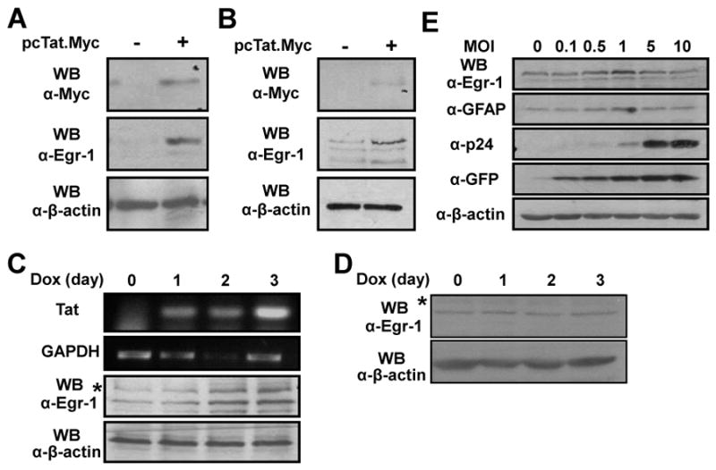
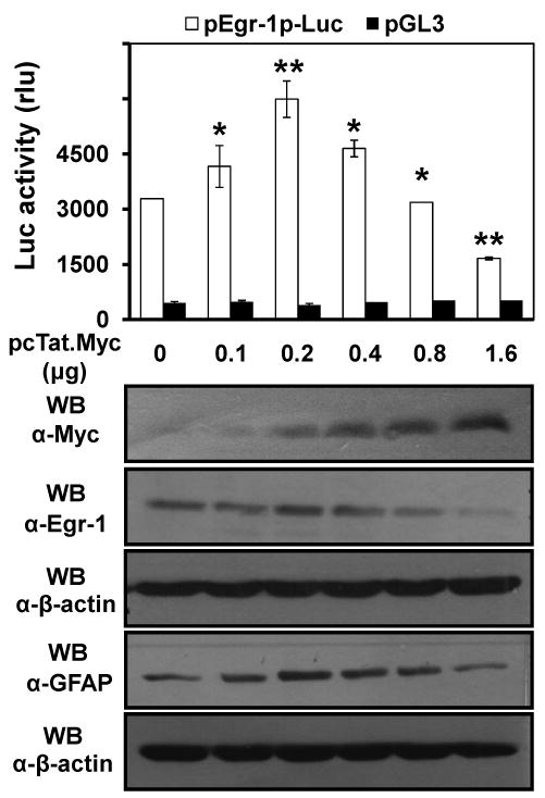
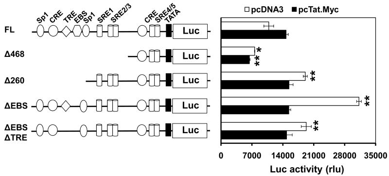
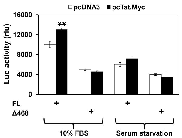
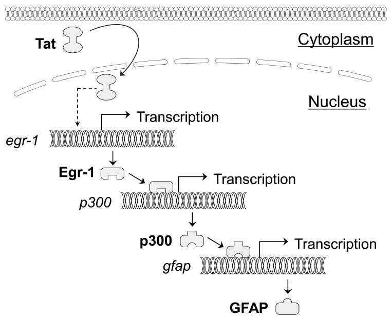
Human immunodeficiency virus type 1 (HIV-1) Tat plays an important role in HIV-associated neuropathogenesis; the underlying mechanisms are still evolving. We have recently shown that HIV-1 Tat induces expression of glial fibrillary acidic protein (GFAP), a characteristic of HIV-1 infection of the central nervous system. We have also shown that the Tat-induced GFAP expression in astrocytes is regulated by p300 and that deletion of the early growth response 1 (Egr-1) cis-transacting element within the p300 promoter abolishes Tat-induced GFAP expression. In this study, we further examined the relationship between Tat and Egr-1 in astrocytes. We found increased Egr-1 protein expression in Tat-expressing human astrocytoma cells and mouse primary astrocytes. Using the Egr-1 promoter-driven firefly luciferase reporter gene assay and the site-directed mutagenesis, we demonstrated that Tat increased Egr-1 expression by transactivating the Egr-1 promoter and involving specific serum response elements within the promoter. Consistent with these data, we showed that Tat transactivation of the Egr-1 promoter was abrogated when astrocytes were cultured in serum-reduced media. Taken together, these results reveal that Tat directly transactivates Egr-1 expression and suggest that Tat interaction with Egr-1 is probably one of the very upstream molecular events that initiate Tat-induced astrocyte dysfunction and subsequent Tat neurotoxicity.
Figures
Similar articles
-
Involvement of p300 in constitutive and HIV-1 Tat-activated expression of glial fibrillary acidic protein in astrocytes.Glia. 2010 Oct;58(13):1640-8. doi: 10.1002/glia.21038. Glia. 2010. PMID: 20578042 Free PMC article.
-
STAT3 and its phosphorylation are involved in HIV-1 Tat-induced transactivation of glial fibrillary acidic protein.Curr HIV Res. 2015;13(1):55-63. doi: 10.2174/1570162x13666150121115804. Curr HIV Res. 2015. PMID: 25613134 Free PMC article.
-
HIV-1 Tat Induces Unfolded Protein Response and Endoplasmic Reticulum Stress in Astrocytes and Causes Neurotoxicity through Glial Fibrillary Acidic Protein (GFAP) Activation and Aggregation.J Biol Chem. 2016 Oct 21;291(43):22819-22829. doi: 10.1074/jbc.M116.731828. Epub 2016 Sep 8. J Biol Chem. 2016. PMID: 27609520 Free PMC article.
-
Astrocyte activation and dysfunction and neuron death by HIV-1 Tat expression in astrocytes.Mol Cell Neurosci. 2004 Nov;27(3):296-305. doi: 10.1016/j.mcn.2004.07.003. Mol Cell Neurosci. 2004. PMID: 15519244
-
Doxycycline-inducible and astrocyte-specific HIV-1 Tat transgenic mice (iTat) as an HIV/neuroAIDS model.J Neurovirol. 2018 Apr;24(2):168-179. doi: 10.1007/s13365-017-0598-9. Epub 2017 Nov 15. J Neurovirol. 2018. PMID: 29143286 Free PMC article. Review.
Cited by
-
HIV-1 Tat Promotes Lysosomal Exocytosis in Astrocytes and Contributes to Astrocyte-mediated Tat Neurotoxicity.J Biol Chem. 2016 Oct 21;291(43):22830-22840. doi: 10.1074/jbc.M116.731836. Epub 2016 Sep 8. J Biol Chem. 2016. PMID: 27609518 Free PMC article.
-
Anti-inflammatory Function of Phyllostachys Edulis Extract in the Hippocampus of HIV-1 Transgenic Rats.J HIV AIDS. 2016 May;2(3):10.16966/2380-5536.126. doi: 10.16966/2380-5536.126. Epub 2016 May 11. J HIV AIDS. 2016. PMID: 27398410 Free PMC article.
-
Exosome-associated release, uptake, and neurotoxicity of HIV-1 Tat protein.J Neurovirol. 2016 Dec;22(6):774-788. doi: 10.1007/s13365-016-0451-6. Epub 2016 May 12. J Neurovirol. 2016. PMID: 27173397 Free PMC article.
-
Roles and functions of HIV-1 Tat protein in the CNS: an overview.Virol J. 2013 Dec 21;10:358. doi: 10.1186/1743-422X-10-358. Virol J. 2013. PMID: 24359561 Free PMC article. Review.
-
Egr-1 is a key regulator of the blood-brain barrier damage induced by meningitic Escherichia coli.Cell Commun Signal. 2024 Jan 17;22(1):44. doi: 10.1186/s12964-024-01488-y. Cell Commun Signal. 2024. PMID: 38233877 Free PMC article.
References
-
- Aicher WK, Sakamoto KM, Hack A, Eibel H. Analysis of functional elements in the human Egr-1 gene promoter. Rheumatol Int. 1999;18:207–14. - PubMed
-
- Bell JE, Anthony IC, Simmonds P. Impact of HIV on regional & cellular organisation of the brain. Curr HIV Res. 2006;4:249–57. - PubMed
-
- Bonifaci N, Sitia R, Rubartelli A. Nuclear translocation of an exogenous fusion protein containing HIV Tat requires unfolding. Aids. 1995;9:995–1000. - PubMed
-
- Bratanich AC, Liu C, McArthur JC, Fudyk T, Glass JD, Mittoo S, Klassen GA, Power C. Brain-derived HIV-1 tat sequences from AIDS patients with dementia show increased molecular heterogeneity [In Process Citation] J Neurovirol. 1998;4:387–93. - PubMed
Publication types
MeSH terms
Substances
Grants and funding
LinkOut - more resources
Full Text Sources
Other Literature Sources
Miscellaneous
