. 2010 Apr 16;141(2):315-30.
doi: 10.1016/j.cell.2010.03.029.

Reconstitution of the RIG-I pathway reveals a signaling role of unanchored polyubiquitin chains in innate immunity

Affiliations

Reconstitution of the RIG-I pathway reveals a signaling role of unanchored polyubiquitin chains in innate immunity

Wenwen Zeng et al. Cell. .

Abstract

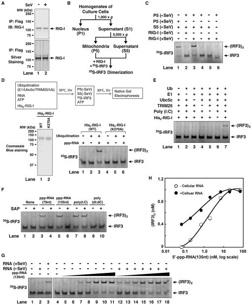
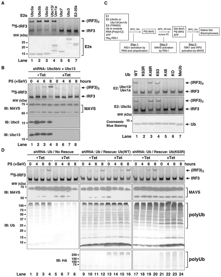
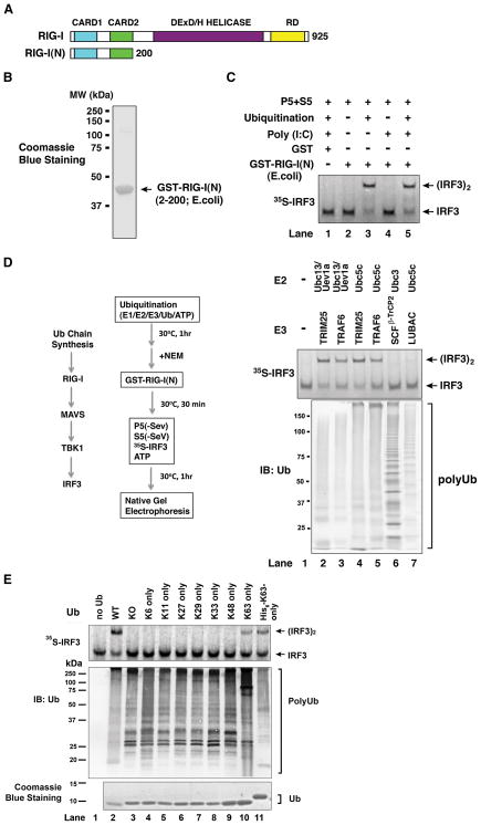
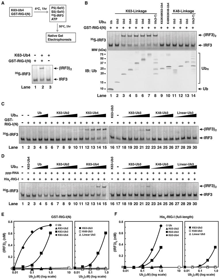
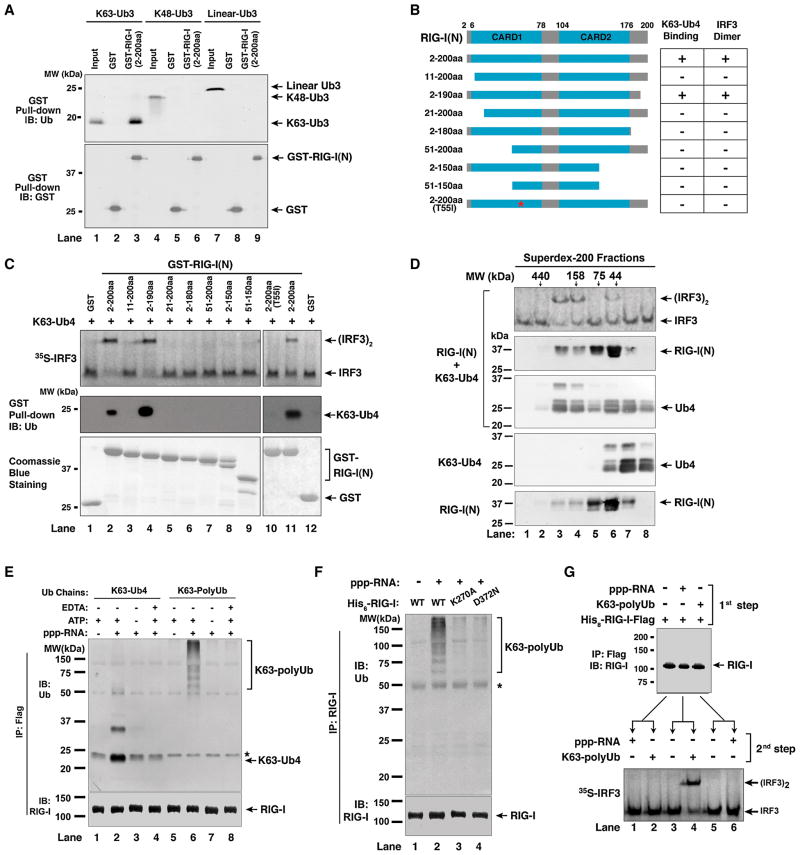
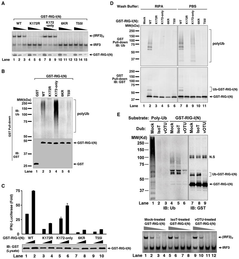
RIG-I detects invading viral RNA and activates the transcription factors NF-kappaB and IRF3 through the mitochondrial protein MAVS. Here we show that RNA bearing 5'-triphosphate strongly activates the RIG-I-IRF3 signaling cascade in a reconstituted system composed of RIG-I, mitochondria, and cytosol. Activation of RIG-I requires not only RNA but also polyubiquitin chains linked through lysine 63 (K63) of ubiquitin. RIG-I binds specifically to K63-polyubiquitin chains through its tandem CARD domains in a manner that depends on RNA and ATP. Mutations in the CARD domains that abrogate ubiquitin binding also impair RIG-I activation. Remarkably, unanchored K63-ubiquitin chains, which are not conjugated to any target protein, potently activate RIG-I. These ubiquitin chains function as an endogenous ligand of RIG-I in human cells. Our results delineate the mechanism of RIG-I activation, identify CARD domains as a ubiquitin sensor, and demonstrate that unanchored K63-polyubiquitin chains are signaling molecules in antiviral innate immunity.


Figures

Figure 1
Figure 1. In Vitro Reconstitution of the RIG-I Pathway and Regulation of RIG-I by RNA- and Ubiquitination
(A) Purification of RIG-I protein from Sendai virus-infected (+SeV) or untreated (−SeV) HEK293T cells stably expressing RIG-I containing a C-terminal Flag epitope. (B) Procedures for isolation of crude mitochondria (P5) and cytosol (S5) by differential centrifugation. (C) Virus-activated RIG-I induced IRF3 dimerization in vitro. The reconstitution reaction contained mitochondria (P5), RIG-I isolated from virus-infected or untreated cells, cytosolic extracts (S5) from uninfected cells, 35S-IRF3 and ATP. Dimerization of IRF3 was analyzed by native gel electrophoresis. (D) In vitro activation of RIG-I by 5′-pppRNA and ubiquitination. His6-tagged RIG-I (wild-type or WT) or its ATPase mutant (K270A) was purified from Sf9 cells (lower left panel), then incubated with 5′-pppRNA (79 nucleotides), ATP, and ubiquitination enzymes as outlined in the diagram. After incubation, aliquots of the reaction mixtures were further incubated with mitochondria (P5) and cytosol (S5) from uninfected cells together with 35S-IRF3 and ATP, and then IRF3 dimerization was analyzed by native gel electrophoresis. (E) In vitro activation of RIG-I by poly(I:C) and ubiquitination. Similar to (D), except that poly(I:C) was used and the dependency on ubiquitin and ubiquitination enzymes was tested. (F) The role of 5′-triphosphate for RNA to activate RIG-I in vitro. Similar to (D) except that the RNA was pretreated with or without shrimp alkaline phosphatase (SAP). (G) 5′-pppRNA and viral RNA are potent activators of the RIG-I pathway. Total RNA was extracted from HEK293T cells from viral-infected or untreated HEK293T cells, then incubated with RIG-I as in (D), followed by IRF3 dimerization assay (lanes 1–3). To measure the potency of 5′-pppRNA in RIG-I activation, increasing amounts of the RNA (135nt) (0.07 to 70 nM, at 3-fold increment) were incubated with RIG-I in the presence (lanes 12–18) or absence of cellular RNA from uninfected cells (lanes 5–11), then IRF3 dimerization assay was performed. (H) IRF3 dimer shown in (G) was quantified with ImageQuant, then plotted against the concentration of 5′-pppRNA. See also Figure S1.
Figure 2
Figure 2. K63 polyubiquitination is Essential for RIG-I Activation
(A) Ubc5 and Ubc13/Uev1a activate RIG-I in vitro. RIG-I was incubated with E1, different E2s as indicated, TRIM25, ubiquitin, RNA and ATP, followed by IRF3 dimerization assay as described in Figure 1D. The E2 proteins (2 μg) were analyzed by Coomassie Blue staining (lower panel). (B) Ubc5 and Ubc13 are required for viral activation of MAVS in the mitochondria. U2OS cells stably integrated with tetracycline-inducible shRNA against Ubc5b/c and Ubc13 were treated with or without tetracycline (Tet). After viral infection for the indicated time, mitochondrial fraction (P5) was prepared and the MAVS activity was measured by IRF3 dimerization assay. (C) K63 of ubiquitin is essential for RIG-I activation in vitro. RIG-I and IRF3 activation assays were performed using Ubc5c (upper panel) or Ubc13/Uev1a (lower panel) as the E2, and various ubiquitin mutants as indicated. KO, lysine-less mutant; MeUb, methylated ubiquitin. (D) K63 polyubiquitination is essential for viral activation of MAVS in the mitochondria. U2OS cells stably integrated with tetracycline-inducible shRNA against endogenous ubiquitin genes and a rescue expression vector for wild-type or K63R mutant of ubiquitin were grown in the presence or absence of tetracycline. After infection by Sendai virus, mitochondria (P5) were prepared to measure MAVS activation. The expression of MAVS and ubiquitin was analyzed by immunoblotting (lower panels). The HA antibody detects HA-Ub(WT) or HA-Ub(K63R) expressed from the transgene rescue vector. See also Figure S2.
Figure 3
Figure 3. Activation of RIG-I N-terminus by K63 Polyubiquitin Chains
(A) Depiction of RIG-I functional domains. (B) Purification of recombinant GST-RIG-I(N) protein from E. coli. (C) Activation of RIG-I(N) by ubiquitination. GST or GST-RIG-I(N) was incubated with ubiquitination reaction (E1, Ubc5, TRIM25 and ubiquitin) and/or poly(I:C) as indicated, then an aliquot of the reaction was further incubated with mitochondria (P5) and cytosol (S5) to measure IRF3 dimerization. (D) K63 polyUb chain synthesis, but not ubiquitination of RIG-I(N), is required for RIG-I activation in vitro. Ubiquitination reactions containing different combinations of E2s and E3s as indicated were carried out before N-ethylmaleimide (NEM) was added to inactivate E1 and E2s. An aliquot of the reaction mixture was incubated with GST-RIG-I(N), then further incubated with mitochondria (P5) and cytosol (S5) to measure IRF3 dimerization. Another aliquot of the reaction was analyzed by immunoblotting with a ubiquitin antibody (lower panel). SCFβ-TrCP2: an E3 complex containing Skp1, Cul1, Rbx1 and β-TrCP2; LUBAC: an E3 complex containing HOIL-1L and HOIP. (E) Similar to D, except that ubiquitination reactions were carried out in the presence of Ubc5c, TRIM25 and various ubiquitin mutants as indicated. See also Figure S3.
Figure 4
Figure 4. Short, Unanchored, K63-Ubiquitin Chains Potently Activate RIG-I
(A) K63-Ub4 activates RIG-I(N). GST-RIG-I(N) (0.2 μM) was incubated with or without K63-Ub4 (0.3 μM), followed by IRF3 dimerization assay as outlined. (B) K63, but not K48, ubiquitin chains activate RIG-I(N). Ubiquitin chains of defined lengths and linkages were incubated with GST-RIG-I(N), then IRF3 dimerization assay was carried out as in (A). The quality of the ubiquitin chains was evaluated by immunoblotting (lower panel) or silver staining (see Figure S4). (C) K63 ubiquitin chains potently activate RIG-I(N) in a chain length- and linkage-dependent manner. Different concentrations of Ub chains (0.01–1 μM) or ubiquitin (0.1–10 μM) were tested for RIG-I(N) activation using the IRF3 dimerization assay. (D) Similar to (C), except that full-length His6-RIG-I and 5′-pppRNA were used in lieu of RIG-I(N) in the reactions. (E & F) IRF3 dimer in C & D, respectively, was quantified using ImageQuant, then plotted against the concentration of Ub or Ub chains.
Figure 5
Figure 5. RIG-I CARD Domains Bind K63 Ubiquitin Chains and This Binding in Full-Length RIG-I is Regulated by RNA and ATP
(A) RIG-I(N) binds specifically to K63 ubiquitin chains. GST or GST-RIG-I(N) was incubated with Ub3 containing K63, K48, or linear linkage, then pulled down with glutathione-Sepharose, followed by immunoblotting. Input represents 10% of Ub3 used in the pull-down experiments. (B) Diagram of RIG-I N-terminus containing the tandem CARD domains and various deletion and point mutants. The table on the right summarizes the results in panel C. (C) Both CARD domains of RIG-I are required for polyUb binding and IRF3 activation. GST-RIG-I(N) and various mutants were incubated with K63-Ub4. 1-μl aliquot of each mixture was used for IRF3 dimerization assay (upper panel), and the remainder was pulled down with glutathione-Sepharose followed by immunoblotting with a Ub antibody (middle panel). The GST-RIG-I(N) and the mutant proteins (2 μg each) were analyzed by Coomassie Blue staining (lower panel). See also Figure S5. (D) RIG-I(N) and K63-Ub4 form active high molecular weight complex. RIG-I(N) was incubated with K63-Ub4 and then the mixture was fractionated on Superdex-200. Aliquots of the fractions were assayed for their ability to stimulate IRF3 dimerization, whereas other aliquots were subjected to immunoblotting with antibodies against RIG-I and ubiquitin, respectively. K63-Ub4 or RIG-I(N) alone was also analyzed by gel filtration on the same column (lower two panels). (E) Full-length RIG-I binds to ubiquitin chains in a manner that depends on 5′-pppRNA and ATP. His8-RIG-I-Flag was incubated with K63-Ub4 or K63-polyUb chains in the presence or absence of 5′-pppRNA (135nt), ATP or EDTA. Following immunoprecipitation with a Flag antibody, the precipitated proteins were detected with an antibody against Ub or RIG-I. The asterisk indicates a non-specific band. (F) Similar to (E), except that RIG-I ATPase mutants (K270A and D372N) were also tested for binding to K63 polyUb chains. The asterisk indicates a non-specific band. (G) Sequential binding of RIG-I to RNA and polyUb leads to IRF3 activation. His8-RIG-I-Flag was incubated with 5′-pppRNA or K63-polyUb in the first step, then immunopurified using a Flag antibody. The purified RIG-I was incubated with K63-polyUb or 5′-pppRNA in the second step, followed by IRF3 dimerization assay.
Figure 6
Figure 6. Polyubiquitin Binding is Required for RIG-I Activation
(A) Mutations at K172 and T55 impair RIG-I(N) activation in vitro. GST-RIG-I(N) and the indicated mutants were expressed and purified from E. coli, then incubated with K63-Ub4, followed by IRF3 dimerization assay (upper panel). Aliquots of the reaction mixtures were analyzed by immunoblotting with a GST antibody (lower panel). 6KR: GST-RIG-I(N) containing six K>R mutations, including K172R. K172-only: similar to 6KR except that K172 is not mutated. (B) Mutations at K172 and T55 impair polyUb binding by RIG-I(N) in vitro. GST-RIG-I(N) and mutant proteins were incubated with K63 polyUb chains, then pulled down with glutathione-Sepharose and analyzed by immunoblotting. (C) Mutations at K172 and T55 impair RIG-I(N)’s ability to induce IFN-β in cells. Different amounts (30 ng and 100 ng) of mammalian expression vectors for GST-RIG-I(N) and mutants were transfected into HEK293-IFNβ-Luc cells, then luciferase activity was measured. Error bars represent the variation range of duplicate experiments. (D) GST-RIG-I(N) and mutant proteins as described in (C) were pulled down with glutathione-Sepharose beads, washed with PBS or RIPA buffer, then analyzed by immunoblotting. (E) GST-RIG-I(N) was expressed in HEK293T cells, then purified using glutathione-Sepharose. The purified protein was treated with isopeptidase T (IsoT) or viral OTU (vOTU) or mock-treated, then immunoblotted with an antibody against ubiquitin or GST (upper panel; N.S indicates a non-specific band). Aliquots of the treated GST-RIG-I(N) were tested for its ability to promote IRF3 dimerization in the reconstitution assay (lower panel). See also Figure S6.
Figure 7
Figure 7. Endogenous Unanchored K63 Polyubiquitin Chains Activate the RIG-I Pathway
(A) A protocol for isolating functional endogenous polyUb chains in human cells. (B) Endogenous polyUb chains bound to RIG-I can be released by heat treatment and remained functional. GST-RIG-I(N)-K172-only was expressed in HEK293T cells and isolated as described in (A), then heated for 5 minutes at the indicated temperatures. After centrifugation, the supernatant containing heat-resistant ubiquitin chains was incubated with GST-RIG-I(N) expressed and purified from E. coli (lanes 7–12). The activity of GST-RIG-I(N) was then measured by IRF3 dimerization assay. As positive controls, K63 polyUb chains were incubated with GST-RIG-I(N)-K172-only, which was then pulled down and heated in parallel experiments (lanes 1–6). endo. polyUb: endogenous polyUb. (C) Endogenous unanchored polyUb chains activate RIG-I(N). PolyUb chains associated with GST-RIG-I(N)-K172-only were captured and released at 75°C as in (B). The heat-resistant supernatant was incubated with GST-RIG-I(N) followed by IsoT treatment (lane 9), or in reverse order (lane 8). As positive controls, unanchored K63 polyUb chains were incubated with GST-RIG-I(N) and IsoT in sequential orders as indicated. In the right panel, the heat supernatant containing endogenous polyUb from HEK293T cells was incubated with or without IsoT, then analyzed by immunoblotting with a ubiquitin antibody. The arrow denotes a ~40 kDa band that is likely K63-Ub6 (see Figure S7B). (D) Similar to (C), except that the supernatant containing endogenous polyUb chains were treated with CYLD. The ubiquitin chains were detected with a ubiquitin antibody or another antibody specific for the K63 linkage of ubiquitin chains. (E) siRNA oligos targeting GFP (control), TRIM25 or CYLD were transfected into HEK293T cells, which were subsequently transfected with an expression vector encoding GST-RIG-I(N)-K172-only. Endogenous polyUb chains associated with the GST-RIG-I(N) protein were isolated as described in (A), then tested in IRF3 dimerization assay and visualized by immunoblotting with a ubiquitin antibody. The efficiency of RNAi was also confirmed by immunoblotting. (F) Potent activation of RIG-I by endogenous polyUb chains. Different amounts of heat-resistant supernatant containing endogenous polyUb were incubated with GST-RIG-I(N) to measure IRF3 dimerization. The concentration of the ubiquitin chains was estimated by semi-quantitative immunoblotting (Figure S7B). Error bars represent the variation range of duplicate experiments. (G) A proposed mechanism of RIG-I activation by RNA and polyUb (see Results and Discussion).

Comment in

  • Ubiquitin gets CARDed.
    Bennett EJ, Harper JW. Bennett EJ, et al. Cell. 2010 Apr 16;141(2):220-2. doi: 10.1016/j.cell.2010.03.047. Cell. 2010. PMID: 20403317
  • Innate immunity: A chain reaction.
    Minton K. Minton K. Nat Rev Immunol. 2010 Jun;10(6):385. doi: 10.1038/nri2783. Nat Rev Immunol. 2010. PMID: 20514677 No abstract available.

Similar articles

Cited by

References

    1. Akira S, Uematsu S, Takeuchi O. Pathogen recognition and innate immunity. Cell. 2006;124:783–801. - PubMed
    1. Bhoj VG, Chen ZJ. Ubiquitylation in innate and adaptive immunity. Nature. 2009;458:430–437. - PubMed
    1. Cui S, Eisenacher K, Kirchhofer A, Brzozka K, Lammens A, Lammens K, Fujita T, Conzelmann KK, Krug A, Hopfner KP. The C-terminal regulatory domain is the RNA 5′-triphosphate sensor of RIG-I. Mol Cell. 2008;29:169–179. - PubMed
    1. Deng L, Wang C, Spencer E, Yang L, Braun A, You J, Slaughter C, Pickart C, Chen ZJ. Activation of the IkappaB kinase complex by TRAF6 requires a dimeric ubiquitin-conjugating enzyme complex and a unique polyubiquitin chain. Cell. 2000;103:351–361. - PubMed
    1. Frias-Staheli N, Giannakopoulos NV, Kikkert M, Taylor SL, Bridgen A, Paragas J, Richt JA, Rowland RR, Schmaljohn CS, Lenschow DJ, et al. Ovarian tumor domain-containing viral proteases evade ubiquitin- and ISG15-dependent innate immune responses. Cell Host Microbe. 2007;2:404–416. - PMC - PubMed

Publication types

MeSH terms

Substances