. 2010 Jun 17;29(24):3509-18.
doi: 10.1038/onc.2010.108. Epub 2010 Apr 19.

Role of SUMO:SIM-mediated protein-protein interaction in non-homologous end joining

Affiliations

Role of SUMO:SIM-mediated protein-protein interaction in non-homologous end joining

Y-J Li et al. Oncogene. .

Abstract

Although post-translational modifications by the small ubiquitin-like modifiers (SUMO) are known to be important in DNA damage response, it is unclear whether they have a role in double-strand break (DSB) repair by non-homologous end joining (NHEJ). Here, we analyzed various DSB repair pathways upon inhibition of SUMO-mediated protein-protein interactions using peptides that contain the SUMO-interaction motif (SIM) and discriminate between mono- and SUMO-chain modifications. The SIM peptides specifically inhibit NHEJ as shown by in vivo repair assays and radio-sensitivity of cell lines deficient in different DSB repair pathways. Furthermore, mono-SUMO, instead of SUMO-chain, modifications appear to be involved in NHEJ. Immunoprecipitation experiments also showed that the SIM peptide interacted with SUMOylated Ku70 after radiation. This study is the first to show an important role for SUMO:SIM-mediated protein-protein interactions in NHEJ, and provides a mechanistic basis for the role of SIM peptide in sensitizing genotoxic stress of cancer cells.


Figures
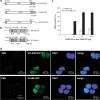
Figure 1
Figure 1
The design of the SIM peptides and their interaction with SUMOylated proteins in cells. (a) A schematic representation of the cDNA fragments inserted into pcDNA3.1 for production of WT (WT-SIM-GFP), SUMO binding-deficient (VI-SIM-GFP) and a deletion mutant (Del-SIM-GFP) of SIM. The peptides are fused with two FLAG-tag repeats, GFP and a nuclear localization sequence. (b) A schematic representation of the construction of the double-SIM peptide (WT-SIM-2r) and SC -SIM control (SC-SIM-2r). The SIM or SC-SIM sequences are separated by the linker (RSPSPPVETSISSTN) and fused with a FLAG tag and a nuclear localization signal. (c) Preferential binding of the double-SIM peptide to poly-SUMO-2/3 chains. GST-fusion WT-SIM-2r was used to pull down poly-SUMO-2/3 (■) or mono-SUMO2/3 (□) at the indicated concentration. The measurement was normalized as 100% (poly-SUMO2/3 at 50 and 100 ng/ml) and control (0 ng/ml) defined as 0%. (d) Localization of WT-SIM-GFP and VI-SIM-GFP (green) in MCF7 cells. PML (red) was visualized by immunocytochemistry and nuclei (blue) were stained by DAPI. DAPI, 4′,6-diamidino-2-phenylindole; GFP, green fluorescent protein; GST, glutathione-S-transferase; PML, SC, scrambled; SIM, SUMO-interaction motif; SUMO, small ubiquitin-like modifiers; WT, wild type.
Figure 2
Figure 2
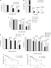
The SIM peptide sensitizes MCF-7 cells to the chemotherapeutic drugs Dox and CPT, and to radiation. (a, b) Viability of MCF-7 cells that stably expressed WT-SIM-GFP and Del-SIM-GFP upon Dox (a) and CPT (b) treatment. The cells were treated with the indicated Dox and CPT concentrations and cell viability, measured by MTS assay, was detected 48 h after treatment. (c) Comparison of the effect of a single-SIM and double-SIM constructs on MCF-7 cell sensitivity to Dox. Cell viability was measured by MTS assay. (d) Cell viability of MCF-7 cell lines stably expressing WT-SIM-GFP and Del-SIM-GFP upon irradiation at the indicated dosages. Cell viability, measured by MTS assay, was detected 48 h after radiation. (e) Comparison of the effect of a single-SIM and double-SIM constructs on MCF-7 cell sensitivity to radiation. Cell viability was measured by MTS assay. Clonogenic assays of cells stably expressing WT-SIM-GFP or the deletion control, Del-SIM-GFP (f), and WT-SIM-2r or the control, SC-SIM-2r (g), after irradiation at the indicated doses. Cell colonies were visualized by crystal violet and counted on day 14 after irradiation. The error bars represent the standard error of the mean of triplicate experiments. P-values are indicated. CPT, camptothecin; Dox, doxorubicin; GFP, green fluorescent protein; SC, scrambled; SIM, SUMO-interaction motif; WT, wild type.
Figure 3
Figure 3
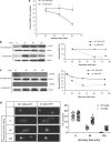
The SIM peptide inhibits DNA repair. (a) γ-H2AX foci formation after irradiation. The graph shows the number of γ-H2AX foci in cells expressing WT-SIM-GFP or VI-SIM-GFP after irradiation (4 Gy) and a 30- to 90-min recovery period. The γ-H2AX foci numbers represent those counted from an average of 40 cells. (b, c) SIM peptide delays γ-H2AX de-phosphorylation in MCF-7 cells after γ-irradiation (4 Gy) (b) or 2 μM Dox treatment (c). γ-H2AX levels were measured at the indicated time points after treatment. γ-H2AX was detected and normalized with H2AX by western blotting and quantified. The western blot images are shown on the left and the quantifications are shown on the right. (d) Comet assays were used to measure the amount of unrepaired DNA damage in cells expressing WT-SIM-GFP and VI-SIM-GFP after γ-radiation (4 Gy) and recovery for 0, 30 or 120 min. Non-irradiated cells were used as controls (−). Representative images are shown on the left. Tail moments (% DNA in the tail × tail length), measured using Cometscore (TriTek), are shown on the left. Every spot represents a single cell, and 30 comet images were measured for each treatment. Dox, doxorubicin; GFP, green fluorescent protein; SIM, SUMO-interaction motif; WT, wild type.
Figure 4
Figure 4
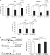
The SIM peptide inhibits NHEJ. (a) The SIM peptide sensitizes WT and BRCA1-deficient (BRCA1−/−) mouse ES cell lines, but not a Ku-deficient (Ku70−/−) cell line, to radiation. Cells were transfected with WT-SIM-2r, SC-SIM-2r or a vector cDNA, and irradiated (10 Gy) or not irradiated. Cell viability was detected 72 h after treatment and normalized to cells transfected with an empty expression vector. (b, c) Schematic representations of fluorescence-based assays for measuring HR or NHEJ DSB repair. (b) After DSBs are generated by I-SceI in cells carrying the DR-GFP reporter, HR uses the downstream GFP sequence (truncated GFP) as a template, which restores the coding sequence of functional GFP. The panel on the right shows the results of repair of DR-GFP in GFP-positive cells from mouse ES cells co-transfected with an I-Sce I expression vector and one of the following: WT-SIM-2R, SC-SIM-2r or expression vector (JS74). (c) EJ5-GFP contains a promoter that is separated from a GFP coding region by a puro gene that is separated by two I-SceI sites. Once DSBs are generated by I-SceI and the puro gene is excised by NHEJ repair, the promoter is joined to the rest of the expression region, leading to restoration of functional GFP. The graph on the upper right panel shows the frequency of repair of EJ5-GFP (resulting in GFP-positive cells) in WT and Ku70−/− ES cells co-transfected with an I-SceI expression vector and one of the following: WT-SIM-2r, SC-SIM-2r or vector control. P-values are indicated. The graph on the lower right panel shows the comparison of the effect of a single-SIM and double-SIM constructs on DNA DSB repair by NHEJ using a GFP-based chromosomal reporter in WT ES cells. (d) The WT-SIM-GFP (WT) or the VI-SIM-GFP (VI) mutant control peptide was immunoprecipitated by an anti-FLAG antibody, followed by western blotting with an anti-Ku70 antibody. A band corresponding to the molecular weight of SUMOylated Ku70 was repeatedly identified. The lower panel shows 10% of the input for immunoprecipitation, stained with Coomassie blue. DSB, double-strand break; ES, embryonic stem; GFP, green fluorescent protein; HR, homologous recombination; NHEJ, non-homologous end joining; SC, scrambled; SIM, SUMO-interaction motif; SUMO, small ubiquitin-like modifiers; WT, wild type.
Figure 4
Figure 4
The SIM peptide inhibits NHEJ. (a) The SIM peptide sensitizes WT and BRCA1-deficient (BRCA1−/−) mouse ES cell lines, but not a Ku-deficient (Ku70−/−) cell line, to radiation. Cells were transfected with WT-SIM-2r, SC-SIM-2r or a vector cDNA, and irradiated (10 Gy) or not irradiated. Cell viability was detected 72 h after treatment and normalized to cells transfected with an empty expression vector. (b, c) Schematic representations of fluorescence-based assays for measuring HR or NHEJ DSB repair. (b) After DSBs are generated by I-SceI in cells carrying the DR-GFP reporter, HR uses the downstream GFP sequence (truncated GFP) as a template, which restores the coding sequence of functional GFP. The panel on the right shows the results of repair of DR-GFP in GFP-positive cells from mouse ES cells co-transfected with an I-Sce I expression vector and one of the following: WT-SIM-2R, SC-SIM-2r or expression vector (JS74). (c) EJ5-GFP contains a promoter that is separated from a GFP coding region by a puro gene that is separated by two I-SceI sites. Once DSBs are generated by I-SceI and the puro gene is excised by NHEJ repair, the promoter is joined to the rest of the expression region, leading to restoration of functional GFP. The graph on the upper right panel shows the frequency of repair of EJ5-GFP (resulting in GFP-positive cells) in WT and Ku70−/− ES cells co-transfected with an I-SceI expression vector and one of the following: WT-SIM-2r, SC-SIM-2r or vector control. P-values are indicated. The graph on the lower right panel shows the comparison of the effect of a single-SIM and double-SIM constructs on DNA DSB repair by NHEJ using a GFP-based chromosomal reporter in WT ES cells. (d) The WT-SIM-GFP (WT) or the VI-SIM-GFP (VI) mutant control peptide was immunoprecipitated by an anti-FLAG antibody, followed by western blotting with an anti-Ku70 antibody. A band corresponding to the molecular weight of SUMOylated Ku70 was repeatedly identified. The lower panel shows 10% of the input for immunoprecipitation, stained with Coomassie blue. DSB, double-strand break; ES, embryonic stem; GFP, green fluorescent protein; HR, homologous recombination; NHEJ, non-homologous end joining; SC, scrambled; SIM, SUMO-interaction motif; SUMO, small ubiquitin-like modifiers; WT, wild type.

Similar articles

Cited by

References

    1. Andrews EA, Palecek J, Sergeant J, Taylor E, Lehmann AR, Watts FZ. Nse2, a component of the Smc5-6 complex, is a SUMO ligase required for the response to DNA damage. Mol Cell Biol. 2005;25:185–196. - PMC - PubMed
    1. Bennardo N, Cheng A, Huang N, Stark JM. Alternative-NHEJ is a mechanistically distinct pathway of mammalian chromosome break repair. PLoS Genet. 2008;4:e1000110. - PMC - PubMed
    1. Bird AW, Yu DY, Pray-Grant MG, Qiu Q, Harmon KE, Megee PC, et al. Acetylation of histone H4 by Esa1 is required for DNA double-strand break repair. Nature. 2002;419:411–415. - PubMed
    1. Carson JP, Zhang N, Frampton GM, Gerry NP, Lenburg ME, Christman MF. Pharmacogenomic identification of targets for adjuvant therapy with the topoisomerase poison camptothecin. Cancer Res. 2004;64:2096–2104. - PubMed
    1. Franken NA, Rodermond HM, Stap J, Haveman J, van Bree C. Clonogenic assay of cells in vitro. Nat Protoc. 2006;1:2315–2319. - PubMed

Publication types