. 2010 Jun 11;285(24):18283-90.
doi: 10.1074/jbc.M109.079137. Epub 2010 Apr 16.

LKB1 suppresses p21-activated kinase-1 (PAK1) by phosphorylation of Thr109 in the p21-binding domain

Affiliations

LKB1 suppresses p21-activated kinase-1 (PAK1) by phosphorylation of Thr109 in the p21-binding domain

Atsuko Deguchi et al. J Biol Chem. .

Abstract

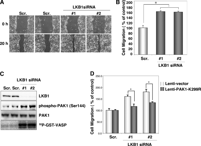
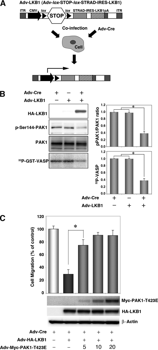
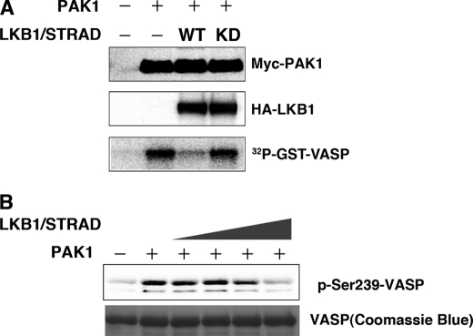
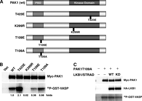
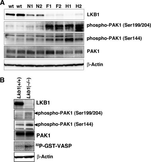
The serine/threonine protein kinase LKB1 is a tumor suppressor gene mutated in Peutz-Jeghers syndrome patients. The mutations are found also in several types of sporadic cancer. Although LKB1 is implicated in suppression of cell growth and metastasis, the detailed mechanisms have not yet been elucidated. In this study, we investigated the effect of LKB1 on cell motility, whose acquisition occurs in early metastasis. The knockdown of LKB1 enhanced cell migration and PAK1 activity in human colon cancer HCT116 cells, whereas forced expression of LKB1 in Lkb1-null mouse embryonic fibroblasts suppressed PAK1 activity and PAK1-mediated cell migration simultaneously. Notably, LKB1 directly phosphorylated PAK1 at Thr(109) in the p21-binding domain in vitro. The phosphomimetic T109E mutant showed significantly lower protein kinase activity than wild-type PAK1, suggesting that the phosphorylation at Thr(109) by LKB1 was responsible for suppression of PAK1. Consistently, the nonphosphorylatable T109A mutant was resistant to suppression by LKB1. Furthermore, we found that PAK1 was activated in the hepatocellular carcinomas and the precancerous liver lesions of Lkb1(+/-) mice. Taken together, these results suggest that PAK1 is a direct downstream target of LKB1 and plays an essential role in LKB1-induced suppression of cell migration.


Figures

FIGURE 1.
FIGURE 1.
Knockdown of endogenous LKB1 increases cell migration and PAK1 activity in HCT116 cells. A, knockdown of LKB1 increases cell migration. HCT116 cells were transfected with two different specific siRNAs against human LKB1. Control cells were transfected with a scrambled (Scr.) siRNA pool. After 24 h of plating, scratches were made with 10-μl pipette tips. Photographs were taken at 0 and 20 h after the wound was made. B, cell migration was normalized so that 100% represented migration distance of control cells. Error bars indicate S.D. The asterisk indicates significant increases compared with control cells (p < 0.001). C, cellular levels of phospho-PAK1 (Ser144), total PAK1 and in vitro PAK1 activity in LKB1 knockdown HCT116 cells. Lysates were prepared from HCT116 cells transfected with the indicated siRNAs. The levels of phospho-PAK1 (Ser144) and PAK1 were determined by Western blotting with the indicated antibodies. Lysates were prepared from HCT116 cells transfected with the indicated siRNAs and immunoprecipitated using an anti-PAK1 antibody. The precipitates were used for in vitro kinase assays using GST-VASP-(158–277) as a substrate. The phosphorylation of GST-VASP was visualized using BAS-5000 Bio-imaging Analyzer. D, induction of PAK1-K299R suppressed the increase in migration in LKB1 knockdown cells. The cells transfected with the indicated siRNAs were then infected with lentivirus lenti-PAK1-K299R or lenti-vector. At 24 h post-infection, cultures were scratches were made with 10-μl pipette tips. Photographs were taken at 0 and 20 h thereafter. Cell migration was normalized so that 100% represented the migration distance of control cells. Error bars indicate S.D. The asterisk represents significant increases compared with control cells (p < 0.001).
FIGURE 2.
FIGURE 2.
Expression of LKB1 in the Lkb1-null MEF cell line (MEF3-2) inhibits cell migration and PAK1 activation. A, schematic structure of the adenoviral construct, adapted from Ref. . ITR, inverted terminal repeat; CMV, human cytomegalovirus immediate early promoter; lox, loxP site sequence; STOP; transcriptional termination cassette; IRES, internal ribosome entry site; pA, polyadenylation signal. Co-infection with Adv-Cre removed the STOP cassette from Adv-LKB1. B, cellular levels of PAK1 autophosphorylation, total PAK1, and in vitro PAK1 activity in LKB1-expressing MEF3-2 cells. MEF 3-2 cells were co-infected with recombinant adenoviruses Adv-Cre and Adv-LKB1. Control cells were infected with Adv-Cre alone. The levels of phospho-PAK1 and PAK1 were determined by Western blotting with anti-phospho-PAK1 (Ser144)/PAK2 (Ser141) and anti-PAK1 antibodies, respectively. Lysates were prepared from MEF3-2 cells infected with the indicated recombinant adenoviruses and immunoprecipitated using an anti-PAK1 antibody. The precipitates were used for in vitro kinase assays with GST-VASP-(158–277) as a substrate. The phosphorylation of GST-VASP was visualized and quantified using BAS-5000 Bio-imaging Analyzer. Error bars indicate S.D. The asterisk represents significant decreases compared with control cells (p < 0.001). C, the constitutively active mutant of PAK1 rescues LKB1-induced suppression of cell migration in MEF3-2 cells. MEF 3-2 cells were co-infected with recombinant adenoviruses Adv-Cre, Adv-LKB1, and Adv-PAK1-T423E at a multiplicity of infection of 5, 10, or 20. Control cells were infected with Adv-Cre alone. Cell migration assays were performed as Fig. 1. Error bars indicate S.D. The asterisk represents significant changes in Adv-Cre/Adv-LKB1/Adv-PAK1-T423E (5) infected cells compared with Adv-Cre/Adv-LKB1-infected cells (p < 0.001). Similar results were obtained in three independent experiments.
FIGURE 3.
FIGURE 3.
LKB1 activity is necessary for suppression of PAK1 activity. A, the wt LKB1, but not the kinase-dead mutant (KD), inhibits PAK1 activity. HEK293T cells were co-transfected with the indicated constructs. At 24 h post-transfection, immunoprecipitates were prepared with an anti-Myc antibody. The precipitates were used for in vitro kinase assays using GST-VASP-(158–277) as a substrate. The kinase activities were visualized using BAS-5000 Bio-imaging Analyzer. Cellular levels of Myc-PAK1 and HA-LKB1 were determined by Western blotting with anti-Myc or anti-HA antibody. B, recombinant LKB1 complex (LKB1-STRAD-MO25) directly inhibits PAK1 activity in vitro in a dose-dependent manner. The LKB1 complex (LKB1-STRAD-MO25) was preincubated with recombinant PAK1 and ATP in the absence of any PAK1 substrates, followed by addition of GST-VASP. The phosphorylated VASP was detected using an anti-phospho-VASP (Ser239) antibody. Coomassie Brilliant Blue was used for detection of GST-VASP-(158–277). Similar results were obtained in three independent experiments.
FIGURE 4.
FIGURE 4.
LKB1 phosphorylates PAK1 in vitro at Thr109. A, schematic presentation of PAK1 and its mutant forms. wt PAK1 contains the p21-binding domain (light gray) and the kinase domain (dark gray). The noncatalytic domains are in white. Thr109 is a possible phosphorylation site for LKB1. B, LKB1 phosphorylates GST-PAK1-PBD at Thr109 in vitro. The recombinant LKB1 complex (LKB1-STRAD-MO25) was incubated with GST-PBD or GST-PBD-T109A. C, LKB1 phosphorylates full-length GST-PAK1-K299R and GST-PAK1-T109A/K299R to a lesser extent. The in vitro kinase assays were performed using the recombinant LKB1 complex with GST-PAK1-K299R or GST-PAK1-T109A/K299R. The phosphorylation of the indicated recombinant proteins was visualized using BAS-5000 Bio-imaging Analyzer. These experiments were repeated at least twice with similar results.
FIGURE 5.
FIGURE 5.
Phosphorylation of PAK1 at Thr109 reduces its activity. A, structures of PAK1 constructs. wt PAK1 contains the p21-binding domain (light gray) and the kinase domain (dark gray). The noncatalytic domains are in white. Also displayed are the constitutively active mutant (T423E), dominant-negative mutant (K299R), phosphorylation-mimicked mutant for Thr109 (T109E), and the nonphosphorylatable mutant for Thr109 (T109A). B, the kinase activities of PAK1 mutants. The HEK293T cells were co-transfected with the indicated constructs. At 24 h post-transfection, the immunoprecipitates were prepared with an anti-Myc antibody. The precipitates were used for in vitro kinase assays using GST-VASP-(158–277) as a substrate. Cellular levels of Myc-PAK1 were determined by Western blotting with anti-Myc antibody. C, LKB1 (wt or kinase-dead (KD)) failed to inhibit the activity of the PAK1-T109A mutant. Cultured HEK293T cells were co-transfected with the indicated constructs. The kinase activities were determined as described under “Experimental Procedures.” Cellular levels of Myc-PAK1 and HA-LKB1 were determined by Western blotting with anti-Myc or anti-HA antibody. Similar results were obtained in three independent experiments. Vec, vector.
FIGURE 6.
FIGURE 6.
Activation of PAK1 in Lkb1(+/−) mouse HCCs and Lkb1(−/−) MEFs. A, activation of PAK1 in Lkb1(+/−) mice HCCs. Cellular levels of phospho-PAK1, PAK1, and β-actin in tissue lysates were determined by Western blotting with anti-phospho-PAK1 (Ser144), anti-phospho-PAK1 (Ser199/204), anti-PAK1, and anti-β-actin. Two wild-type livers (W1 and W2), pairs of tumor (H1 and H2), precancerous lesions (F1 and F2) and adjacent normal tissue (N1 and N2) from two Lkb1(+/−) mice were analyzed. B, increased PAK1 activity in Lkb1(−/−) MEFs. Cellular levels of phospho-PAK1, PAK1, and β-actin in tissue lysates were determined by Western blotting with the indicated antibodies. For in vitro kinase assay, cell lysates were prepared from MEF cells and immunoprecipitated using an anti-PAK1 antibody. The precipitates were used for in vitro kinase assays using GST-VASP-(158–277) as a substrate. The phosphorylation of GST-VASP was visualized using BAS-5000 Bio-imaging Analyzer. Similar results were obtained in three independent experiments.

Similar articles

Cited by

References

    1. Giardiello F. M., Welsh S. B., Hamilton S. R., Offerhaus G. J., Gittelsohn A. M., Booker S. V., Krush A. J., Yardley J. H., Luk G. D. (1987) N. Engl. J. Med. 316, 1511–1514 - PubMed
    1. Hemminki A., Markie D., Tomlinson I., Avizienyte E., Roth S., Loukola A., Bignell G., Warren W., Aminoff M., Höglund P., Järvinen H., Kristo P., Pelin K., Ridanpää M., Salovaara R., Toro T., Bodmer W., Olschwang S., Olsen A. S., Stratton M. R., de la Chapelle A., Aaltonen L. A. (1998) Nature 391, 184–187 - PubMed
    1. Jenne D. E., Reimann H., Nezu J., Friedel W., Loff S., Jeschke R., Müller O., Back W., Zimmer M. (1998) Nat. Genet. 18, 38–43 - PubMed
    1. Launonen V. (2005) Hum. Mutat. 26, 291–297 - PubMed
    1. Alessi D. R., Sakamoto K., Bayascas J. R. (2006) Annu. Rev. Biochem. 75, 137–163 - PubMed

Publication types