

Myosin binding protein-C slow: an intricate subfamily of proteins
- PMID: 20396395
- PMCID: PMC2852610
- DOI: 10.1155/2010/652065
Myosin binding protein-C slow: an intricate subfamily of proteins
Abstract
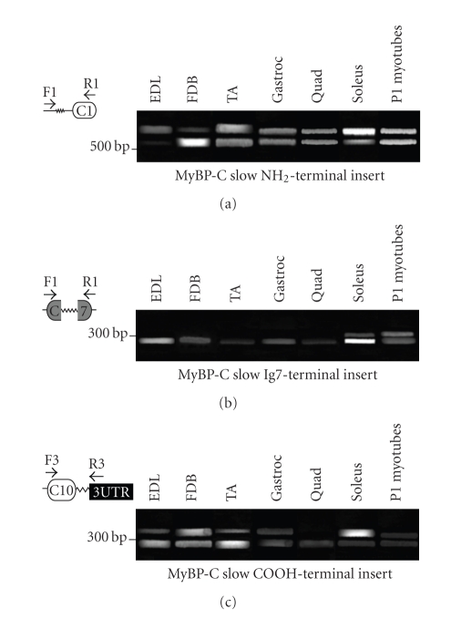
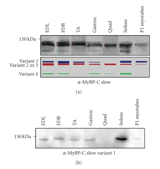
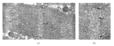
Myosin binding protein C (MyBP-C) consists of a family of thick filament associated proteins. Three isoforms of MyBP-C exist in striated muscles: cardiac, slow skeletal, and fast skeletal. To date, most studies have focused on the cardiac form, due to its direct involvement in the development of hypertrophic cardiomyopathy. Here we focus on the slow skeletal form, discuss past and current literature, and present evidence to support that: (i) MyBP-C slow comprises a subfamily of four proteins, resulting from complex alternative shuffling of the single MyBP-C slow gene, (ii) the four MyBP-C slow isoforms are expressed in variable amounts in different skeletal muscles, (iii) at least one MyBP-C slow isoform is preferentially found at the periphery of M-bands and (iv) the MyBP-C slow subfamily may play important roles in the assembly and stabilization of sarcomeric M- and A-bands and regulate the contractile properties of the actomyosin filaments.
Figures

Similar articles
-
Obscurin interacts with a novel isoform of MyBP-C slow at the periphery of the sarcomeric M-band and regulates thick filament assembly.Mol Biol Cell. 2009 Jun;20(12):2963-78. doi: 10.1091/mbc.e08-12-1251. Epub 2009 Apr 29. Mol Biol Cell. 2009. PMID: 19403693 Free PMC article.
-
Skeletal MyBP-C isoforms tune the molecular contractility of divergent skeletal muscle systems.Proc Natl Acad Sci U S A. 2019 Oct 22;116(43):21882-21892. doi: 10.1073/pnas.1910549116. Epub 2019 Oct 7. Proc Natl Acad Sci U S A. 2019. PMID: 31591218 Free PMC article.
-
Myosin binding protein-C: a regulator of actomyosin interaction in striated muscle.J Biomed Biotechnol. 2011;2011:636403. doi: 10.1155/2011/636403. Epub 2011 Oct 16. J Biomed Biotechnol. 2011. PMID: 22028592 Free PMC article. Review.
-
COOH-terminal truncated human cardiac MyBP-C alters myosin filament organization.C R Acad Sci III. 2001 Mar;324(3):251-60. doi: 10.1016/s0764-4469(00)01292-0. C R Acad Sci III. 2001. PMID: 11291312
-
Cardiac myosin binding protein C.Circ Res. 1999 May 28;84(10):1117-26. doi: 10.1161/01.res.84.10.1117. Circ Res. 1999. PMID: 10347086 Review.
Cited by
-
Thick Filament Protein Network, Functions, and Disease Association.Compr Physiol. 2018 Mar 13;8(2):631-709. doi: 10.1002/cphy.c170023. Compr Physiol. 2018. PMID: 29687901 Free PMC article. Review.
-
Myosin Binding Protein-C Forms Amyloid-Like Aggregates In Vitro.Int J Mol Sci. 2021 Jan 13;22(2):731. doi: 10.3390/ijms22020731. Int J Mol Sci. 2021. PMID: 33450960 Free PMC article.
-
Etiology of genetic muscle disorders induced by mutations in fast and slow skeletal MyBP-C paralogs.Exp Mol Med. 2023 Mar;55(3):502-509. doi: 10.1038/s12276-023-00953-x. Epub 2023 Mar 1. Exp Mol Med. 2023. PMID: 36854776 Free PMC article. Review.
-
The sarcomeric M-region: a molecular command center for diverse cellular processes.Biomed Res Int. 2015;2015:714197. doi: 10.1155/2015/714197. Epub 2015 Apr 15. Biomed Res Int. 2015. PMID: 25961035 Free PMC article. Review.
-
Myosin binding protein-C slow: a multifaceted family of proteins with a complex expression profile in fast and slow twitch skeletal muscles.Front Physiol. 2013 Dec 25;4:391. doi: 10.3389/fphys.2013.00391. eCollection 2013. Front Physiol. 2013. PMID: 24399972 Free PMC article.
References
-
- Clark KA, McElhinny AS, Beckerle MC, Gregorio CC. Striated muscle cytoarchitecture: an intricate web of form and function. Annual Review of Cell and Developmental Biology. 2002;18:637–706. - PubMed
-
- Franzini-Armstrong C. Functional significance of membrane architecture in skeletal and cardiac muscle. Society of General Physiologists series. 1996;51:3–18. - PubMed
-
- Oakley CE, Chamoun J, Brown LJ, Hambly BD. Myosin binding protein-C: enigmatic regulator of cardiac contraction. International Journal of Biochemistry and Cell Biology. 2007;39(12):2161–2166. - PubMed
-
- Starr R, Offer G. Polypeptide chains of intermediate molecular weight in myosin preparations. FEBS Letters. 1971;15(1):40–44. - PubMed
Publication types
MeSH terms
Substances
Grants and funding
LinkOut - more resources
Full Text Sources
Medical
Miscellaneous
