. 2010 Jul;84(13):6515-26.
doi: 10.1128/JVI.02722-09. Epub 2010 Apr 14.

Caveolin-1 modulates HIV-1 envelope-induced bystander apoptosis through gp41

Affiliations

Caveolin-1 modulates HIV-1 envelope-induced bystander apoptosis through gp41

Xiao Mei Wang et al. J Virol. 2010 Jul.

Abstract

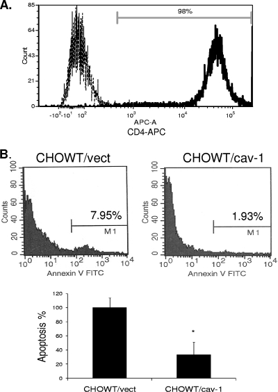
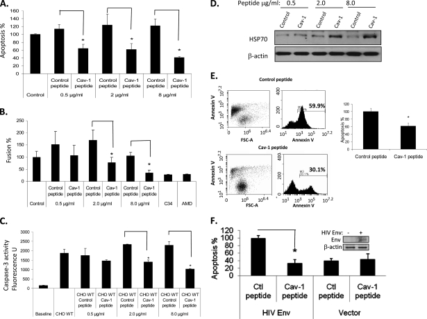
Human immunodeficiency virus (HIV) envelope (Env)-mediated bystander apoptosis is known to cause the progressive, severe, and irreversible loss of CD4(+) T cells in HIV-1-infected patients. Env-induced bystander apoptosis has been shown to be gp41 dependent and related to the membrane hemifusion between envelope-expressing cells and target cells. Caveolin-1 (Cav-1), the scaffold protein of specific membrane lipid rafts called caveolae, has been reported to interact with gp41. However, the underlying pathological or physiological meaning of this robust interaction remains unclear. In this report, we examine the interaction of cellular Cav-1 and HIV gp41 within the lipid rafts and show that Cav-1 modulates Env-induced bystander apoptosis through interactions with gp41 in SupT1 cells and CD4(+) T lymphocytes isolated from human peripheral blood. Cav-1 significantly suppressed Env-induced membrane hemifusion and caspase-3 activation and augmented Hsp70 upregulation. Moreover, a peptide containing the Cav-1 scaffold domain sequence markedly inhibited bystander apoptosis and apoptotic signal pathways. Our studies shed new light on the potential role of Cav-1 in limiting HIV pathogenesis and the development of a novel therapeutic strategy in treating HIV-1-infected patients.


Figures

FIG. 1.
FIG. 1.
Interaction of Cav-1 and gp41. (A) Cav-1 and V5-gp41 from NL4-3 (lanes 1 and 2) or AD8 (lane 3) were cotransfected into 293T cells. (Top) After 48 h, the cells were harvested and subjected to immunoprecipitation (IP) using an antibody against V5 followed by immunoblotting (IB) using anti-Cav-1. (Middle) Alternatively, immunoprecipitation was performed with anti-Cav-1 antibody and immunoblots were performed by using anti-V5 antibody. (Bottom) Total Cav-1 or V5-gp41 in the cell lysate served as the loading control. (B) Immunoprecipitation followed by immunoblots of HeLa cells as described above (A). (C) CHOWT (WT) and CHOEE (EE) cells transfected with Cav-1 cDNA were serum starved overnight. The cells were harvested and subjected to immunoprecipitation of anti-gp41 or anti-Cav-1 antibody followed by immunoblotting for Cav-1 or gp41. Total Cav-1 and gp41 in the cell lysate served as the loading control. (D) CHOWT and CHOEE cells were serum starved overnight. The cells were harvested and subjected to fractionation by sucrose density ultracentrifugation. Ten fractions were obtained and analyzed by immunoblotting. The levels of Cav-1 and gp41 in different fractions are shown. Flotillin-1 served as a lipid raft marker. (E) Fraction 4 was obtained and subjected to immunoprecipitation of gp41 followed by immunoblotting for Cav-1 and vice versa. Total gp41 and Cav-1 in fraction 4 served as the loading control.
FIG. 2.
FIG. 2.
Cav-1 modulates HIV-1 Env-induced bystander apoptosis in SupT1 cells. (A) SupT1 cells were cocultured with CHOWT/Cav-1 cells and CHOWT/vector cells for 24 h. The suspended cells were collected and subjected to annexin V FITC staining and flow cytometry analysis. Representative clones are shown. Cav-1 expression was determined by immunoblotting analysis of the six individual clones. Coomassie blue stain served as the loading control. (B) Overexpression of Cav-1 does not affect apoptosis in CHOEE cells. SupT1 cells were cocultured with CHOEE cells stably transfected with the pCZ-cav-1 or pCZ vector for 24 h. The experiments were performed as described above (A). (C) siRNA targeting Cav-1 or control siRNA was transfected into CHOWT cells. After 48 h, the cells were harvested and subjected to immunoblotting to determine Cav-1 expression levels. Coomassie blue stain served as the loading control. CTL, control. (D) CHOWT cells transfected with Cav-1 or control siRNA were cocultured with SupT1 cells for 24 h. The suspended cells were harvested and subjected to annexin V FITC staining and flow cytometry analysis. Apoptosis is expressed as annexin V staining relative to that of the control siRNA transfection group. (E) THP-1 macrophages cotransfected with Env-expressing plasmid along with pCZ-cav-1 or pCZ and used as effector cells with SupT1 as targets. Apoptosis is expressed as annexin V staining relative to that of cells receiving vector alone (pCZ). Results are expressed as the means ± standard deviations (SD) from three determinations. *, P < 0.05.
FIG. 3.
FIG. 3.
Cav-1 modulates HIV-1 Env-induced bystander apoptosis in peripheral blood CD4+ T cells. (A) CD4+ T cells isolated from peripheral blood were subjected to CD4-allophycocyanin (APC) staining and flow cytometry analysis. (B) CD4+ T cells were cocultured with CHOWT/Cav-1 and CHOWT/vector cells for 24 h. The suspended cells were collected and subjected to annexin V staining and flow cytometry analysis. Apoptosis is expressed as annexin V staining relative to that of the vector group. Results are expressed as the means ± SD from three determinations. *, P < 0.05.
FIG. 4.
FIG. 4.
Mutation of the Cav-1 binding site in HIV-1 Env gp41. (A) HIV-1 Env containing mutant gp41 (MT) or wild-type gp41 (WT) from NL4-3 was cotransfected with the Cav-1 expression construct into 293T cells. The cells were harvested and subjected to immunoprecipitation of Cav-1 followed by immunoblotting for gp41 and vice versa. Total Cav-1 or gp41 in the cell lysate served as the loading control. (B) SupT1 cells were cocultured with CHO cells stably transfected with the wild-type envelope and vector, the wild-type envelope and Cav-1, mutant envelope and vector, or mutant envelope and Cav-1 for 24 h. The suspension cells were collected and subjected to annexin V FITC staining and flow cytometry analysis. Apoptosis is expressed as annexin V staining relative to those of the WTEnv and vector transfection group. Results are expressed as the means ± SD of three determinations. *, P < 0.05. (C) Expression levels of Cav-1 and gp41. β-Actin served as a loading control.
FIG. 5.
FIG. 5.
Cav-1 modulates gp41-induced membrane fusion and hemifusion. (A) The gp41 sequence is shown with the Cav-1 binding site as well as T20, C34, 2F5, and 4E10 targeting sites. Tryptophan 111 changed to alanine is shown in boldface type. (B) CHOWT/Cav-1 or CHOWT/vector cells were stained with the membrane dye Dil and subsequently cocultured with SupT1 cells for 24 h with or without C34 or AMD3100. The suspended cells were harvested and subjected to flow cytometry analysis. (C) Fusion is expressed as Dil staining in SupT1 cells relative to that of CHOWT/vector cells. Results are expressed as the means ± SD from three determinations. *, P < 0.05. (D) Similar experiments were done with the staining of CMTMR. Positive staining is SupT1 cells stained directly with CMTMR. (E) CHO cells stably transfected with wild-type envelope and vector, wild-type envelope and Cav-1, mutant envelope and vector, or mutant envelope and Cav-1 were stained with Dil and subsequently cocultured with SupT1 cells for 24 h. Fusion is expressed as Dil staining in SupT1 cells relative to that of the wild-type envelope and vector group. Results are expressed as the means ± SD from three determinations. *, P < 0.05.
FIG. 6.
FIG. 6.
Cav-1 modulation of apoptotic signal pathways. SupT1 cells were cocultured with CHOWT cells, stably transfected with the pCZ-cav-1 or pCZ vector, for 24 h. (A and B) The suspended cells were collected and subjected first to a caspase-3 activity assay (A) and second to an ROS production assay (B). Results are expressed as the means ± SD from three determinations. *, P < 0.05. SupT1 cell lysates served as the control (CTL). (C) SupT1 cells under the same conditions as those described above (A and B) were also harvested and subjected to immunoblot analysis with Hsp70. β-Actin served as the loading control. (D) CHOWT cells transfected with Hsp70 siRNA or control siRNA for 48 h. These cells were cocultured with SupT1 cells for 24 h. The suspended cells were collected and subjected to a caspase-3 activity assay. Results are expressed as the means ± SD from three determinations. *, P < 0.05.
FIG. 7.
FIG. 7.
The Cav-1 peptide modulates HIV-1 Env-induced bystander apoptosis. SupT1 cells were cocultured with CHOWT cells for 24 h in the presence of control and Cav-1 peptides at a concentration of 0.5 μg/ml, 2 μg/ml, or 8 μg/ml. (A to D) The suspended cells were harvested and subjected to annexin V-APC staining and flow cytometry analysis (A), a fusion assay (B), a caspase activity assay (C), and Hsp70 immunoblotting (D). (E) CD4+ T cells isolated from peripheral blood were cocultured with CHOWT cells transfected with vector or the Cav-1 gene for 24 h. The suspended cells were collected and subjected to annexin V-APC staining and flow cytometry analysis. Apoptosis is expressed as annexin V staining relative to that of the vector group. (F) Jurkat cells were transfected with an Env expression or vector-alone plasmid construct and were used as effector cells. These transfected cells were cocultured with SupT1 cells for 24 h in the presence of the control or Cav-1 peptide at a concentration of 0.8 μg/ml. SupT1 cells were isolated by using magnetic beads and anti-CD8 antibody. Apoptosis is expressed as annexin V staining relative to that of the vector group. Results are expressed as the means ± SD from three determinations. *, P < 0.05.
FIG. 8.
FIG. 8.
The Cav-1 peptide inhibits HIV production. The efficacy of the Cav-1 peptide was tested in a limiting single-round infection using NL4-3 RE luc HIV. NL4-3 RE luc and pCI-NL4-3-Env (Env-expressing construct) were cotransfected into 293T cells. Supernatants harvested from transfected 293T cells were used to infect SupT1 cells. To determine infection levels, lysates from infected cells were used to measure luciferase activity. The level of luciferase activity is expressed relative to that of the control peptide (Ctl). Results are expressed as the means ± SD from three determinations. *, P < 0.05.

Similar articles

Cited by

References

    1. Algeciras, A., D. H. Dockrell, D. H. Lynch, and C. V. Paya. 1998. CD4 regulates susceptibility to Fas ligand- and tumor necrosis factor-mediated apoptosis. J. Exp. Med. 187:711-720. - PMC - PubMed
    1. Ameisen, J. C. 1998. HIV. Setting death in motion. Nature 395:117-119. - PubMed
    1. Ameisen, J. C., and A. Capron. 1991. Cell dysfunction and depletion in AIDS: the programmed cell death hypothesis. Immunol. Today 12:102-105. - PubMed
    1. Ameisen, J. C., J. Estaquier, T. Idziorek, and F. De Bels. 1995. Programmed cell death and AIDS pathogenesis: significance and potential mechanisms. Curr. Top. Microbiol. Immunol. 200:195-211. - PubMed
    1. Anderson, R. G. 1993. Caveolae: where incoming and outgoing messengers meet. Proc. Natl. Acad. Sci. U. S. A. 90:10909-10913. - PMC - PubMed

Publication types

Substances