. 2010 Jun;30(12):2850-61.
doi: 10.1128/MCB.01202-09. Epub 2010 Apr 12.

Serine dephosphorylation of receptor protein tyrosine phosphatase alpha in mitosis induces Src binding and activation

Affiliations

Serine dephosphorylation of receptor protein tyrosine phosphatase alpha in mitosis induces Src binding and activation

Andrei M Vacaru et al. Mol Cell Biol. 2010 Jun.

Abstract

Receptor protein tyrosine phosphatase alpha (RPTPalpha) is the mitotic activator of the protein tyrosine kinase Src. RPTPalpha serine hyperphosphorylation was proposed to mediate mitotic activation of Src. We raised phosphospecific antibodies to the two main serine phosphorylation sites, and we discovered that RPTPalpha Ser204 was almost completely dephosphorylated in mitotic NIH 3T3 and HeLa cells, whereas Ser180 and Tyr789 phosphorylation were only marginally reduced in mitosis. Concomitantly, Src pTyr527 and pTyr416 were dephosphorylated, resulting in 2.3-fold activation of Src in mitosis. Using inhibitors and knockdown experiments, we demonstrated that dephosphorylation of RPTPalpha pSer204 in mitosis was mediated by PP2A. Mutation of Ser204 to Ala did not activate RPTPalpha, and intrinsic catalytic activity of RPTPalpha was not affected in mitosis. Interestingly, binding of endogenous Src to RPTPalpha was induced in mitosis. GRB2 binding to RPTPalpha, which was proposed to compete with Src binding to RPTPalpha, was only modestly reduced in mitosis, which could not account for enhanced Src binding. Moreover, we demonstrate that Src bound to mutant RPTPalpha-Y789F, lacking the GRB2 binding site, and mutant Src with an impaired Src homology 2 (SH2) domain bound to RPTPalpha, illustrating that Src binding to RPTPalpha is not mediated by a pTyr-SH2 interaction. Mutation of RPTPalpha Ser204 to Asp, mimicking phosphorylation, reduced coimmunoprecipitation with Src, suggesting that phosphorylation of Ser204 prohibits binding to Src. Based on our results, we propose a new model for mitotic activation of Src in which PP2A-mediated dephosphorylation of RPTPalpha pSer204 facilitates Src binding, leading to RPTPalpha-mediated dephosphorylation of Src pTyr527 and pTyr416 and hence modest activation of Src.


Figures
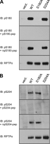
FIG. 1.
FIG. 1.
Specificity of anti-pS180 and anti-pS204 antibodies. HEK293 cells were transiently transfected with empty vector, WT HA-RPTPα, HA-RPTPα S180A, or HA-RPTPα S204A. The cells were lysed, and the lysates were fractionated on 7.5% SDS-polyacrylamide gels, transferred to polyvinylidene difluoride (PVDF) membranes, and immunoblotted with anti-pS180 (A) or anti-pS204 (B) antibodies. As indicated, the antibodies were used alone or together with the phospho- or nonphosphopeptides against which the antibodies were raised. The total levels of HA-RPTPα in the lysates were probed with anti-RPTPα antibody and shown in the bottom panels (A and B). IB, immunoblot.
FIG. 2.
FIG. 2.
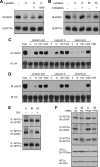
Mitotic dephosphorylation of RPTPα. (A) Endogenous RPTPα was immunoprecipitated from NIH 3T3 lysates (0.5 mg total protein) from unsynchronized (U), mitotic (M), and replated cells after mitotic arrest (1 h, 3 h, and 5 h). The immunoprecipitates were boiled in reducing sample buffer, and the samples were run on a 7.5% SDS-polyacrylamide gel. The proteins were transferred to PVDF membranes, and the membranes were probed with anti-pS204 antibody and subsequently, after stripping, with anti-pS180, anti-pY789, and anti-RPTPα. The cell cycle distribution (percent) of the NIH 3T3 cells used to prepare each sample is shown in the table beneath. (B) Unsynchronized NIH 3T3 cells and cells obtained by mitotic shake-off after nocodazole and paclitaxel treatment were lysed, and endogenous RPTPα was immunoprecipitated from approximately 1 mg total protein/sample. The samples were processed as described for panel A. The blots were quantified, and the phosphorylation levels were normalized for the total amount of RPTPα (bottom panel) and expressed as the ratio to the phosphorylation levels in the unsynchronized cells. The values are presented under each lane. Western blot quantification was performed using Quantity One software (Bio-Rad). All the experiments were repeated at least three times with similar results, and representative experiments are presented in this figure. IP, immunoprecipitation; IB, immunoblot.
FIG. 3.
FIG. 3.
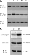
Mitotic phosphorylation and activation of Src. (A) A fraction of the lysates used for RPTPα immunoprecipitation (Fig. 2A) was boiled in SDS sample buffer, and the samples were run on a 7.5% SDS-polyacrylamide gel. The proteins were transferred to PVDF membranes, and the membranes were probed with anti-pY416 antibody and subsequently, after stripping, with anti-npY527 and anti-Src. (B) Endogenous Src was immunoprecipitated with cross-linked antibodies to protein A beads from unsynchronized and mitotic NIH 3T3 cells. Half of the immunoprecipitate was subjected to an in vitro kinase assay, using enolase as substrate. The other half was used for immunoblotting with anti-Src antibody followed by enhanced chemiluminescence (ECL) (bottom panel). The amount of incorporated phosphate was visualized by autoradiography (top panel). The positions of enolase and Src are indicated by arrows. WCLs, whole-cell lysates; IB, immunoblot; IP, immunoprecipitation.
FIG. 4.
FIG. 4.
RPTPα and Src phosphorylation in HeLa cells. Unsynchronized (U) and mitotic (M) HeLa cell lysates (1 mg total protein) were split in half. One half was used to immunoprecipitate endogenous RPTPα (A). The samples and the Western blots were prepared as described for Fig. 2A. (B) The second half was used for Src immunoprecipitation. Cross-linked Src antibodies were used for this purpose. The immunoprecipitates were fractionated by 7.5% SDS-PAGE, and after transfer the membranes were probed with anti-pY416 antibody and subsequently, after stripping, with anti-npY527 and anti-Src. The experiment was performed at least three times with similar results, and a representative experiment is shown here. IP, immunoprecipitation; IB, immunoblot.
FIG. 5.
FIG. 5.
PP2A dephosphorylates RPTPα in vitro and in vivo. (A to D) Endogenous RPTPα was immunoprecipitated from unsynchronized NIH 3T3 cells (A and B). The immunoprecipitates were incubated with fresh lysates of unsynchronized (U) or mitotic (M) NIH 3T3 cells in the presence of orthovanadate (V), sodium fluoride (NaF), or a cocktail of PP1/PP2A inhibitors containing okadaic acid, calyculin A, and tautomycin (100 nM each). After the reactions were terminated, the proteins were separated by 7.5% SDS-PAGE and transferred to PVDF membranes. The blots were probed with anti-pS204 antibody and subsequently, after stripping, with anti-RPTPα antibody. HA-RPTPα overexpressed in COS1 cells was immunoprecipitated and incubated with unsynchronized (C) or mitotic (D) NIH 3T3 lysates in the presence of increasing amounts of the PP1/PP2A inhibitors as indicated. As a control, immunoprecipitated HA-RPTPα was incubated with cell lysis buffer (CLB). The lysates were removed, and the samples were processed as for panel A. Finally, the blots were probed with anti-pS204 antibody and subsequently, after stripping, with anti-HA tag antibody. (E) NIH 3T3 cells arrested with nocodazole were treated with 100 nM okadaic acid (OA). RPTPα was immunoprecipitated and blotted, and the blots were probed with pS180, pS204, and RPTPα antibodies. These experiments have been done at least three times, and representative blots are shown. (F) siRNA-mediated knockdown of PP2Acα in unsynchronized (U) or mitotic (M) NIH 3T3 cells was done as described in Materials and Methods. Efficiency of knockdown was monitored by blotting using a PP2Acα-specific antibody and equal loading using an actin antibody (bottom two panels). RPTPα was immunoprecipitated, blotted, and probed using pS180, pS204, and RPTPα antibodies, as indicated. IP, immunoprecipitation; IB, immunoblot.
FIG. 6.
FIG. 6.
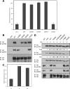
RPTPα catalytic activity is not significantly influenced by serine phosphorylation. (A) WT HA-RPTPα, HA-RPTPα C433S, and serine mutants of HA-RPTPα (S180A, S204A, and S204D) were immunoprecipitated from transfected COS1 cells, and their ability to release [32P]phosphate from phosphorylated MBP was detected. The combined results from three independent experiments are depicted here. (B) For one of the experiments, the serine phosphorylation levels for each overexpressed protein are shown. (C) Endogenous RPTPα was immunoprecipitated from lysates of unsynchronized (U) and mitotic (M) NIH 3T3 cells (1 mg total protein). For the negative control (N), the anti-RPTPα antibody was not added during immunoprecipitation. The ability to dephosphorylate MBP is depicted in the graph. Each bar represents the average of three independent experiments ± standard deviation. The phosphatase activity of WT HA-RPTPα (A) and endogenous RPTPα from unsynchronized cells (C) was set to 100%, and the other data were calculated relative to these values. (D) HA-tagged RPTPα WT and the mutants indicated were immunoprecipitated from transfected COS1 cells and incubated with Src immunoprecipitated from transfected SYF cells. After incubation the samples were boiled in SDS sample buffer and resolved by 7.5% SDS-PAGE. The blots were probed for Src phosphorylation as well as for the amounts of HA-RPTPα present in each reaction. Ser phosphorylation levels of RPTPα are also shown. Src phosphorylation levels were quantified and normalized for the total amount of Src and expressed as the ratio to the phosphorylation levels of Src incubated with immunoprecipitates from empty-vector-transfected cells. The values are presented under each corresponding sample. IP, immunoprecipitation; IB, immunoblot.
FIG. 7.
FIG. 7.
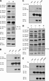
Src binding to RPTPα is induced in mitosis by RPTPα pSer204 dephosphorylation, independently of GRB2. (A) Unsynchronized (U) and mitotic (M) cells were lysed (3 mg total protein), and endogenous RPTPα was immunoprecipitated. The immunoprecipitates were fractionated on a 12.5% SDS-polyacrylamide gel and tested for coimmunoprecipitated Src and GRB2. Phosphorylation of immunoprecipitated RPTPα was also probed. Src and the heavy chain (HC) of the antibody are indicated by arrows. The amount of coimmunoprecipitated GRB2 was quantified and normalized for the total amount of GRB2 in the lysates. The input levels of Src and GRB2 in the lysates (3% of the total lysate run on the same gel) are shown. (B) Src and RPTPα WT, Y789F mutant, or empty vector were cotransfected into SYF cells. The cells were lysed, and Src was immunoprecipitated with cross-linked anti-Src antibodies. The samples were fractionated on a 7.5% SDS-polyacrylamide gel, transferred to PVDF membranes, and immunoblotted with anti-RPTPα serum (top panel) and anti-Src monoclonal antibody (MAb) 327 (middle panel). Whole-cell lysates were monitored for HA-RPTPα expression (bottom panel). (C) As in panel B, except that mutant Src R175L, with a mutation that impairs the ability of the SH2 domain to bind to pTyr residues in target proteins, was cotransfected. (D) Unsynchronized (U) and mitotic (M) NIH 3T3 cells were treated with 100 nM okadaic acid (OA) or left untreated. The samples were processed as for panel A. To reduce the signal from the antibody heavy chain, the blot showing the amount of coimmunoprecipitated Src was probed with horseradish peroxidase (HRP)-coupled protein A, which was less sensitive and hence did not allow detection of the slower-migrating Src band. (E) SYF cells were cotransfected with Src and HA-RPTPα WT, mutants (S204A and S204D), or empty vector (vect.). Coimmunoprecipitation of (mutant) RPTPα with Src was detected as described for panel B. The experiments were repeated at least three times with similar results, and representative experiments are shown here. WCLs, whole-cell lysates; IP, immunoprecipitation; IB, immunoblot.
FIG. 8.
FIG. 8.
RPTPα-mediated activation of Src in mitosis. RPTPα is phosphorylated constitutively on Ser180, Ser204, and Tyr789. At interphase, phosphorylation of Ser204 prohibits binding of Src, and as a result, Src is not activated. In mitosis, pSer204 is dephosphorylated due to activation of PP2A. Nonphospho-Ser204 binds Src through yet-to-be-identified regions in Src and RPTPα while remaining phosphorylated on Tyr789 and bound to the adaptor protein GRB2. Src binding to RPTPα in mitosis leads to dephosphorylation of pTyr527 and pTyr416 in Src, resulting in modest activation of Src kinase activity. Following release from mitosis, Ser204 is rapidly phosphorylated and Src phosphorylation reverts.

Similar articles

Cited by

References

    1. Abell, C. W., and T. M. Monahan. 1973. The role of adenosine 3′,5′-cyclic monophosphate in the regulation of mammalian cell division. J. Cell Biol. 59:549-558. - PMC - PubMed
    1. Alonso, A., J. Sasin, N. Bottini, I. Friedberg, A. Osterman, A. Godzik, T. Hunter, J. Dixon, and T. Mustelin. 2004. Protein tyrosine phosphatases in the human genome. Cell 117:699-711. - PubMed
    1. Andersen, J. N., A. Elson, R. Lammers, J. Romer, J. T. Clausen, K. B. Moller, and N. P. Moller. 2001. Comparative study of protein tyrosine phosphatase-epsilon isoforms: membrane localization confers specificity in cellular signalling. Biochem. J. 354:581-590. - PMC - PubMed
    1. Bagrodia, S., I. Chackalaparampil, T. E. Kmiecik, and D. Shalloway. 1991. Altered tyrosine 527 phosphorylation and mitotic activation of p60c-src. Nature 349:172-175. - PubMed
    1. Bhandari, V., K. L. Lim, and C. J. Pallen. 1998. Physical and functional interactions between receptor-like protein-tyrosine phosphatase alpha and p59fyn. J. Biol. Chem. 273:8691-8698. - PubMed

Publication types

Substances