. 2010 May 28;285(22):16789-97.
doi: 10.1074/jbc.M110.101501. Epub 2010 Apr 2.

Multiple molecules of Hsc70 and a dimer of DjA1 independently bind to an unfolded protein

Affiliations

Multiple molecules of Hsc70 and a dimer of DjA1 independently bind to an unfolded protein

Kazutoyo Terada et al. J Biol Chem. .

Abstract

Protein folding is a prominent chaperone function of the Hsp70 system. Refolding of an unfolded protein is efficiently mediated by the Hsc70 system with either type 1 DnaJ protein, DjA1 or DjA2, and a nucleotide exchange factor. A surface plasmon resonance technique was applied to investigate substrate recognition by the Hsc70 system and demonstrated that multiple Hsc70 proteins and a dimer of DjA1 initially bind independently to an unfolded protein. The association rate of the Hsc70 was faster than that of DjA1 under folding-compatible conditions. The Hsc70 binding involved a conformational change, whereas the DjA1 binding was bivalent and substoichiometric. Consistently, we found that the bound (14)C-labeled Hsc70 to the unfolded protein became more resistant to tryptic digestion. The gel filtration and cross-linking experiments revealed the predominant presence of the DjA1 dimer. Furthermore, the Hsc70 and DjA1 bound to distinct sets of peptide array sequences. All of these findings argue against the generality of the widely proposed hypothesis that the DnaJ-bound substrate is targeted and transferred to Hsp70. Instead, these results suggest the importance of the bivalent binding of DjA1 dimer that limits unfavorable transitions of substrate conformations in protein folding.


Figures
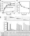
FIGURE 1.
FIGURE 1.
Refolding activity of the Hsc70 system. A, refolding of chemically denatured firefly luciferase. The protein was refolded in the mixture without (open circle) or with 10 mg/ml of the cytosolic extract (filled circle). For comparison purposes, the luciferase was refolded in the reticulocyte lysate (40%, open triangle) or the combination of purified components (filled triangle, 200 μg/ml Hsc70, 20 μg/ml DjA2, and 20 μg/ml Bag1). Similar results were obtained with DjA1. B, refolding of denatured luciferase (5.7 nm) with variable amounts of chaperone components. The activity was monitored after 10 (open bars) and 60 min (solid or hatched bars). Hsc70 was added at 200 μg/ml (+), serial dilution by a factor of 3 (hatched box) and 0. DjA2 was added at 20 μg/ml (+), serial dilution by a factor of 3 (hatched box) and 0. Bag1 at 20 μg/ml (+), serial dilution by a factor of 3 (hatched box) and 0. The molar ratios of chaperone component and the luciferase are shown on the bottom of the graph. C, refolding of luciferase with delayed addition of the chaperone combination. The denatured luciferase was added into the refolding buffer without chaperones. The three chaperone components were added at the indicated times and allowed to refold for 60 min.
FIGURE 2.
FIGURE 2.
Binding of chaperones to the coupled and unfolded luciferase. A and B, binding of Hsc70 (200 μg/ml or 2.8 μm) and DjA1 (20 μg/ml or 0.22 μm as a dimer) to various amounts of coupled luciferase. C, the molar ratios for binding of chaperones to the luciferase on sensor chips. The bound Hsc70 (A) and DjA1 (B) at the ends of association phases were plotted against amount of the luciferase. D and E, sensorgrams for delayed injections of Hsc70 (80 μg/ml) and DjA1 (20 μg/ml). The luciferase on the chip was unfolded prior to chaperone injection (arrow) and washed by the running buffer for the indicated times. F, decrease of chaperone binding capacity. The bound chaperones at the ends of association phases (D and E) were plotted versus the delayed injection times.
FIGURE 3.
FIGURE 3.
Binding of Hsc70 and DjA1 to coupled luciferase. A and B, sensorgrams (colored curves) for various amounts of the respective chaperones. The running buffer was supplemented with 1 mm ATP. The model equations used to fit the model are shown. The residual plots (top panels) were also provided to give a graphical indication of experimental data deviation from the fitted curves (black). C and D, simulation of the sensorgrams with longer time periods (30 min of association and 30 min of dissociation). The curves were obtained from the kinetics parameters in Table 1 using the BIAsimulation software program. The bulk refractive index was subtracted from the total data. E, tryptic digestion of Hsc70. The 14C-methylated Hsc70 was allowed to bind to the unfolded luciferase beads (lanes 1–4). After the incubation for 3 or 40 min, the bound Hsc70 was separated and treated with trypsin for 5 min (lanes 3 and 4). The [14C]Hsc70 with a nucleotide was also treated (lanes 6 and 7). The blue triangles represent bands found only in [14C]Hsc70 samples without unfolded substrate. The red triangle represents the band with the bound [14C]Hsc70 samples (lanes 3 and 4). The amount of samples: 5% (lanes 1 and 2); 50% (lanes 3, 4, 6, and 7); 0.2 μg of [14C]Hsc70 (lane 5). F, detection of DjA1 dimer. The DjA1 protein was eluted mainly at a position corresponding to 100,000 by a gel filtration experiment (red arrow, left panel). The DjA1 protein was cross-linked with dimethyl suberimidate, separated by SDS-PAGE, and stained with Coomassie Brilliant Blue (right panel).
FIGURE 4.
FIGURE 4.
Simultaneous addition of chaperone components. A, sensorgrams of the interaction between chaperone combinations and the luciferase (thick curves). ATP, together with the chaperone components, was only added during the association phase. B, differences between the combinational and individual chaperon binding. C, refolding of luciferase with a higher amount of DjA1. The refolding mixtures contained Hsc70 (2.8 μm) and variable amount of DjA1 (0–7.9 μm) with (closed circles) or without (open circles) Bag1 (0.77 μm). The activity was monitored after 60 min. D, refolding of the denatured luciferase on the sensor chip. The coupled luciferase on the sensor chip was manually processed for the unfolding procedure. The chip was immersed in the HKMB-ATP solution with or without the chaperone combination for 30 min and soaked into the luciferase assay buffer to check the activity.
FIGURE 5.
FIGURE 5.
Chaperone binding to cellulose-bound peptide scan. A and B, a peptide scan derived from sequences of luciferase was screened for Hsc70 (A) and DjA1 (B) binding. The last spots of rows (right) and the N-terminal residues of peptides of the first spots of rows (left) are indicated. C and D, differential binding of Hsc70 and DjA1 to peptides. Peptides that bind to Hsc70 contain several large hydrophobic and aromatic residues (Leu, Ile, Phe, Val, Trp, and Tyr; black boxes) with basic residues (Arg and Lys; blue circles). Peptides that bind to DjA1 contain large hydrophobic and aromatic residues with multiple acidic residues (Glu and Asp; yellow circles). E, ribbon and space filling representations of the structure of native luciferase from the x-ray structure (Protein Data Bank entry 1LCI) (43) using the RasMol 2.7 software program. The cyan and yellow regions indicate the binding sites for Hsc70 (C) and DjA1 (D), respectively. Lysine residues in the Hsc70 binding sites are indicated by sticks. The other lysine residues (31 of 40 residues) are blue sticks (bottom).
FIGURE 6.
FIGURE 6.
Model for refolding of luciferase by the Hsc70 system. Step 1, multiple Hsc70 proteins and a dimer of DjA1 independently bind to the proper binding sites on an unfolded substrate protein. A, Hsc70 only. Step 2, the bound Hsc70-ATP molecules suppress hydrophobic interactions among the binding sites. Conversion to the ADP form is slow. The complex undergoes multiple transitions in an energy landscape. The complex constraints on the transitions or gets stuck into the local minima of the landscape. B, DjA1 only. Step 2, a U-shaped dimer of DjA1 binds to two distant sites on a substrate and reduces a considerable range of the conformational transitions. The DjA1 binding prevents some hydrophobic interactions, but DjA1 dissociation results in eventual aggregation. C, the complete Hsc70 system. Step 0, the Hsc70-ATP and free DjA1 are in equilibrium in the solution. Step 2. The bound Hsc70-ATP suppresses the unfavorable interactions, whereas the bound DjA1 reduces the range of transitions. Some of the bound DjA1 accelerates ATP hydrolysis of the bound Hsc70. A free DnaJ protein in solution may act on the other bound Hsc70-ATP. A nucleotide exchange factor facilitates dissociation of the bound Hsc70-ADP.

Similar articles

Cited by

References

    1. Bukau B., Horwich A. L. (1998) Cell 92, 351–366 - PubMed
    1. Hartl F. U., Hayer-Hartl M. (2002) Science 295, 1852–1858 - PubMed
    1. Parsell D. A., Lindquist S. (1993) Annu. Rev. Genet. 27, 437–496 - PubMed
    1. Terada K., Kanazawa M., Bukau B., Mori M. (1997) J. Cell Biol. 139, 1089–1095 - PMC - PubMed
    1. Terada K., Mori M. (2000) J. Biol. Chem. 275, 24728–24734 - PubMed

Publication types

LinkOut - more resources