. 2010 Mar 23;5(3):e9819.
doi: 10.1371/journal.pone.0009819.

Induction of autophagy by cystatin C: a mechanism that protects murine primary cortical neurons and neuronal cell lines

Affiliations

Induction of autophagy by cystatin C: a mechanism that protects murine primary cortical neurons and neuronal cell lines

Belen Tizon et al. PLoS One. .

Abstract

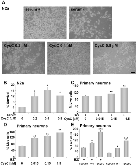
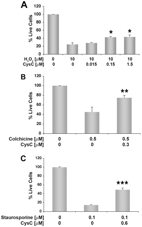
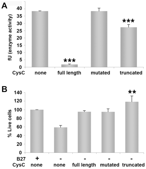
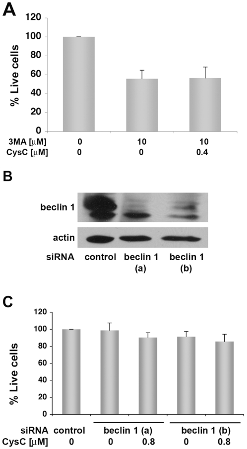
Cystatin C (CysC) expression in the brain is elevated in human patients with epilepsy, in animal models of neurodegenerative conditions, and in response to injury, but whether up-regulated CysC expression is a manifestation of neurodegeneration or a cellular repair response is not understood. This study demonstrates that human CysC is neuroprotective in cultures exposed to cytotoxic challenges, including nutritional-deprivation, colchicine, staurosporine, and oxidative stress. While CysC is a cysteine protease inhibitor, cathepsin B inhibition was not required for the neuroprotective action of CysC. Cells responded to CysC by inducing fully functional autophagy via the mTOR pathway, leading to enhanced proteolytic clearance of autophagy substrates by lysosomes. Neuroprotective effects of CysC were prevented by inhibiting autophagy with beclin 1 siRNA or 3-methyladenine. Our findings show that CysC plays a protective role under conditions of neuronal challenge by inducing autophagy via mTOR inhibition and are consistent with CysC being neuroprotective in neurodegenerative diseases. Thus, modulation of CysC expression has therapeutic implications for stroke, Alzheimer's disease, and other neurodegenerative disorders.


Conflict of interest statement

Competing Interests: The authors have declared that no competing interests exist.

Figures

Figure 1
Figure 1. In vitro neuroprotection by either extracellular or endogenouse human CysC.
A. Light microscopy pictures of N2a cells incubated for 48 hours in medium containing serum or in serum-free medium in the absence or presence of different concentrations of CysC. Scale bar represents 100 µm. B. Neuronal survival was measured by counting live cells, and expressed as percentage of neuronal survival in cultures incubated in serum-containing medium. Data are the mean and SEM of 4 experiments. C. D. Primary rat cortical neurons were cultured in neurobasal medium in the presence (C) or absence (D) of B27-supplement and different concentrations of human CysC for 24 hours. Cell survival analyzed by the MTS assay is expressed as percentage of live cells in cultures incubated in B27-supplemented medium without CysC. Data are the mean and SEM of 3 experiments. F and P values determined by one way ANOVA for (C) were 85.09 and <0.0001 and for (D) 34.00 and <0.0001. E Primary cortical neurons isolated from brains of CysC knockout (CysCko), transgenic mice overexpressing human CysC (CysCtg), or wild type (WT) mice were incubated in B27-supplement containing or lacking media. Survival is expressed as percentage of live cells in wild type cultures incubated in B27-supplemented medium. For groups incubated with B27 the F and P values determined by one way ANOVA were 20.60 and 0.0007 and for groups incubated without B27 were 68.93 and <0.0001.
Figure 2
Figure 2. Neuroprotection of primary rat cortical neurons from a variety of insults.
Primary rat cortical neurons were cultured in B27-supplemented neurobasal medium in the presence of 10 µM H2O2 (A), 0.5 µM colchicine (B), or 0.1 µM staurosporine (C) and different concentrations of human CysC for 24 hours. Cell survival analyzed by the MTS assay is expressed as percentage of live cells in cultures incubated in B27-supplemented medium without CysC. Data are the mean and SEM of 3 experiments. F and P values determined by one way ANOVA for (A) were 7.45 and 0.01. Differences between CysC treated and untreated samples were calculated by Student's t-test for B and C.
Figure 3
Figure 3. CysC forms lacking cathepsin B inhibitory activity are protective against nutrition-deprivation-induced cell death.
A. In vitro analysis of cathepsin B specific activity measured in the presence of 1.7 µM of three different forms of CysC: recombinant full length human CysC; recombinant full length CysC mutated in the inhibitory active site (mutated); and N-terminally truncated human urinary CysC (truncated). Cathepsin B inhibitory activity was calculated as activity per mg of total protein and normalized for cathepsin B protein level, presented as fluorescent units (fU). Data are the mean and SEM of 3 experiments. F and P values determined by one way ANOVA were 147.6 and <0.0001. B. Cortical primary neurons derived from embryos of CysC knockout mice were incubated in neurobasal medium either containing or lacking B27, in the presence of 0.8 µM of the three forms of CysC. Cell survival analyzed by the MTS assay is expressed as percentage of live cells in cultures incubated in supplemented media without CysC. Data are the mean and SEM of 3 experiments. F and P values determined by one way ANOVA were 9.39 and 0.005.
Figure 4
Figure 4. CysC does not protect neuronal cells from toxicity when autophagy is inhibited.
A. N2a cells were incubated for 44 hours in serum-free medium with 3MA, an inhibitor of autophagy, in the presence or absence of 0.4 µM CysC. Neuronal survival was measured by counting live cells, and expressed as percentage of neuronal survival in cultures incubated in serum-deprived medium. Data are the mean and SEM of 4 experiments. There were no significant differences between samples containing 3MA with or without CysC. B. N2a cells were treated with two forms of beclin 1 siRNA (a and b). Western blot analysis with anti-beclin 1 antibody shows inhibition of beclin 1 expression in treated cells. C. Beclin 1 siRNA treatment of N2a cells attenuates the protective effect of CysC from serum-deprivation induced death. Cell survival was measured by the MTS assay. Data are the mean and SEM of 3 experiments. No significant difference between CysC treated and non- treated cells was observed.
Figure 5
Figure 5. CysC induces cleavage of LC3 and the generation of vesicles stained for LC3-II, associated with autophagic vesicles.
A. N2a neuroblastoma cells were incubated for 18 hours in serum containing or serum-free media containing the indicated concentrations of CysC. Cells were stained with an anti-LC3 antibody enriched for LC3-II and visualized in a fluorescence microscope. Scale bar represents 10 µm. BE. N2a cells were incubated in serum-supplemented or serum-free media containing different concentrations of CysC for 12 hours. Cell lysate proteins were analyzed by Western blot using anti-LC3 antibodies. Representative images of Western blot analyses of cell lysate proteins in serum-free medium (B) and in serum-supplemented medium (D) are shown. The intensity of the LC3-I and LC3-II bands was measured and results are expressed as the ratio of LC3-II to total LC3 (LC3-I + LC3-II) values (C and E). Data are the mean and SEM of 4 experiments. F and P values determined by one way ANOVA for (E) were 5.11 and 0.02 (F and G) Western blots with anti-LC3 antibodies of primary cortical neurons isolated from CysC knockout mice incubated for 12 hours in media containing (F) or lacking (G) B27-supplement in the presence of different concentrations of CysC.
Figure 6
Figure 6. CysC induces an increase in the number of autophagic vesicles (AVs) in neuronal cells.
Electron micrographs of primary rat cortical neurons (A) incubated for 12 hours in either B27-supplemented or deprived medium in the absence or presence of 0.75 µM CysC. The total number of AVs per cell was counted for at least 20 cells/condition, and the average number of vesicles per cell is shown (B). Data are the mean and SEM. Differences between CysC treated and untreated samples were calculated by Student's t-test.
Figure 7
Figure 7. CysC enhances total lysosomal-dependent protein degradation in serum-deprived neuronal cells.
A. Effect of increasing concentrations of CysC on total rates of protein degradation. N2a cells were labeled for 2 days with [3H]-leucine. After extensive washing, cells were incubated in serum-containing or serum-free media. Removal of serum maximally activates lysosomal degradation. The cells maintained in serum-free media were supplemented or not with increasing concentrations of CysC as labeled. The rate of total protein degradation at the indicated times was calculated as the percentage of total radiolabeled protein transformed into soluble amino acids. B. Effect of inhibition of lysosomal proteolysis on the CysC-induced increase in protein degradation. N2a cells were labeled as in A and then maintained in serum-free media and supplemented or not with CysC. Where indicated 20 mM NH4Cl and 100 µM leupeptin were added to inhibit lysosomal proteolysis. Protein degradation was calculated as in A. C. Effect of CysC on macroautophagy-dependent proteolysis. N2a cells labeled as in A and maintained in serum-free media were supplemented or not with CysC. Half of the cells were treated with 10 mM 3MA to inhibit macroautophagy. The percentage of lysosomal degradation that results from autophagic degradation (3MA sensitive), in the presence or absence of CysC was calculated. Values are mean and SED of triplicate wells in 3–4 different experiments. One way ANOVA for differences between CysC treated and untreated samples were significant for *p = 0.05; **p = 0.001 and between control and ammonium chloride treated samples were significant for + p = 0.01.
Figure 8
Figure 8. CysC induces autophagy via the mTOR signaling pathway in either serum-containing or serum-free medium.
A. N2a cells were incubated for 12 hrs in serum-containing or serum-free medium in the presence or absence of CysC. Cell lysate proteins were separated by gel electrophoresis and blotted with antibodies to P70S6 kinase, p-P70S6 kinase (Thr389), or β-tubulin. Representative images of Western blot analysis are presented. B. The intensity of the bands was measured, and p-P70S6 protein levels were calculated relative to total P70S6 values showing a decrease in the level of P70S6 kinase phosphorylation. Data are the mean and SEM of 6 experiments. For serum containing groups the F and P values determined by one way ANOVA were 9.07 and 0.006 and for serum deprived groups were 10.18 and 0.005.

Similar articles

Cited by

References

    1. Abrahamson M, Alvarez-Fernandez M, Nathanson CM. Cystatins. Biochem Soc Symp. 2003;70:179–199. - PubMed
    1. Bernstein HG, Kirschke H, Wiederanders B, Pollak KH, Zipress A, et al. The possible place of cathepsins and cystatins in the puzzle of Alzheimer disease: a review. Mol Chem Neuropathol. 1996;27:225–247. - PubMed
    1. Levy E, Jaskolski M, Grubb A. The role of cystatin C in cerebral amyloid angiopathy and stroke: cell biology and animal models. Brain Pathol. 2006;16:60–70. - PMC - PubMed
    1. Pawlik M, Sastre M, Calero M, Mathews PM, Schmidt SD, et al. Overexpression of human cystatin C in transgenic mice does not affect levels of endogenous brain amyloid β peptide. J Mol Neurosci. 2004;22:13–18. - PubMed
    1. Huh CG, Hakansson K, Nathanson CM, Thorgeirsson UP, Jonsson N, et al. Decreased metastatic spread in mice homozygous for a null allele of the cystatin C protease inhibitor gene. Mol Pathol. 1999;52:332–340. - PMC - PubMed

Publication types

MeSH terms