. 2010;10(1):74-84.
doi: 10.1159/000276895. Epub 2010 Mar 26.

Conservation of the TGFbeta/Labial homeobox signaling loop in endoderm-derived cells between Drosophila and mammals

Affiliations

Conservation of the TGFbeta/Labial homeobox signaling loop in endoderm-derived cells between Drosophila and mammals

Gwen A Lomberk et al. Pancreatology. 2010.

Abstract

Background/aims: Midgut formation in Drosophila melanogaster is dependent upon the integrity of a signaling loop in the endoderm which requires the TGFbeta-related peptide, Decapentaplegic, and the Hox transcription factor, Labial. Interestingly, although Labial-like homeobox genes are present in mammals, their participation in endoderm morphogenesis is not clearly understood.

Methods: We report the cloning, expression, localization, TGFbeta inducibility, and biochemical properties of the mammalian Labial-like homeobox, HoxA1, in exocrine pancreatic cells that are embryologically derived from the gut endoderm.

Results: HoxA1 is expressed in pancreatic cell populations as two alternatively spliced messages, encoding proteins that share their N-terminal domain, but either lack or include the homeobox at the C-terminus. Transcriptional regulatory assays demonstrate that the shared N-terminal domain behaves as a strong transcriptional activator in exocrine pancreatic cells. HoxA1 is an early response gene for TGFbeta(1) in pancreatic epithelial cell populations and HoxA1 protein co-localizes with TGFbeta(1) receptors in the embryonic pancreatic epithelium at a time when exocrine pancreatic morphogenesis occurs (days E16 and E17).

Conclusions: These results report a role for HoxA1 in linking TGFbeta-mediated signaling to gene expression in pancreatic epithelial cell populations, thus suggesting a high degree of conservation for a TGFbeta/labial signaling loop in endoderm-derived cells between Drosophila and mammals. and IAP.


Figures
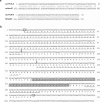
Fig. 1
Fig. 1
Expression of labial-like genes in rat pancreas containing the highly conserved homeobox domain. a A pancreas cDNA library was used as a template in a PCR amplification using degenerate primers for the highly conserved homeobox domain encoding for the sequences EKEFHFN and IWFQRRMK, which resulted in the amplification of two different fragments encoding the homeobox motifs for the Labial-like rat HoxA1 and HoxB1 paralogs. The sequences obtained from the PCR-amplified cDNAs, rat PCR-A and rat PCR-B, were used for FASTA analysis against sequences deposited in the GenBank database using GCG analysis software. Since the homeobox domain of many members of this family is highly conserved, the sequences were compared at the DNA level to make more meaningful comparisons. Note that rat PCR-A compared closely (98%) with the homeobox-encoding domain of the murine HoxA1 and that rat PCR-B was almost identical to the human HoxB1. b Nucleotide and deduced amino acid sequences of the rat HoxA1 gene is depicted. The full-length coding sequence of rat HoxA1 was obtained by pancreas cDNA and genomic library screening. Nucleotides are numbered at left, amino acids are numbered at right, and the initiator and termination codons are boxed. The homeobox motif is boxed. Arrows represent the donor and acceptor sites for an alternative splicing event which leads to a frameshift in translation and a premature termination (represented in bold type, see fig. 4). The peptide region used to generate a polyclonal antibody against the homeobox-containing HoxA1 is underlined with asterisks. Overall, rat HoxA1 is 96% similar to the mouse homolog at the DNA level and almost identical (99%) at the protein level as determined using FASTA analysis (GCG, Madison, Wisc., USA).
Fig. 2
Fig. 2
Expression of HoxA1 is found in adult rat tissues and exocrine pancreatic cell lines. a Northern blot analysis was performed on total cellular RNA (20 μg) isolated from the poorly differentiated ductular-like rat exocrine pancreatic cell line, AR4IP, and from the adult rat tissues, pancreas, brain, heart, kidney, liver, lung, and testis. GAPDH was used to normalize loading. Note that HoxA1 was enriched in the pancreatic cell populations. In addition, high levels of HoxA1 expression were detected in lung. b PCR amplification of HoxA1 from five human ductular-like pancreatic cell lines, PANC-1, AsPC-1, BxPC-3, HS 766T, and Capan-1. Note that HoxA1 was expressed in each of these pancreatic cell lines. c Recombinant GST-HoxA1 was purified from bacteria, and 5 mg of the fusion protein was used as a positive control for a polyclonal HoxA1 antibody derived from a mouse HoxA1 peptide sequence by Western blot analysis. The anti-HoxA1 antibody recognized this purified GST fusion protein at its anticipated MW of 66 kDa. Western blot analysis of homogenates (50 μg) from whole rat pancreas and the exocrine pancreatic cell line AR4IP detects a single polypeptide which has a MW of 47 kDa. Western blot analysis using pre-adsorbed antisera was negative (data not shown). d Indirect immunofluorescence of HoxA1 in AR4IP cells. Cells were plated on coverslips and processed for immunofluorescence using HoxA1 antisera as a primary antibody. Note that HoxA1 localized to the nucleus in the exocrine pancreatic cell line. AR4IP. This localization was not detected in cells stained with pre-immune serum (data not shown).
Fig. 3
Fig. 3
HoxA1 is expressed in the developing rat pancreas. Immunohistochemistry of developing rat pancreas at embryonic day 16 (E16) (a, c, e, g) or embryonic day 17 (b, d, f, h). Immunolocalization of HoxA1 was performed using HoxA1 antisera (a, b). The exocrine and endocrine portions of the gland were detected using anti-α-amylase and anti-insulin, respectively (c, d), while control staining was performed using the HoxA1 antisera pre-adsorbed with purified GST-HoxA1 fusion protein (e, f). Note that HoxA1 was detected at both E16 and E17 in the ductular-like pancreatic precursor cells (arrows), which give rise to exocrine pancreatic cell populations, and that this immunoreactivity was abolished upon pre-adsorption of the antibody (e, f). Co-localization of the TGFβ receptor type I (data not shown) and II (g, h) showed staining in the exocrine pancreas in a similar distribution as HoxA1. Bars = 100 μm.
Fig. 4
Fig. 4
Amplification of alternatively spliced forms of HoxA1 from pancreatic populations. a To better determine the size of the HoxA1 transcript, 40 μg of total RNA from AR41P cells were separated further by agarose gel electrophoresis and used for Northern blot analysis. HoxA1 was expressed in these cells as a 2.2-kb and a 2.0-kb transcript. b PCR analysis of alternatively spliced forms of HoxA1 from pancreatic populations. Primers were designed against the rat HoxA1, which flank the intron/exon boundaries within the gene and used in PCR amplification against cDNA derived from the full-length rat HoxA1 cDNA, AR4IP cells, and adult rat pancreas. The upper arrow indicates the expected size product of a full-length homeobox-encoding mRNA, while the lower arrow designates the expected size of an alternatively spliced message previously reported by LaRosa and Gudas [33]. Note that the pancreatic populations, AR41P and adult rat pancreas, express both the full-length rat HoxA1 (A) and an alternatively spliced transcript (B). c Schematic diagram of the alternatively spliced forms of HoxA1. The cDNAs amplified in b were cloned, sequenced and shown to encode a homeobox-containing (HoxA1/box+) (‘A’) and a homeobox-less (HoxA1/box) (‘B’) protein. This latter product is a result of a frameshift which occurs at the site of splicing, causing a premature termination of the protein sequence (also see fig. 1).
Fig. 5
Fig. 5
Regulation of HoxA1 expression in response to TGFβ1 in pancreatic cell populations. a, b AR4IP cells were treated with 5 ng/ml TGFβ1 for 0, 30, 60, 120, or 240 min in the absence (a) or presence (b) of the protein synthesis inhibitor, cycloheximide. RNA was extracted, separated on a 1.5% gel, and used for Northern blot analysis for HoxA1 gene expression. Note that a significant increase in both HoxA1 mRNAs occurred within 1 h of growth factor stimulation. This upregulation was independent of protein synthesis, indicating that HoxA1 is an early response gene for this peptide growth factor in exocrine pancreatic cell populations. c Pancreata were isolated from day 17 prenatal rats, minced, and treated with or without 10 ng/ml TGFβ1 for 30 min. RNA was extracted and used for Northern blot analysis for HoxA1 gene expression. Note that an increase in both HoxA1 mRNAs occurred within 30 min of growth factor stimulation.
Fig. 6
Fig. 6
A transcriptional activation domain is located within the HoxA1 N-terminal shared domain. a Diagram of the DNA constructs and experimental strategy for determination of the transactivation properties of HoxA1. These distinct effector plasmids were co-transfected into the pancreatic cell line, PANC-1, along with the reporter plasmid carrying five GAL4 recognition sites upstream of the CAT gene. As a control for basal transcriptional activity, the CAT reporter plus the GAL4 DNA-binding motif alone (GAL4 DBM) effector plasmid was used. b Histogram of the CAT activity using the reporter constructs with the various GAL4 DBM-HoxA1 effector plasmids. The GAL4 DNA-binding domain alone (Control) had a basal transcriptional activity of 1.0 ± 0.1, the entire N-terminal 245 amino acids of HoxA1/box+, GAL4-HoxA1(1–245), stimulated transcription 54 ± 2.4-fold over basal activity, the shared domain ‘SD’ between HoxA1/box+ and HoxA1/box, GAL4-HoxA1 (1–122), activated transcription 48 ± 2.1-fold above the control value, GAL4-HoxA1 (1–74) activated transcription 4.88 ± 0.6, and GAL4-HoxA1 (123–245), the residues unique to HoxA1/box+, activated transcription only 1.9 ± 0.1-fold over control. Note that the GAL4-HoxA1 shared domain (amino acids 1–122) behaved as a transcriptional activator, as effectively as the entire N-terminal region of HoxA1/box+, while the most N-terminal region of the shared domain (1–74), as well as the region unique to HoxA1/box+ (123–245) showed transactivation properties with less than 10% of this value. However, the second portion of the shared domain, GAL4-HoxA1 (75–122), exhibited ∼65% transactivating properties of the entire shared domain. Further division of this region (amino acids 75–100 and 101–122) showed a significant reduction in activity, indicating that the transactivating properties of this protein reside within a 47 amino acid region (amino acids 75–122), which is shared between both alternative spliced variants of this gene product. CAT activity was determined using an ELISA assay (Roche). A plasmid carrying β-galactosidase, pSV-β-galactosidase (Promega) was also co-transfected in order to normalize values to β-galactosidase activity measured using a colorimetric enzyme assay system (Promega). Each experiment was performed independently at least two times in triplicate. Bars represent SEM.

Similar articles

Cited by

References

    1. Manak JR, Scott MP. A class act: conservation of homeodomain protein functions. Dev Suppl. 1994:61–77. - PubMed
    1. Botas J. Control of morphogenesis and differentiation by HOM/HOX genes. Curr Opin Cell Biol. 1993;5:1015–1022. - PubMed
    1. Kappen C, Ruddle FH. Evolution of a regulatory gene family: HOM/HOX genes. Curr Opin Genet Dev. 1993;3:931–938. - PubMed
    1. Diederich RJ, Merrill VK, Pultz MA, Kaufman TC. Isolation, structure, and expression of labial, a homeotic gene of the antennapedia complex involved in Drosophila head development. Genes Dev. 1989;3:399–414. - PubMed
    1. Merrill VK, Diederich RJ, Turner FR, Kaufman TC. A genetic and developmental analysis of mutations in labial, a gene necessary for proper head formation in Drosophila melanogaster. Dev Biol. 1989;135:376–391. - PubMed

Publication types

MeSH terms