. 2010 May;84(10):5336-50.
doi: 10.1128/JVI.02494-09. Epub 2010 Mar 17.

Macropinocytotic uptake and infection of human epithelial cells with species B2 adenovirus type 35

Affiliations

Macropinocytotic uptake and infection of human epithelial cells with species B2 adenovirus type 35

Stefan Kälin et al. J Virol. 2010 May.

Abstract

Human adenovirus serotype 35 (HAdV-35; here referred to as Ad35) causes kidney and urinary tract infections and infects respiratory organs of immunocompromised individuals. Unlike other adenoviruses, Ad35 has a low seroprevalence, which makes Ad35-based vectors promising candidates for gene therapy. Ad35 utilizes CD46 and integrins as receptors for infection of epithelial and hematopoietic cells. Here we show that infectious entry of Ad35 into HeLa cells, human kidney HK-2 cells, and normal human lung fibroblasts strongly depended on CD46 and integrins but not heparan sulfate and variably required the large GTPase dynamin. Ad35 infections were independent of expression of the carboxy-terminal domain of AP180, which effectively blocks clathrin-mediated uptake. Ad35 infections were inhibited by small chemicals against serine/threonine kinase Pak1 (p21-activated kinase), protein kinase C (PKC), sodium-proton exchangers, actin, and acidic organelles. Remarkably, the F-actin inhibitor jasplakinolide, the Pak1 inhibitor IPA-3, or the sodium-proton exchange inhibitor 5-(N-ethyl-N-isopropyl) amiloride (EIPA) blocked endocytic uptake of Ad35. Dominant-negative proteins or small interfering RNAs against factors driving macropinocytosis, including the small GTPase Rac1, Pak1, or the Pak1 effector C-terminal binding protein 1 (CtBP1), potently inhibited Ad35 infection. Confocal laser scanning microscopy, electron microscopy, and live cell imaging showed that Ad35 colocalized with fluid-phase markers in large endocytic structures that were positive for CD46, alphanu integrins, and also CtBP1. Our results extend earlier observations with HAdV-3 (Ad3) and establish macropinocytosis as an infectious pathway for species B human adenoviruses in epithelial and hematopoietic cells.


Figures
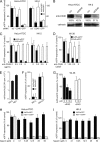
FIG. 1.
FIG. 1.
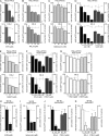
CD46 and αν integrins are required for Ad35 infection, HSPG is not. (A) HeLa-ATCC (left) or HK-2 (right) cells were transfected with 20 nM siRNAs against CD46, GFP, or nonsilencing scrambled siRNA (scr) for 72 h and infected with Ad35-eGFP (MOI of 5) or Ad5-eGFP (MOI of 5) for 8 h, followed by Safire fluorescence analysis. (B) Quantification of knockdown efficiency by Western blots against CD46, normalized against calnexin. (C and D) HeLa-ATCC (left) or HK-2 (right) cells (C) or Wi-38 cells (D) were preincubated with an inhibiting antibody against CD46 (MEM-258) in the cold for 30 min and infected for 8 h or 15 h, respectively. (E) 3H-Ad35 was bound to human melanoma M21L cells lacking αν integrin or to M21 αν integrin-positive cells. (F) M21L or M21 cells were infected with Ad35-eGFP or Ad5-eGFP and analyzed by Safire fluorescence analyses. (G) Wi-38 cells were preincubated with cRGD or cRAD peptides in the cold for 30 min and infected with Ad35-eGFP for 15 h. (H and I) Ad35-eGFP (MOI of 5) or HSV-1-eGFP (MOI of 5) were preincubated for 30 min with different concentrations of heparin as indicated, followed by inoculation of HeLa-ATCC or HK-2 cells for 8 h, and infection analyses were done with a Safire2 plate reader after normalization to the cell numbers determined by 4′,6-diamidino-2-phenylindole (DAPI) staining (see Materials and Methods). Note that Ad35-eGFP transduction was not affected whereas HSV-1-eGFP transduction was strongly reduced in both cell lines.
FIG. 2.
FIG. 2.
Cell type-specific requirements of dynamin. HeLa-ATCC or HeLa-K cells were transfected with dominant-negative K44A-Dyn2-mRFP, wild-type Dyn2-mRFP, or mRFP for 24 h and infected with Ad35-eGFP or Ad5-eGFP (MOI of 5) for 8 h. (A) Representative images of transfected and infected HeLa-ATCC cells, fixed and recorded for mRFP and GFP fluorescence in a wide-field microscope. (B) NIH ImageJ quantification of total fluorescence intensities. (C) HeLa-K cells transfected as for panel A and in addition with the carboxy-terminal domain of AP180, infected and assayed by fluorescence-activated cell sorting. Dominant-negative K44A-Dyn2 inhibited both Ad35-eGFP and Ad5-eGFP, whereas the carboxy-terminal domain of AP180 had no effects on Ad35-eGFP but inhibited Ad5-eGFP by 40%. (D) Dynasore-independent transduction of Wi-38 cells by Ad35-eGFP (left) and dynasore-dependent transduction by Ad5-eGFP (right panel) in the absence of cell toxicity, indicated by cell number measurements shown with gray line graphs, with 100% representing noninfected non-drug-treated conditions.
FIG. 3.
FIG. 3.
Inhibitors against Pak1, PKC, sodium/proton exchanger, and actin reduce Ad35-eGFP transduction. HeLa-ATCC and HK-2 cells were preincubated with the indicated concentrations of drugs for 30 min and infected with Ad35-eGFP for 8 h. Cells were analyzed on a Safire2 plate reader, and eGFP intensities were normalized to the DAPI signal of the cell nuclei, representing cell numbers. Results for EIPA are shown in panel A, NH4Cl in B, bafilomycin (Baf) in C, jasplakinolide (Jas) and cytochalasin D (CytD) in D, the PKC inhibitor Gö6976 in E, and the Pak1 inhibitor IPA-3 and its inactive derivative PIR3.5 in F and G for both Ad35-eGFP and Ad5-eGFP, respectively. Panels H to K show results with macropinocytic interference in Wi-38 cells transduced with Ad35-eGFP or Ad5-eGFP. Note the absence of significant cell toxicity, as indicated by cell number measurements shown with gray line graphs, with 100% representing noninfected non-drug-treated conditions.
FIG. 4.
FIG. 4.
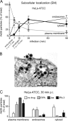
EM analysis of Ad35 entry into HeLa-ATCC cells. (A) Cells on glass coverslips were incubated with doubly CsCl-purified Ad35 particles for 90 min in the cold at high a MOI of 5,000, washed extensively, warmed to 37°C in growth medium containing 0.2% BSA for the indicated times, fixed, and processed for quantitative transmission EM analyses of virus particles at the plasma membrane, in endosomes, and in the cytosol. Half-maximal time points for endocytic uptake and penetration into the cytosol were 7 and 15 min, respectively. (B) Electron micrograph of an Ad35-infected HeLa-ATCC cell 30 min p.i. Stars depict virus particles in the cytosol, arrows depict particles in endosomes, and arrowheads depict particles at the plasma membrane. (C) The same experiment was repeated with cells treated or not treated with the sodium/proton inhibitor EIPA (50 μM), the F-actin stabilizer Jas (300 nM), or the Pak1 inhibitor IPA-3 (25 μM) and infected for 30 min.
FIG. 5.
FIG. 5.
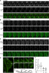
Ad35-TR is associated with GFP-CD46 clusters early in infection. Stably transfected CHO-GFP-CD46 cells were infected with Ad35-TR (1 μg/ml) for 5 min and recorded in a spinning disc confocal microscope equipped with a warm chamber at 37°C from 5 min to 15 min p.i. at an acquisition frequency of 0.06 Hz. (A) Control cell with extended wobbling of GFP-CD46 membrane domains and frequently overlapping signals of CD46 and Ad35-TR. B Cell pretreated with 300 nM jasplakinolide (Jas) for 30 min. (C) Cell pretreated with 25 μM IPA-3. Time stamps are in min (′). (D) Analyses of GFP-CD46 wobbling ruffles, with schematic drawing shown in panel E of an untreated, infected cell with two zoom-in views marked “*” and “**” with the corresponding time stamp min (′) and seconds (′′) p.i. (F) Quantitative box plot analyses of wobbling ruffles in control noninfected cells, infected cells, and infected cells treated with Jas or IPA-3.
FIG. 6.
FIG. 6.
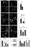
Rac1, Pak1, and CtBP1 are required for Ad35-eGFP transduction of HeLa-ATCC cells. Cells were transfected with the indicated constructs for 24 h and infected for 8 h, fixed, recorded in a wide-field microscope, and analyzed with ImageJ. (A) Representative images of dominant-negative mRFP-T17N-Rac1- or Rac-wt-transfected cells. (B) Quantification of the mean fluorescence intensity per cell with indicated number of cells (n). (C and D) Representative images of Pak1 autoinhibitory domain (AID)-transfected cells infected with Ad35-eGFP or Ad5-eGFP (C) and quantification of the mean fluorescence intensity per cell (D). (E) Representative images of phosphorylation-defective CtBP1-S147A-transfected cells infected with Ad35-eGFP or Ad5-eGFP. (F) Quantification of the mean fluorescence intensities of GFP expression per cell in wild-type (wt) CtBP1 and the S147A and S147D CtBP1 mutants. (G) Cells were transfected with 50 nM siRNA against indicated targets for 72 h, infected for 8 h, and analyzed with Safire2. Cell numbers were normalized to the DAPI signal. (H) Knockdown quantification of Pak1 and CtBP1 by Western blotting, normalized against calnexin. (I) A549 cells were transfected with 20 nM siRNA against CtBP1, Pak1, or dynamin2 for 48 h, infected with Ad35-eGFP (16 h), and analyzed by flow cytometry. K562 cells were transfected with 20 nM siRNA against CtBP1 or clathrin heavy-chain (CHC) as described previously (3), infected with Ad35-eGFP (16 h), and analyzed by flow cytometry.
FIG. 7.
FIG. 7.
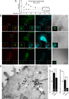
Ad35-TR induces fluid phase uptake and colocalizes with CD46, integrins, and CtBP1 in dextran-filled macropinosomes. (A) Ad35 (2 mg/ml) was cold bound to HeLa-ATCC cells for 1 h. Cells were washed, pulsed with dextran-FITC (0.5 mg/ml) in warm medium (37°C) containing BSA 5 min before the indicated time points, and prepared for flow cytometric analysis. (B) Cells were cold bound with Ad35-TR (1 μg/ml) for 1 h, washed, and incubated at 37°C in the presence of dextran-FITC (0.5 mg/ml) for 10 min, fixed, stained for the indicated antigens, and analyzed by confocal laser scanning microscopy with corresponding differential interference contrast (DIC) images. Fluorescence images represent single sections, and insets are magnifications of the white boxed areas. (C) Electron micrograph of an Ad35-infected HeLa-ATCC cell (cold synchronized infection at a MOI of 5,000), pulsed in the presence of BSA-nanogold for 10 min. Arrowheads indicate BSA-gold particles, black arrows show Ad35 in BSA-gold-positive endosomes, white arrows show virus particles in endosomes without BSA-gold, small triangles indicate viruses at the plasma membrane, and stars depict Ad35 particles in the cytosol. (D) Quantification of panel C, including HeLa-ATCC cells inoculated with Ad2-ts1, which is defective in endosomal escape. “N” indicates the number of virus particles, and “n” indicates the number of BSA-gold particles.
FIG. 8.
FIG. 8.
Schematic model of infectious macropinocytosis of Ad35 in human epithelial cells. Ad35 binds to CD46 and αν integrins independently of heparan sulfate proteoglycans (HSPGs). Ad35 is internalized in an actin-, Rac1-, Pak1-, CtBP1-, and PKC-dependent manner and localizes to dextran-filled vesicles with its receptors CD46, αν integrins, and also CtBP1. Ad35 escapes to the cytosol by an unknown mechanism and traffics to the nucleus for infection. dn, dominant negative; si, small interfering.

Similar articles

Cited by

References

    1. Albert, M. L. 2004. Death-defying immunity: do apoptotic cells influence antigen processing and presentation? Nat. Rev. Immunol. 4:223-231. - PubMed
    1. Albinsson, B., and A. H. Kidd. 1999. Adenovirus type 41 lacks an RGD alpha(v)-integrin binding motif on the penton base and undergoes delayed uptake in A549 cells. Virus Res. 64:125-136. - PubMed
    1. Amstutz, B., M. Gastaldelli, S. Kälin, N. Imelli, K. Boucke, E. Wandeler, J. Mercer, S. Hemmi, and U. F. Greber. 2008. Subversion of CtBP1 controlled macropinocytosis by human adenovirus serotype 3. EMBO J. 27:956-966. - PMC - PubMed
    1. Amyere, M., B. Payrastre, U. Krause, P. V. Smissen, A. Veithen, and P. J. Courtoy. 2000. Constitutive macropinocytosis in oncogene-transformed fibroblasts depends on sequential permanent activation of phosphoinositide 3-kinase and phospholipase C. Mol. Biol. Cell 11:3453-3467. - PMC - PubMed
    1. Araki, N., M. T. Johnson, and J. A. Swanson. 1996. A role for phosphoinositide 3-kinase in the completion of macropinocytosis and phagocytosis by macrophages. J. Cell Biol. 135:1249-1260. - PMC - PubMed

Publication types