. 2010 May 15;341(2):444-58.
doi: 10.1016/j.ydbio.2010.03.001. Epub 2010 Mar 11.

Zebrafish chordin-like and chordin are functionally redundant in regulating patterning of the dorsoventral axis

Affiliations

Zebrafish chordin-like and chordin are functionally redundant in regulating patterning of the dorsoventral axis

Amanda M Branam et al. Dev Biol. .

Abstract

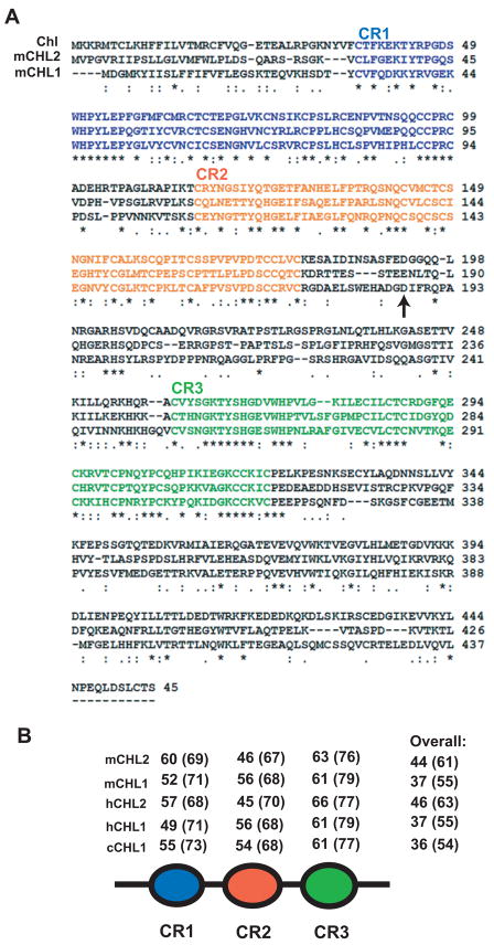
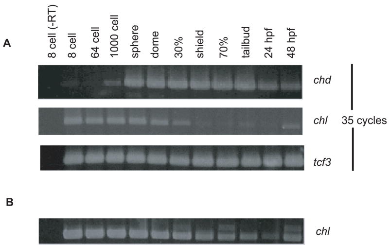
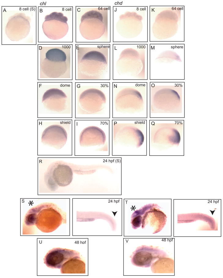
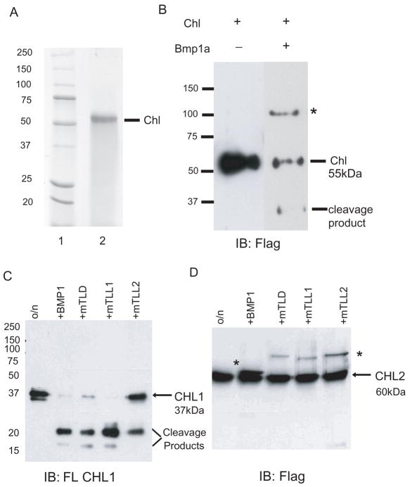
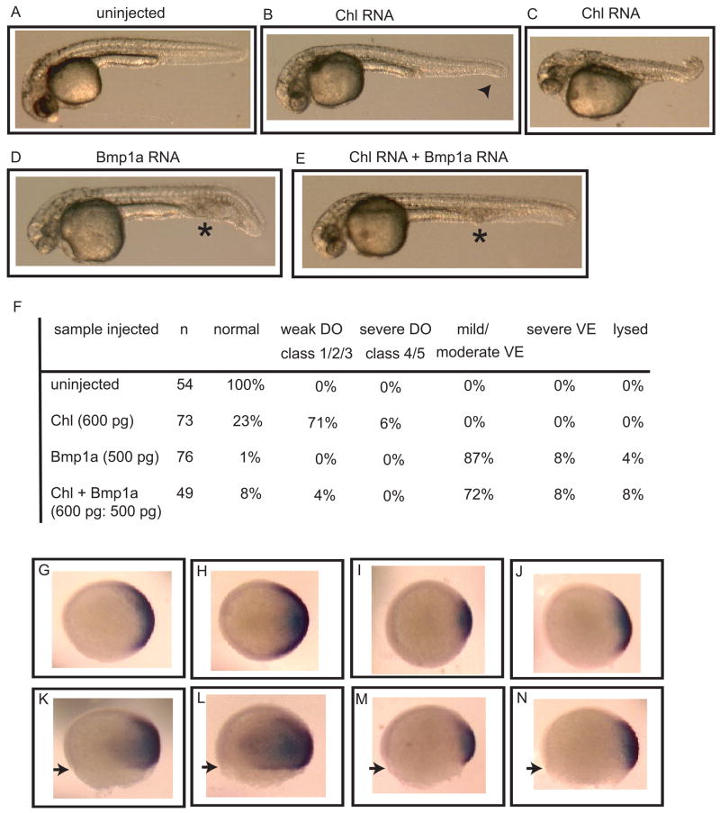
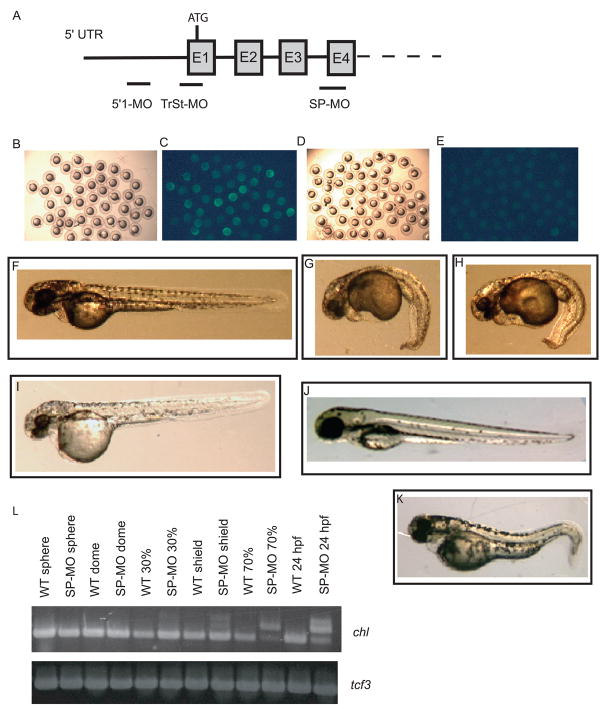
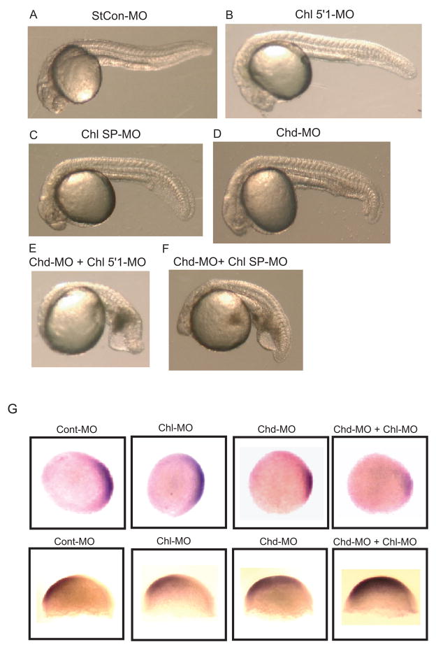
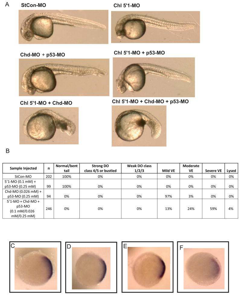
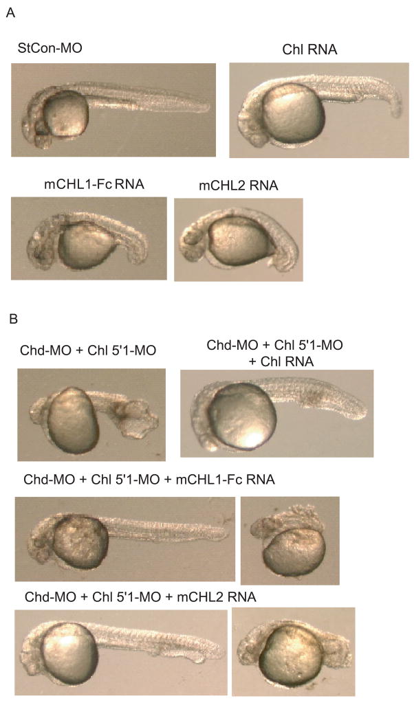
Chordin is the prototype of a group of cysteine-rich domain-containing proteins that bind and modulate signaling of various TGFbeta-like ligands. Chordin-like 1 and 2 (CHL1 and 2) are two members of this group that have been described in human, mouse, and chick. However, in vivo roles for CHL1 and 2 in early development are unknown due to lack of loss-of-function analysis. Here we identify and characterize zebrafish, Danio rerio, CHL (Chl). The chl gene is on a region of chromosome 21 syntenic with the area of murine chromosome 7 bearing the CHL2 gene. Inability to identify a separate zebrafish gene corresponding to the mammalian CHL1 gene suggests that Chl may serve roles in zebrafish distributed between CHL1 and CHL2 in other species. Chl is a maternal factor that is also zygotically expressed later in development and has spatiotemporal expression patterns that differ from but overlap those of zebrafish chordin (Chd), suggesting differences but also possible overlap in developmental roles of the two proteins. Chl, like Chd, dorsalizes embryos upon overexpression and is cleaved by BMP1, which antagonizes this activity. Loss-of-function experiments demonstrate that Chl serves as a BMP antagonist with functions that overlap and are redundant with those of Chd in forming the dorsoventral axis.


Figures

Fig. 1
Fig. 1
Alignment of Chl and murine CHL deduced amino acid sequences. (A) Alignment of the Chl amino acid sequence with those of murine (m) CHL1 and CHL2. An arrow denotes the site at which CHL1 is cleaved by BMP1/Tolloid-like proteinases. (B) Schematic of shared CHL protein domain structure and comparison of percents identity and similarity (parentheses) between CRs of Chl and murine (m), human (h) and chick (c) CHL1 and CHL2. Alignment was with CLUSTAL W. Sequences of the longer/ventroptin-α form of CHL1 (Nakayama et al., 2001; Sakuta et al., 2001) and form of CHL2 of similar length (Nakayama et al., 2004), both of which are similar in length to Chl, were used for the alignment.
Fig. 2
Fig. 2
Comparison of the temporal distributions of Chl and Chd expression during embryogenesis. (A) RT-PCR analysis of Chl and Chd RNA levels was performed for harvested embryos from 8-cell to 48 hpf stages. PCR was for 35 cycles. Expression levels of transcription factor 3 (tcf3) RNA are shown as a loading control. (B) RT-PCR was performed using 40 cycles to demonstrate presence of Chl RNA transcripts from shield stage through 24 hpf.
Fig. 3
Fig. 3
Comparison of the spatial distributions of Chl and Chd expression during embryogenesis. Whole-mount in situ hybridization compares the spatial distribution patterns of Chl (B–I) and Chd expression (J–Q) in 8-cell stage through 70% epiboly embryos. (A) 8-cell embryo hybridized with a sense Chl control probe. Anti-sense probes were hybridized to (B and J) 8-cell, (C and K) 64-cell, (D and L) 1000-cell, (E and M) sphere stage, (F and N) dome stage, (G and O) 30% epiboly, (H and P) shield stage, and (I and Q) 70% epiboly embryos. (R) 24 hpf embryo hybridized with a sense Chl control probe. Signal resulting for hybridization to anti-sense probes for Chl (S and U) and Chd (T and V) is shown for 24 hpf (S and T) and the head regions of 48 hpf (U and V) embryos. Arrowheads mark overlapping Chl and Chd signals in 24 hpf tailbud. Asterisks mark midbrain and hindbrain structures enriched for both Chl (S) and Chd (T) signal at 24 hpf. Similar overlapping signals for Chl (U) and Chd (V) are evident in the brains of 48 hpf embryos.
Fig. 4
Fig. 4
Chl and CHL1 are substrates for BMP1/Tolloid-like proteinases. (A) A Coomassie stained SDS-PAGE gel is shown of protein molecular weight markers (lane 1) and of ~55-kDa secreted and purified Chl (lane 2). (B–D) Immunoblotting with anti-FLAG antibody (B and D) or anti-CHL1 antibody (C) is shown for in vitro assays in which recombinant Chl was incubated in the absence (−) or presence (+) of Bmp1a (B), or in which recombinant short, neuralin form of CHL1 (C) or CHL2 (D) was incubated in the absence (o/n) or presence of mammalian BMP1, mTLD, mTLL1, or mTLL2. Positions of the ~30-kDa Chl (B) and ~20-kDa and smaller CHL1 (C) cleavage products are shown. In B and D, asterisks mark the positions of FLAG-tagged BMP1 and related proteinases. Molecular masses (in kDa) are indicated for protein standards.
Fig. 5
Fig. 5
Effects on dorsoventral patterning by overexpression of Chl in the presence or absence of Bmp1a. Phenotypes are shown of an uninjected 24 hpf control embryo (A), 24 hpf embryos with mild dorsalizaiton (B) or moderate dorsalization (C) after injection with 600 pg Chl RNA, and 24 hpf embryos injected with 500 pg Bmp1a RNA (D), or a combination of 600 pg Chl and 500 pg Bmp1a RNA (E). An arrowhead marks partial tail fin loss, asterisks mark blood pooling; signs of mild dorsalization or ventralization, respectively. (F) Quantification of phenotypes at 24 hpf. Severity of dorsalization (DO) is numerically classified as per Mullins et al. (Mullins et al., 1996). Degree of ventralization (VE) was not numerically classified. Whole-mount in situ hybridization is shown (animal view, panels G–J; lateral view, panels K–N) for expression of the dorsal marker Chd at 70% epiboly for uninjected embryos (G and K) or embryos injected, as above, with RNA for Chl alone (H and L), Bmp1a alone (I and M), or both Chl and Bmp1a (J and N). Arrowheads mark extent of epiboly. All embryos in panels G–N are from the same clutch of eggs and same developmental stage.
Fig. 6
Fig. 6
Treatment with Chl-specific anti-sense MOs. (A) A Schematic is shown representing MOs targeting the Chl translation start site (TrSt-MO), or a non-overlapping region of the chl 5′UTR (5′1-MO), or the acceptor splice site of chl exon 4 (E4). Brightfield (B and D) and darkfield (C and E) exposures are shown of 70% epiboly embryos injected at the one cell stage with 1 ng Chl-GFP RNA and 0.5 mM of either standard control (StCon) MO (B and C) or 5′1-MO (D and E). A comparison at 48 hpf is shown of embryos treated with 0.5 mM standard control (StCon) MO (F), 0.5 mM 5′1-MO (G), 0.5 mM 5′1-MO plus 0.25 mM p53-MO (H), 0.1 mM 5′1-MO (I), or 0.5 mM SP-MO (K), or with. (L) RT-PCR demonstrates the efficacy of SP-MO in inhibiting chl RNA splicing. Inhibition of splicing leading to accumulation of unspliced RNA, represented by one to two lower mobility bands, is clearly seen for shield stage, 70% epiboly, and 24 hpf embryos. Faint bands corresponding to unspliced RNA may also be discerned at 30% epiboly, especially upon comparison of lanes for samples from untreated (WT 30%) and splice site MO-treated (SP-MO 30%) embryos. Expression levels of transcription factor 3 (tcf3) RNA are shown as a loading control.
Fig. 7
Fig. 7
Effects in embryos co-injected with MOs against both Chl and Chd demonstrate overlapping roles for these genes in dorsoventral patterning. A comparison is shown at 24 hpf of embryos treated with 0.5013 mM StCon-MO (A), 0.1 mM Chl 5′1-MO plus 0.0013 mM StCon-MO (B), 0.5 mM Chl SP-MO plus 0.0013 mM StCon-MO (C), or 0.026 mM Chd-MO alone (D), or with combinations of 0.1 mM Chl 5′1-MO plus 0.026 mM Chd-MO (E), or 0.5 mM Chl SP-MO plus 0.026 mM Chd-MO (F). Single injections of 5′1-MO (B) and SP-MO (C) had added StCon-MO to match total amounts of MOs used in the co-injection experiments (E and F). (G) In situ hybridization shows altered expression patterns at 30% epiboly for the dorsal marker Chd (top panel) and at 70% epiboly for the ventral marker Gata2 (bottom panel) in response to treatment of embryos with the Chd MO (CHD-MO) and Chl 5′1′MO (Chl-MO), in comparison to embryos treated with StCon-MO (Cont-MO). Concentrations of MOs used were the same as those used in panel A. Note the increased reduction of the Chd expression domain and enlargement of the Gata2 expression domain in response to co-injection with the Chd and Chl MOs.
Fig. 8
Fig. 8
Synergistic effects of Chl and Chd MOs in inducing ventralization does not involve p53-mediated off target effects. (A) A comparison is shown of 24 hpf embryos treated with 0.38 mM StCon-MO, 0.1 mM Chl 5′1-MO, 0.026 mM Chd-MO plus 0.25 mM p53-MO, 0.1 mM Chl 5′1-MO plus 0.026 mM Chd-MO, or with 0.1 mM Chl 5′1 plus 0.026 mM Chd-MO and 0.25 mM p53-MO. (B) Quantification of phenotypes at 24 hpf. Severity of dorsalization (D) is numerically classified as per Mullins et al. (Mullins et al., 1996). Degree of ventralization (VE) was not numerically classified. (C–D) Whole mount in situ hybridization is shown for expression of the dorsal marker Chl at 30% epiboly for embryos treated with 0.38 mM StCon-MO (C), 0.1 mM Chl 5′1-MO plus 0.25 mM p53-MO (D), 0.026 mM Chd-MO plus 0.25 mM p53-MO (E), or 0.1 mM Chl 5′1 plus 0.026 mM Chd-MO and 0.25 mM p53-MO.
Fig. 9
Fig. 9
Chl, and murine CHL1 and CHL2 can rescue embryos ventralized by co-injection of Chl and Chd MOs. (A) Embryos were dorsalized by injection of RNA encoding Chl, murine CHL1 fused to an Fc domain (mCHL1-Fc), or CHL2 (mCHL2) (1 ng, 80 pg, or 1 pg, respectively). Also shown, for comparison, is a representative embryo injected with 0.1 mM of StCon-MO. (B) Embryos co-injected with Chd-MO and Chl 5′1-MO are for the most part moderately to severely ventralized, whereas overexpression of Chl RNA rescues embryos co-injected with Chd-MO and Chl 5′1-MO, such that many are only mildly ventralized (shown) or weakly or strongly dorsalized (not shown). Overexpression of murine CHL1-Fc (mCHL1-Fc) or murine CHL2 (mCHL2) also rescued embryos co-injected with Chd-MO and Chl 5′1-MO, resulting in a range of phenotypes that included mildly ventralized [left] and severely dorsalized [right].

Similar articles

Cited by

References

    1. Bachiller D, Klingensmith J, Kemp C, Belo JA, Anderson RM, May SR, McMahon JA, McMahon AP, Harland RM, Rossant J, De Robertis EM. The organizer factors Chordin and Noggin are required for mouse forebrain development. Nature. 2000;403:658–61. - PubMed
    1. Blader P, Rastegar S, Fischer N, Strahle U. Cleavage of the BMP-4 antagonist chordin by zebrafish tolloid. Science. 1997;278:1937–40. - PubMed
    1. Coffinier C, Tran U, Larrain J, De Robertis EM. Neuralin-1 is a novel Chordin-related molecule expressed in the mouse neural plate. Mech Dev. 2001;100:119–22. - PubMed
    1. Dal-Pra S, Furthauer M, Van-Celst J, Thisse B, Thisse C. Noggin1 and Follistatin-like2 function redundantly to Chordin to antagonize BMP activity. Dev Biol. 2006;298:514–26. - PubMed
    1. De Robertis EM, Kuroda H. Dorsal-ventral patterning and neural induction in Xenopus embryos. Annu Rev Cell Dev Biol. 2004;20:285–308. - PMC - PubMed

Publication types

MeSH terms

Substances

LinkOut - more resources