. 2010 May 7;285(19):14088-100.
doi: 10.1074/jbc.M109.017129. Epub 2010 Mar 10.

Induction of neutrophil gelatinase-associated lipocalin expression by co-stimulation with interleukin-17 and tumor necrosis factor-alpha is controlled by IkappaB-zeta but neither by C/EBP-beta nor C/EBP-delta

Affiliations

Induction of neutrophil gelatinase-associated lipocalin expression by co-stimulation with interleukin-17 and tumor necrosis factor-alpha is controlled by IkappaB-zeta but neither by C/EBP-beta nor C/EBP-delta

Joachim R Karlsen et al. J Biol Chem. .

Abstract

Neutrophil gelatinase-associated lipocalin (NGAL) is a siderophore-binding antimicrobial protein that is up-regulated in epithelial tissues during inflammation. We demonstrated previously that the gene encoding NGAL (LCN2) is strongly up-regulated by interleukin (IL)-1beta in an NF-kappaB-dependent manner but not by tumor necrosis factor (TNF)-alpha, another potent activator of NF-kappaB. This is due to an IL-1beta-specific synthesis of the NF-kappaB-binding co-factor IkappaB-zeta, which is essential for NGAL induction. We demonstrate here that NGAL is strongly induced by stimulation with TNF-alpha in the presence of IL-17, a pro-inflammatory cytokine produced by the newly discovered subset of CD4(+) T helper cells, T(H)-17. In contrast to the murine NGAL orthologue, 24p3/lipocalin 2, we found no requirement for C/EBP-beta or C/EBP-delta for NGAL induction by IL-17 and TNF-alpha as neither small interfering RNAs against the two C/EBP mRNAs nor mutation of the C/EBP sites in the LCN2 promoter abolished IL-17- and TNF-alpha-induced up-regulation of NGAL. NGAL induction is governed solely by NF-kappaB and its co-factor IkappaB-zeta. This was demonstrated by a pronounced reduction in the amount of NGAL mRNA and NGAL protein synthesized in cells treated with small interfering RNA against IkappaB-zeta and a total lack of activation of an LCN2 promoter construct with a mutated NF-kappaB site. As IL-17 stimulation stabilizes the IkappaB-zeta transcript, we propose a model where TNF-alpha induces activation and binding of NF-kappaB to the promoters of both NFKBIZ and LCN2 genes but induce only transcription of IkappaB-zeta. Co-stimulation with IL-17 leads to accumulation of IkappaB-zeta mRNA and IkappaB-zeta protein, which can bind to NF-kappaB on the LCN2 promoter and thus induce NGAL expression.


Figures

FIGURE 1.
FIGURE 1.
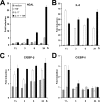
Induction of NGAL synthesis by co-stimulation with IL-17 and TNF-α. A, cells were harvested at time “0” and at 24 h after addition of fresh medium without cytokines or supplemented with IL-1β (100 pg/ml), TNF-α (20 ng/ml), IL-17 (200 ng/ml), or IL17 and TNF-α (200 and 20 ng/ml). RNA was isolated and hybridized to 32P-labeled probes as indicated. B, schematic representation of the fold-induction of NGAL mRNA in A following normalization to β-actin intensities. The fold-induction is shown relative to the amount measured at time 0. C and D, the amount of NGAL and IL-8 in the medium of A549 cells was determined at the indicated time points after addition of fresh medium without cytokines or supplemented with IL-17 and/or TNF-α in the concentrations given. Data from one of two independent experiments showing essentially the same results are presented. Data are presented as the mean ± S.D.
FIGURE 2.
FIGURE 2.
Time courses of NGAL, IL-8, C/EBP-β, and C/EBP-δ mRNAs following stimulation with IL-17 and/or TNF-α. The amount of NGAL (A), IL-8 (B), C/EBP-β (C), and C/EBP-δ (D) mRNA was determined by real time PCR at the indicated time points after addition of fresh medium without cytokines or supplemented with TNF-α, IL-17, or IL-17 and TNF-α at the concentrations indicated in the legend to Fig. 1. Expression of the transcripts is shown as fold-induction relative to expression measured in unstimulated cells at time 0. Data are presented as the mean ± S.D.
FIGURE 3.
FIGURE 3.
Knockdown of C/EBP-β expression does not affect NGAL expression. Untransfected A549 cells (−) or cells transfected with 40 nmol of two different siRNAs against C/EBP-β (siB1 and siB2) or control siRNA (cntl) and stimulated with IL-17 (200 ng/ml) and TNF-α (20 ng/ml) for 2 h were analyzed for mRNA (A) and protein expression (B). A, the amount of C/EBP-β mRNA, as determined by real time PCR, showed a 2–2½-fold induction for untransfected and Cntl siRNA-transfected cells relative to unstimulated cells. In contrast, cells treated with siB1 and siB2 showed a C/EBP-β mRNA level about 20% of that found in cells with Cntl siRNA after stimulation with the two cytokines. B, analysis of total cell lysates showed a similar strong reduction in the amount of C/EBP-β protein in siB1- and siB2-transfected cells. Both the 35- and 20-kDa forms of C/EBP-β was affected. C, a new series of experiments again showed a significant reduction of C/EBP-β mRNA as well as of the IL-6 mRNA in cells treated with siB1 and stimulated for 2 h with IL-17 and TNF-α, whereas the amounts of C/EBP-δ and IL-8 mRNAs were unaffected. D, at 24 h post-stimulation the levels of C/EBP-β and IL-6 mRNAs was still reduced in siB1-transfected cells, whereas the transcripts for C/EBP-δ, IL-8, and NGAL were unaffected by the treatment. E, this observation also holds true for NGAL, IL-6, and IL-8 also when measuring the amount of these three proteins in the medium of the cytokine-stimulated cells, as comparable levels were found for NGAL and IL-8 irrespective of whether the cells were transfected with siRNA or not, whereas the amount of secreted IL-6 was strongly diminished from siB1-treated cells. The real time PCR data are shown relative to the most highly expressed transcript in each experiment. Data are presented as the mean ± S.D. GAPDH, glyceraldehyde-3-phosphate dehydrogenase.
FIGURE 4.
FIGURE 4.
Knockdown of C/EBP-δ expression does not affect NGAL expression. Untransfected A549 cells (−) or cells transfected with 40 nmol of two different siRNAs against C/EBP-δ (siD1 and siD2) or control siRNA (cntl) and stimulated with IL-17 (200 ng/ml) and TNF-α (20 ng/ml) for 2 h were analyzed for mRNA (A) and protein expression (B). A, the amount of C/EBP-δ mRNA was strongly induced after 2 h stimulation with IL-17 and TNF-α in untransfected and Cntl siRNA-transfected cells relative to unstimulated cells (as seen also in Fig. 2). However, if the stimulated cells were transfected with siD1 and siD2, the level was 4 to 5 times lower. B, transfection with siD1 or siD2 in the same manner affected the amount of C/EBP-δ protein measured in total cell lysates after a 2-h stimulation. C, transfection with siD1 and stimulation with the two cytokines for 2 h reduced the levels of C/EBP-δ and IL-6 mRNAs without having any effect on the levels of the C/EBP-β and IL-8 trancripts. D, when analyzing the cells after 24 h of stimulation with IL-17 and TNF-α, the amount of C/EBP-δ and IL-6 mRNAs was still lower in siD1-treated cells, whereas the levels of C/EBP-β, IL-8, and NGAL mRNAs were very similar in all cytokine-treated cells. E, the amount of secreted NGAL, IL-6, and IL-8 from the cells were affected in the same manner as their cognate transcripts by siD1 treatment compared with Cntl siRNA-transfected and untransfected cells. The real time PCR data are shown relative to the most highly expressed transcript in each experiment. Data are presented as the mean ± S.D. GAPDH, glyceraldehyde-3-phosphate dehydrogenase.
FIGURE 5.
FIGURE 5.
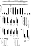
Activity of the LCN2 promoter in stimulated A549 cells. Top, DNA sequence and putative regulatory consensus elements of the −200 to 1 region of the LCN2 gene (6). The underlined sequences denote putative transcription factor recognition sites and the numbers above the sequence show the end points of the −183, −153, and −120 deletion mutants. The transcriptional start site is indicated with a dot. The extent of the substituted sequences Sub C1, C2, and C1-C2 are shown with bars under the sequence. A, fold-induction of the 1695-bp LCN2 promoter following stimulation with IL-17, TNF-α, and IL-17 + TNF-α shown relative to the activity from unstimulated cells (−). B, LCN2 promoter with deletions ranging from −1695 to −120, as denoted under the columns, was transfected into A549 cells and stimulated with IL-17 (200 ng/ml) and TNF-α (20 ng/ml) for 24 h. The fold-induction following stimulation is shown. Deleting the region below −183 abolished the ability of the promoter to be induced by the cytokines. C, relative CAT activities of promoter constructs in unstimulated cells with point mutations or substitutions of different transcription factor binding sites. The activities are shown relative to the wild type −1695 promoter, which was given the value “1.” D, fold-induction of the point mutations and substitutions following stimulation with IL-17 and TNF-α for 24 h. Fold-induction in CAT activity of pCAT3-promoter (SV40) and the same construct with four tandem repeats of the NF-κB sequence (4 × κB) from the IL8 promoter (E) or IL6 promoter (F) inserted upstream of the SV40 basal promoter. The fold-induction was determined for IL-1β, TNF-α, and IL-17-stimulated cells relative to the promoter activities of the unstimulated SV40 and “4 × κB” constructs, respectively. G, relative firefly luciferase activities of the 24p3–282-luc (wt), 24p3–282-κBm-luc (κBm), and 24p3–282-C/EBPm-luc (C/EBPm) promoter constructs in unstimulated cells. The activities are shown relative to the wild type Lcn2 (24p3) promoter, which was given the value 1. H, fold-induction in firefly luciferase activity of the wt, κBm, and C/EBPm promoter constructs following stimulation with IL-17 and TNF-α or IL-1β relative to the promoter activities of the unstimulated promoter constructs. All results are the mean ± S.D. of three independent transfections. The CAT activities were normalized to the firefly luciferase activity from the co-transfected vector Rous sarcoma virus-Luc. The firefly luciferase activities of the Lcn2 promoter constructs were normalized to the Renilla luciferase activity of the co-transfected vector pGL4.74. Data are presented as the mean ± S.D.
FIGURE 6.
FIGURE 6.
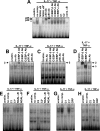
Binding of nuclear complexes to the κB and C/EBP elements of the LCN2 promoter. Nuclear extracts from A549 cells that were either not stimulated (unstim.) or stimulated with IL-17 (200 ng/ml) and TNF-α (20 ng/ml) for 1½ h (IL-17 + TNF-α) were used for EMSA. A, a 32P-labeled probe containing the κB element of the LCN2 promoter was incubated with either no competitor (no comp.) or a 250-fold excess of an unlabeled oligo, which was identical in sequence to the probe (NGAL κB), only differed by a mutated κB element (NGAL κB*), contained sequences of the C/EBP-1 (C1) or C/EBP-2 (C2) elements of the LCN2 promoter, or carried a consensus κB sequence (cons. κB). Antibodies against p50, p65, C/EBP-β, C/EBP-δ, and C/EBP-ϵ were tested for their ability to cause a supershift of the nuclear complex associated with the probe. Supershifts with the p50 and p65 antibodies are shown with an arrow. The specific band formed on the NGAL-κB probe with the nuclear extract from cytokine-stimulated cells is indicated with an asterisk. B, a probe containing the C/EBP-1 element of the LCN2 promoter (C1) was incubated without antibody (no Ab) or with antibodies against C/EBP-β, C/EBP-δ, C/EBP-ϵ, p50, or p65. Supershift with anti-C/EBP-β is indicated with an arrow. C, an experiment similar to that in B was performed with a probe containing the C/EBP-2 element of the LCN2 promoter (C2). A supershift band was also in this case seen with anti-C/EBP-β (arrow). D, a control for supershift using the CRP oligo as probe. Supershift was observed for anti-C/EBP-β and anti-C/EBP-δ. E, the C1 probe was incubated with either no competitor (no comp.) or a 250-fold excess of an unlabeled oligo, which was identical in sequence to the probe (C1), had a 2-base pair mutation in the C/EBP-binding element (C1*), or contained the wild-type (NGAL κB) or mutated (NGAL κB*) NGAL-κB sequence. F, identical to the experiment in E except for C1 being exchanged by C2. G and H, identical to the experiments in E and F except for the NGAL-κB oligoes being substituted by the C/EBP-containing oligo CPR.
FIGURE 7.
FIGURE 7.
Induction of NGAL mRNA synthesis by co-stimulation with IL-17 and TNF-α requires de novo protein synthesis. Cells were harvested at the indicated time points after addition of fresh medium without cytokines or supplemented with IL-17 (200 ng/ml) and TNF-α (20 ng/ml) in the presence or absence of 10 μg/ml of cycloheximide to abolish synthesis of protein. Cycloheximide was added 30 min prior to stimulation with the cytokines. RNA was isolated and analyzed by real time PCR for transcripts of NGAL, IκB-ζ, and IL-8. A significantly higher level of NGAL mRNA was observed in cytokine-stimulated cells without cycloheximide at 5 and 8 h post-stimulation than in cytokine-stimulated cells also receiving cycloheximide; *, p = 0.047 and **, p = 0.031. Errors bars show the S.D. for each experiment.
FIGURE 8.
FIGURE 8.
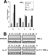
Time courses of IκB-ζ mRNA and protein levels following stimulation with IL-17 and/or TNF-α. The amount of IκB-ζ mRNA (A) and protein (B) was determined by real time PCR (A) or Western blot (B) at the indicated time points after addition of fresh medium without cytokines or supplemented with TNF-α, IL-17, or IL-17 and TNF-α in the concentrations indicated in the legend to Fig. 1. The expression of the transcripts is shown as fold-induction relative to the expression measured in unstimulated cells at time 0. A control (a lysate from cells stimulated 1½ h with IL-17 + TNF-α) was included on the blot for unstimulated and TNF-α-stimulated cells to ensure that the IκB-ζ protein could be detected under the experimental conditions employed (data not shown). Real time data are presented as the mean ± S.D.
FIGURE 9.
FIGURE 9.
Knockdown of IκB-ζ expression causes a decrease in NGAL expression. Untransfected A549 cells (−) or cells transfected with 40 nmol of IκB-ζ-siRNA (siκB) or control siRNA (cntl) were grown in medium with or without IL-17 (200 ng/ml) and TNF-α (20 ng/ml) for 2 (A and B) or 24 h (C and D) and then harvested for total RNA and protein isolation. The siRNA against IκB-ζ has been used previously (18). A, the amount of IκB-ζ and IL-8 mRNA was determined by real time PCR and demonstrated a reduction in IκB-ζ in siκB-treated cells. B, protein (whole cell lysates) from cells treated as above was analyzed by Western blot with antibodies against IκB-ζ and glyceraldehyde-3-phosphate dehydrogenase (GAPDH) and demonstrated a significant knockdown of IκB-ζ in siκB-treated cells. C, the amount of NGAL and IL-8 mRNA in cells stimulated for 24 h was determined by real time PCR and demonstrated a strong reduction of NGAL mRNA in cells that had received siκB. D, analogous to the mRNA profiles a significant drop of NGAL was measured in the medium of stimulated cells containing siκB. Treatment with siκB did not affect the amount of IL-8 secreted to the medium. All real time PCR data are shown relative to the most highly expressed transcript in each experiment. Data are presented as the mean ± S.D.
FIGURE 10.
FIGURE 10.
Model for up-regulation of NGAL by co-stimulation with IL-17 and TNF-α. Stimulation of the TNF-α receptor (TNF-R) induces activation and translocation of NF-κB to the promoters of the NFKBIZ (IκB-ζ) and LCN2 (NGAL) genes. Binding of NF-κB to the NFKBIZ promoter induces IκB-ζ transcription but the IκB-ζ mRNA is unstable and rapidly degraded. Binding of NF-κB to the LCN2 promoter is not sufficient to initiate transcription. Stimulation of the IL-17 receptor (IL-17-R) generates an intracellular signal that stabilizes the IκB-ζ transcript and allows translation of IκB-ζ protein. The newly synthesized IκB-ζ is then able to translocate and bind to NF-κB on the LCN2 promoter and thereby initiate transcription of the gene. Ligation of the IL-1 receptor (IL-1-R) generates signals that both activate NF-κB and stabilize the IκB-ζ mRNA. This explains why NGAL can be induced by stimulation with IL-1β alone.

Similar articles

Cited by

References

    1. Cowland J. B., Sørensen O. E., Sehested M., Borregaard N. (2003) J. Immunol. 171, 6630–6639 - PubMed
    1. Sørensen O. E., Cowland J. B., Theilgaard-Mönch K., Liu L., Ganz T., Borregaard N. (2003) J. Immunol. 170, 5583–5589 - PubMed
    1. Sørensen O. E., Thapa D. R., Rosenthal A., Liu L., Roberts A. A., Ganz T. (2005) J. Immunol. 174, 4870–4879 - PubMed
    1. Kao C. Y., Chen Y., Thai P., Wachi S., Huang F., Kim C., Harper R. W., Wu R. (2004) J. Immunol. 173, 3482–3491 - PubMed
    1. Kjeldsen L., Bainton D. F., Sengeløv H., Borregaard N. (1994) Blood 83, 799–807 - PubMed

Publication types

MeSH terms