. 2010 May 1;21(9):1470-81.
doi: 10.1091/mbc.e09-06-0486. Epub 2010 Mar 10.

Dissecting the M phase-specific phosphorylation of serine-proline or threonine-proline motifs

Affiliations

Dissecting the M phase-specific phosphorylation of serine-proline or threonine-proline motifs

Chuan Fen Wu et al. Mol Biol Cell. .

Abstract

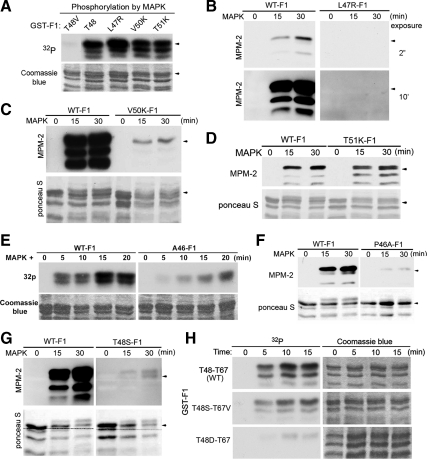
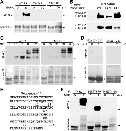
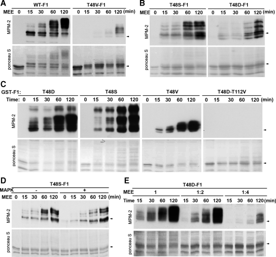
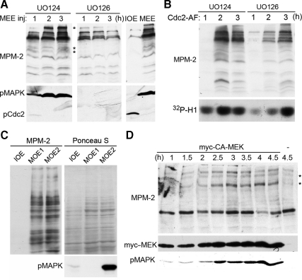
M phase induction in eukaryotic cell cycles is associated with a burst of protein phosphorylation, primarily at serine or threonine followed by proline (S/TP motif). The mitotic phosphoprotein antibody MPM-2 recognizes a significant subset of mitotically phosphorylated S/TP motifs; however, the required surrounding sequences of and the key kinases that phosphorylate these S/TP motifs remain to be determined. By mapping the mitotic MPM-2 epitopes in Xenopus Cdc25C and characterizing the mitotic MPM-2 epitope kinases in Xenopus oocytes and egg extracts, we have determined that phosphorylation of TP motifs that are surrounded by hydrophobic residues at both -1 and +1 positions plays a dominant role in M phase-associated burst of MPM-2 reactivity. Although mitotic Cdk and MAPK may phosphorylate subsets of these motifs that have a basic residue at the +2 position and a proline residue at the -2 position, respectively, the majority of these motifs that are preferentially phosphorylated in mitosis do not have these features. The M phase-associated burst of MPM-2 reactivity can be induced in Xenopus oocytes and egg extracts in the absence of MAPK or Cdc2 activity. These findings indicate that the M phase-associated burst of MPM-2 reactivity represents a novel type of protein phosphorylation in mitotic regulation.


Figures

Figure 1.
Figure 1.
The MAPK phosphorylation site T48 resides in a strong MPM-2 epitope. (A) Diagrams of GST-tagged xCdc25C fragments used as substrates in phosphorylation. (B) GST-tagged N and C fragments of xCdc25C were simultaneously phosphorylated with 1:5-diluted MEE for 30 min, washed, gel-separated, and immunoblotted with MPM-2. Total proteins were stained by Ponceau S. (C) GST-tagged F1, F2, and F3 fragments of xCdc25C were treated as described for B. (D) The wild type (WT) and the T48V mutant form of GST-F1 were phosphorylated with activated recombinant MAPK in the presence of [γ-32P]ATP, and washed substrates were gel-separated and then subjected to autoradiography or immunoblotted with MPM-2 and anti-GST antibodies. (E) The activated recombinant MAPK and semirecombinant Cdc2/cyclin B were adjusted to similar levels of MBP-phosphorylating activity as determined by 32P incorporation. (F) The wild type (WT) and the T48V mutant form of GST-xCdc25C were phosphorylated with activated recombinant MAPK and semirecombinant Cdc2/cyclin B used in E in the presence of [γ-32P]ATP, and washed substrates were gel-separated and the subjected to autoradiography or immunoblotted with MPM-2.
Figure 2.
Figure 2.
A phosphorylated TP motif that is surrounded by hydrophobic residues at both −1 and +1 positions generates strong MPM-2 reactivity. (A) The wild type (WT) and indicated mutant forms of GST-F1 were phosphorylated with activated recombinant MAPK for 20 min in the presence of [γ-32P]ATP, and washed substrates were gel-separated, stained with Coomassie blue, and subjected to autoradiography. (B) The wild type (WT) and the L47R mutant form of GST-F1 were phosphorylated with activated recombinant MAPK for indicated minutes, and washed substrates were gel-separated and immunoblotted with MPM-2. (C) The wild type (WT) and the V50K mutant form of GST-F1 were treated as described for B. Total proteins were stained by Ponceau S. (D) The wild type (WT) and the T51K mutant form of GST-F1 were treated as described for C. (E) The wild type and the P46A mutant forms of GST-F1 were phosphorylated with activated recombinant MAPK in the presence of [γ-32P]ATP, and washed substrates were gel-separated, stained with Coomassie blue and subjected to autoradiography. (F) The wild type (WT) and the P46A mutant forms of GST-F1 were treated as described for C. (G) The wild type (WT) and the T48S mutant form of GST-F1 were treated as described for C. (H) The wild type (WT) and indicated mutant forms of GST-F1 were treated as described for E.
Figure 3.
Figure 3.
The novel phosphorylation site T112 also resides in a strong MPM-2 epitope. (A) The wild type (WT) and indicated mutant forms of GST-F1 were phosphorylated with activated recombinant MAPK for indicated minutes, and washed substrates were gel-separated and immunoblotted with MPM-2. Total proteins were stained by Ponceau S. (B) Xenopus oocytes were injected with mRNAs for the wild type (WT) or the T48D or T48V mutant form of myc-xCdc25C. Extracts of mature oocytes were immunoprecipitated with MPM-2 or anti-myc antibodies, and the immunocomplexes were immunoblotted with anti-myc antibodies. (C) The wild type (WT) and T48S mutant form of GST-F1 were phosphorylated with 1:5-diluted MEE for indicated minutes, and the washed and gel-separated substrates were immunoblotted with MPM-2 after protein staining with Ponceau S. (D) GST-F2 and GST-F3 were phosphorylated with 1:5-diluted MEE for 2 h or were mock-treated and washed substrates were gel-separated and immunoblotted with MPM-2 after protein staining with Ponceau S. (E) F1 contains two additional TP motifs in addition to the T48-containing TP motif. (F) The wild type (WT) and T67V or T112V mutant form of T48S-F1 were treated as described for D.
Figure 4.
Figure 4.
Phosphorylation of the T112-containing MPM-2 epitope is facilitated by the prior phosphorylation at T48. (A) The wild type (WT) and the T48V mutant form of GST-F1 were phosphorylated with 1:5-diluted MEE for indicated minutes, and washed substrates were gel-separated and immunoblotted with MPM-2 after protein staining with Ponceau S. (B) The T48S and the T48D mutant forms of GST-F1 were treated as described for A. (C) Indicated mutant forms of GST-F1 were phosphorylated with undiluted MEE for indicated minutes, and washed and gel-separated substrates were immunoblotted with MPM-2 after protein staining with Ponceau S. (D) The T48S mutant form of GST-F1 was first phosphorylated with recombinant MAPK or mock-treated, and then phosphorylated with 1:4-diluted MEE for indicated minutes. The washed and gel-separated substrates were immunoblotted with MPM-2 after protein staining with Ponceau S. (E) The T48D mutant form of GST-F1 was phosphorylated with indicated dilutions of MEE for indicated minutes, and washed substrates were gel-separated and immunoblotted with MPM-2 after protein staining with Ponceau S.
Figure 5.
Figure 5.
Neither MAPK nor Cdc2 is responsible for the phosphorylation of the T112-containing MPM-2 epitope. (A) Xenopus oocytes cultured in the absence or continued presence of UO126 were first injected with a nondegradable cyclin B and then stimulated by progesterone. Extracts of oocytes collected at the indicated hours after progesterone stimulation were immunoblotted with antibodies for activated-MAPK (pMAPK), total MAPK, and inactive Cdc2 (pCdc2). UO126-treated mature oocyte extracts (MOE1) and non-UO126-treated mature oocyte extracts (MOE2) were used for phosphorylation in B. (B) The T48D mutant form of GST-F1 was phosphorylated with MOE1 and MOE2 described in A for the indicated minutes, and the washed substrates were gel-separated and immunoblotted with MPM-2 after protein staining with Ponceau S. (C) Undiluted MEE was incubated with DMSO, 300 nM RO-3306, or 60 μM roscovitine (ROSCO) for indicated minutes, and histone H1 kinase activity was determined by 32P incorporation. (D) The T48D mutant form of GST-F1 was phosphorylated with undiluted MEE supplemented with DMSO, 300 nM RO-3306 or 60 μM roscovitine (ROSCO) for indicated minutes. The washed substrates were gel-separated and immunoblotted with MPM-2 after protein staining with Ponceau S.
Figure 6.
Figure 6.
MAPK-catalyzed phosphorylation of the MPM-2 epitope does not play a major role in the M phase–associated burst of MPM-2 reactivity. (A) Xenopus oocytes cultured in the continued presence of UO126 or UO124 were injected with MEE, and extracts of oocytes collected at the indicated hours after the injection were immunoblotted with MPM-2 and antibodies that recognize phosphorylated/activated MAPK (pMAPK) or phosphorylated/inactivated Cdc2 (pCdc2). Asterisk indicates the MPM-2–reactive proteins inhibited by UO126. (B) Xenopus oocytes cultured in the continued presence of UO126 or UO124 were injected with mRNA for Cdc2-AF, and extracts of oocytes collected at the indicated hours after the injection were immunoblotted with MPM-2 and assayed for H1 kinase activity by 32P incorporation. (C) Xenopus oocytes were treated as described for Figure 5A, and IOE, MOE1, and MOE2 were immunoblotted with MPM-2 and antibodies that recognize phosphorylated/activated MAPK (pMAPK) after total proteins were stained with Ponceau S. (D) Extracts of oocytes collected at the indicated hours after injection of below M phase–inducing levels of CA-MEK were immunoblotted with MPM-2 and antibodies that recognize phosphorylated/activated MAPK (pMAPK) or myc-epitope tag. Asterisk, the MPM-2–reactive proteins induced by activated MAPK.
Figure 7.
Figure 7.
The Cdc2-catalyzed phosphorylation of the MPM-2 epitope does not play a major role in the M phase–associated burst of MPM-2 reactivity. (A) Extracts of oocytes collected at indicated hours after injection of Xenopus Wee1 or Xp95 (control) RNA were immunoblotted with MPM-2, anti-xCdc25C antibodies or antibodies that recognize phosphorylated/inactivated Cdc2 (pCdc2). Asterisk indicates an MPM-2–reactive band inhibited by Wee1. (B) After IE was diluted with an equal volume of EB or XB and incubated with OA for indicated minutes, samples were immunoblotted along with MEE by MPM-2 and anti-xCdc25C antibodies. Arrow, the position of interphase xCdc25C; asterisk, gel mobility–shifted xCdc25; and ×, a nonspecific band recognized by anti-xCdc25C antibodies. (C) After MEE was mixed with DMSO, RO-3306, or roscovitine (ROSCO), histone H1 kinase activity was determined by 32P incorporation. (D) After IE was mixed with DMSO, RO-3306, or roscovitine (ROSCO), it was diluted with EB and incubated with OA for the indicated minutes. Samples were gel-separated and immunoblotted with MPM-2 after protein staining with Ponceau S. (E) After the T48D mutant form of GST-F1 was incubated with control buffer, MEE, or IE treated with EB plus OA, washed substrates were gel-separated and immunoblotted with MPM-2 after protein staining with Ponceau S. (F) After IE was incubated with XB or EB plus OA for 30 min, samples were immunoblotted with MPM-2 and determined for phosphorylation of histone H1 by 32P incorporation. (G) Indicated reagents were added to XB-diluted IE, and samples collected at 30 min were immunoblotted with MPM-2 in parallel with MEE.

Similar articles

Cited by

References

    1. Alvarez E., Northwood I. C., Gonzalez F. A., Latour D. A., Seth A., Abate C., Curran T., Davis R. J. Pro-Leu-Ser/Thr-Pro is a consensus primary sequence for substrate protein phosphorylation. Characterization of the phosphorylation of c-myc and c-jun proteins by an epidermal growth factor receptor threonine 669 protein kinase. J. Biol. Chem. 1991;266:15277–15285. - PubMed
    1. Brittle A. L., Ohkura H. Centrosome maturation: Aurora lights the way to the poles. Curr. Biol. 2005;15:R880–R882. - PubMed
    1. Campbell L. E., Proud C. G. Differing substrate specificities of members of the DYRK family of arginine-directed protein kinases. FEBS Lett. 2002;510:31–36. - PubMed
    1. Cano E., Mahadevan L. C. Parallel signal processing among mammalian MAPKs. Trends Biochem. Sci. 1995;20:117–122. - PubMed
    1. Che S., El-Hodiri H. M., Wu C. F., Nelman-Gonzalez M., Weil M. M., Etkin L. D., Clark R. B., Kuang J. Identification and cloning of xp95, a putative signal transduction protein in Xenopus oocytes. J. Biol. Chem. 1999;274:5522–5531. - PubMed

Publication types

MeSH terms