. 2010 May;151(5):2361-72.
doi: 10.1210/en.2009-1338. Epub 2010 Mar 9.

Activity of retinoic acid receptor-alpha is directly regulated at its protein kinase A sites in response to follicle-stimulating hormone signaling

Affiliations

Activity of retinoic acid receptor-alpha is directly regulated at its protein kinase A sites in response to follicle-stimulating hormone signaling

Nadine C Santos et al. Endocrinology. 2010 May.

Abstract

Retinoic acid receptor-alpha (RARA) is crucial for germ cell development in the testis, as shown by the degenerated testis in Rara gene knockout mice, which are sterile. Similarly, FSH is known to regulate Sertoli cell proliferation and differentiation, indirectly controlling the quantity of the spermatogenic output. Interestingly, FSH inhibited, via activation of FSH receptor, cAMP, and protein kinase A (PKA), the nuclear localization and transcriptional activity of RARA. Given that retinoic acid, the ligand for RARA, is known to regulate cell proliferation and differentiation, we investigated whether FSH regulates RARA by a direct posttranslational phosphorylation mechanism. Mutagenesis of serine 219 (S219) and S369 at the PKA sites on RARA to either double alanines or double glutamic acids showed that both PKA sites are important for RARA activity. The negative charges at the PKA sites, whether they are from glutamic acids or phosphorylation of serines, decreased the nuclear localization of RARA, heterodimerization with retinoid X receptor-alpha, and the transcriptional activity of the receptor. On the other hand, the double-alanine mutant that cannot be phosphorylated at the 219 and 369 amino acid positions did not respond to cAMP and PKA activation. Wild-type and double-mutant RARA interacted with PKA, but only in the presence of cAMP or FSH. These results together suggest that FSH may regulate cell proliferation and differentiation of Sertoli cells, at least partially, by directly affecting the PKA sites of RARA and controlling the transcriptional function of the receptor.


Figures
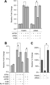
Figure 1
Figure 1
FSH effect on endogenous RARA transcriptional activity is via FSHR in primary Sertoli cells. A and B, Cultured primary Sertoli cells were transfected with a RARE-tk-Luc reporter, β-galactosidase plasmid, and FSHRi or control RNAi (A) or transfected with a RARE-tk-Luc reporter and β-galactosidase plasmid (B). Transfected primary Sertoli cells were allowed to recover for 24 h and treated for 24 h with ATRA (1 μm), Am580 (0.1 μm), rhFSH (25 μm), H-89 (10 μm), or Ro 41-5253 (5 μm), as indicated. C, Real-time RT-PCR to quantify Fshr mRNA levels in untransfected primary Sertoli cells or cells transfected with Fshr siRNA (FSHRi) or control RNAi (control RNAi). The relative percent change in the expression level of Fshr was evaluated using the 2−ΔΔCt method (38,39), using a housekeeping gene, ribosomal S2, as the internal control. Fshr expression in untransfected cells was set to 100%. It is not known why the Fshr mRNA level actually increased to more than 100% in the presence of control RNAi. Each bar is the mean from three different cell samples with luciferase and PCR assays conducted in triplicate for each sample. *, Significant differences between means ± sem at P ≤ 0.05; a line over two bars without an asterisk is not significant at P ≤ 0.05.
Figure 2
Figure 2
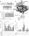
RARA PKA sites and the COS-7 cell system. Panel A, Schematic diagram showing the modular domains (A–F) of RARA and two predicted PKA consensus sequences including S219 and S369 sites with their scores. Both sites are located in the LBD in the E region and are found conserved among species (data not shown). AF-1 and -2, Activation domain 1 and 2; DBD, DNA-binding domain. Panel B, Tertiary structure rendering of E domains from RARA (dark) and RXRA (light). The ATRA ligand (stick), the S219 and S369 sites (spheres), and helices 4–10 and 12 (H4–H10 and H12) of the LBD are labeled. Panels C and D, COS-7 cells were transfected with pFLAG-RARA-WT and pcDNA-Rxra-WT cDNA at a 1:1 ratio, and time-dependent expression of RARA and RXRA examined by immunoblotting (IB) with anti-RARA (1:300 dilution), anti-FLAG (1:1000 dilution), anti-RXRA (1:300 dilution), and anti-GAPDH (1:1000 dilution) antibodies (panel C) or cell extracts were used to conduct immunoprecipitation (IP) with anti-RXRA antibody, followed by IB with anti-RARA and anti-RXRA antibodies (panel D). GAPDH protein levels were used as loading control in panel C. Protein collected from a mouse Sertoli cells line (MSC-1), cultured primary Sertoli cells (pSC), and rat testis served as a negative control for FLAG tag expression (panel C). Three times as much volume of input was loaded to compare to the amount of immunoprecipitate eluates in panel D. Experiments for panels C and D were performed three times. Panel E, The dose-dependent response of RARA transcriptional activity in COS-7 cells transfected with RARE-tk-Luc reporter, β-galactosidase plasmid, and pFLAG-RARA-WT or pFLAG empty vector. In the presence of ATRA (1 μm) or Am580 (0.1 μm), cells were treated with either increasing concentrations of db-cAMP (0.1, 0.25 or 0.5 mm) and H-89 (5.0, 10.0 or 20.0 μm) or one concentration of db-cAMP (0.25 mm). Panel F, RARA transcriptional activity in the presence of RARE-tk-Luc reporter, β-galactosidase plasmid, and pFLAG-RARA-WT or pFLAG empty vector and increasing amounts of pcDNA-Rxra-WT cDNA (0.1, 0.2, 0.3, or 0.4 μg/well) in COS-7 cells. Cells were treated for 24 h with vehicle (NT), ATRA (1 μm), or Ro 41-5253 (5 μm). Each bar in panels E and F corresponds to the mean of three different cell samples, and luciferase assays were conducted in triplicate for each sample. *, Significant differences between means ± sem at P ≤ 0.05; a line over two bars without an asterisk is not significant at P ≤ 0.05.
Figure 3
Figure 3
PKA regulation of RARA is via combinatory action on RARA PKA sites (S219 and S369) in the presence of RXRA-WT and RXRA-S27A mutant. A and B, RARA transcriptional activity after transfection of COS-7 cells with RARE-tk-Luc reporter, β-galactosidase plasmid, and pFLAG empty vector (A and B) or pFLAG-RARA-WT (A and B), -S219/369A (A), -S219/369E (A), -S369A (B), or -S369E (B) cDNA in the presence of ATRA (1 μm), db-cAMP (0.25 mm), and H-89 (10 μm), as indicated. C and D, RARA transcriptional activity after transfection of COS-7 cells with RARE-tk-Luc reporter, β-galactosidase plasmid, and pFLAG empty vector (C and D) or pFLAG-RARA-WT (C and D), -S219/369A (D), or -S219/369E (D) and increasing amounts (0.1, 0.2, 0.3, or 0.4 μg/well) of CMX-HisRxra-S27A cDNA (C) or 1:1 ratio of CMX-HisRxra-S27A cDNA to pFLAG-RARA (D), in the presence of vehicle (NT), ATRA, db-cAMP, and H-89, as indicated. Each bar is the mean from three different cell samples with luciferase assays conducted in triplicate for each sample. * or †, Significant differences between means ± sem with P ≤ 0.05; a line over two bars without an asterisk or dagger is not significant at P ≤ 0.05.
Figure 4
Figure 4
RARA transcriptional activity is regulated by PP1 and PP2A. A and B, RARA transcriptional activity after transfection of COS-7 cells with RARE-tk-Luc reporter, β-galactosidase plasmid, and pFLAG-RARA-WT, -S219/369A, or -S219/369E cDNA (A) and endogenous RARA transcriptional activity in cultured primary Sertoli cells transfected with RARE-tk-Luc reporter and β-galactosidase plasmid (B) in the presence of ATRA, db-cAMP, rhFSH, PP1-specific inhibitor TC (5 μm), or PP2A-specific inhibitor OkAc (2 nm), as indicated. Luciferase assays were conducted on cells under various concentrations of OkAc and TC to determine the optimal dose (data not shown). At higher concentrations of OkAc, PP1A can be inhibited. Each bar is the mean from three different cell samples with luciferase assays conducted in triplicate for each sample. Significant differences between means ± sem are indicated: *, P ≤ 0.05; **, P ≤ 0.1; a line over two bars without an asterisk is not significant at P ≤ 0.05.
Figure 5
Figure 5
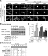
Nuclear localization of RARA-glutamic acid double mutants is less effective. A, COS-7 cells were transfected with pFLAG-RARA-WT, -S219/369A, or -S219/369E cDNA and, at 24 h after transfection, treated for 1 h with vehicle, ATRA, db-cAMP, H-89, or PP2A-specific inhibitor, as indicated, and fixed for immunofluorescence analysis with anti-FLAG antibody (a–r). If applicable, cells were pretreated for 30 min with db-cAMP. The number of cells with RARA exclusively nuclear or with RARA localized to both the nucleus and cytoplasm were counted. Images that are representative of 60% of cells (d, f, g, and i) are indicated with a diamond (⋄); otherwise, images are representative of 80–100% of cells. B–D, Nuclear lysates were collected from COS-7 cells transfected with pFLAG-RARA-WT, -S219/369A, or -S219/369E cDNA and treated with ATRA and/or db-cAMP. Expression of RARA protein was analyzed by IB with anti-FLAG antibody (B). Band intensity in B was measured and normalized to Coomassie-stained bands in the same lane, and average normalized results of three separate blots were graphed as relative fold change (C). The result with no treatment was normalized to 1.0. Coomassie staining of the membrane was used for loading control (B), and IB with anti-actin (1:200) was used as controls for cytoplasmic contamination (D). All experiments were repeated at least three times. *, Significant differences between mean ± sem at P ≤ 0.05; a line over two bars without an asterisk is not significant at P ≤ 0.05.
Figure 6
Figure 6
PKA interacts with WT RARA in the presence of rhFSH or db-cAMP. A, Cell lysates of cultured primary Sertoli cells (pSC) and COS-7 cells transfected with pFLAG-RARA-WT and pcDNA-Rxra-WT were subjected to IB using anti-PKA-RI (1:300) and anti-GAPDH antibodies. GAPDH protein levels were used as loading control. B and C, COS-7 cells were transfected with pcDNA-Rxra-WT and pFLAG-RARA-WT, -S219/369A, or -S219/369E cDNA and, at 24 h after transfection, treated for 1 h with vehicle, ATRA, db-cAMP, and H-89, as indicated. Primary Sertoli cells were treated for 1 h on the third day of culture with vehicle, ATRA, rhFSH, and H-89, as indicated, and the lysate was collected in appropriate buffer (B). Cell lysates were then subjected to IP with human and mouse anti-RARA antibody and IB with anti-RARA and anti-PKA-RI antibody. Experiments were conducted three times.
Figure 7
Figure 7
RARA interaction with RXRA is hindered when RARA carries negative charges at S219 and S369 PKA sites. A, COS-7 cells were transfected with pcDNA-Rxra-WT and pFLAG-RARA-WT, -S219/369A, or -S219/369E cDNA and, at 24 h after transfection, treated for 1 h with vehicle, ATRA, db-cAMP, and H-89, as indicated. Cell lysates were then subjected to IP with human and mouse anti-RARA antibody and IB with anti-RARA, anti-RXRA, and anti-PPP2R1A/B (1:200) antibodies. B, Expressed RARA-S219E or -S369E in COS-7 cells was subjected to IP with anti-RARA antibody and IB with anti-RARA, anti-RXRA antibodies. C, Primary Sertoli cells (pSC) were treated for 1 h on the third day of culture with vehicle, ATRA, and rhFSH, as indicated, and lysate was collected in appropriate buffer. Endogenous RARA in primary Sertoli cells was subjected to IP using anti-RARA antibody and IB with anti-RARA, anti-RXRA, and anti-PPP2R1A/B antibodies. Experiments were conducted three times.

Similar articles

Cited by

References

    1. Griswold MD 1998 The central role of Sertoli cells in spermatogenesis. Semin Cell Dev Biol 9:411–416 - PubMed
    1. Sharpe RM 1994 Regulation of spermatogenesis. In: Knobil E, Neill JD, eds. The physiology of reproduction. 2nd ed. New York: Raven Press; 1363–1434
    1. Kerr J 1999 Male reproductive system. In: Kerr JB, ed. Atlas of functional histology. London: Mosby; 339–353
    1. Russell LD, Ettlin RA, SinhaHikim AP, Clegg ED 1990 Mammalian spermatogenesis. In: Russell LD, Ettlin RA, SinhaHikim AP, Clegg ED, eds. Histological and histopathological evaluation of the testis. Clearwater: Cache River Press; 1–38
    1. Orth J, Christensen AK 1977 Localization of 125I-labeled FSH in the testes of hypophysectomized rats by autoradiography at the light and electron microscope levels. Endocrinology 101:262–278 - PubMed

Publication types

MeSH terms