. 2010 Apr 1;123(Pt 7):1099-107.
doi: 10.1242/jcs.059469. Epub 2010 Mar 9.

Sip1, the Drosophila orthologue of EBP50/NHERF1, functions with the sterile 20 family kinase Slik to regulate Moesin activity

Affiliations

Sip1, the Drosophila orthologue of EBP50/NHERF1, functions with the sterile 20 family kinase Slik to regulate Moesin activity

Sarah C Hughes et al. J Cell Sci. .

Abstract

Organization of the plasma membrane in polarized epithelial cells is accomplished by the specific localization of transmembrane or membrane-associated proteins, which are often linked to cytoplasmic protein complexes, including the actin cytoskeleton. In this study, we identified Sip1 as a Drosophila orthologue of the ezrin-radixin-moesin (ERM) binding protein 50 (EBP50; also known as the Na(+)/H(+) exchanger regulatory factor NHERF1). In mammals, EBP50/NHERF1 is a scaffold protein required for the regulation of several transmembrane receptors and downstream signal transduction activity. In Drosophila, loss of Sip1 leads to a reduction in Slik kinase protein abundance, loss of Moesin phosphorylation and changes in epithelial structure, including mislocalization of E-cadherin and F-actin. Consistent with these findings, Moesin and Sip1 act synergistically in genetic-interaction experiments, and Sip1 protein abundance is dependent on Moesin. Co-immunoprecipitation experiments indicate that Sip1 forms a complex with both Moesin and Slik. Taken together, these data suggest that Sip1 promotes Slik-dependent phosphorylation of Moesin, and suggests a mechanism for the regulation of Moesin activity within the cell to maintain epithelial integrity.


Figures

Fig. 1.
Fig. 1.
A comparison of the domain composition of the Drosophila Sip1 (NP_524712) protein with human EBP50/NHERF1 (NP_004243) and NHERF2 (NP_001123484). Drosophila Sip1 (CG10939) contains only a single N-terminal PDZ domain (shaded region), which is most similar to the second PDZ domain in human EBP50/NHERF1. The percentage amino acid identity and similarity are indicated between the PDZ domains by vertical arrows. The percentage identity and similarity between the EB domain of Sip1 and human EBP50/NHERF1 is indicated by vertical arrows. The C-terminal FERM-binding domain also appears to be conserved. The interacting domain as determined by overlapping clones from the two-hybrid interaction of Sip1 and Moesin is indicated by the dark horizontal line (Y2H).
Fig. 2.
Fig. 2.
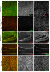
Sip1 protein localization. Wild type (A-C) and Sip1 mutant (Sip106373, D-F) embryos (12-14 hours after egg laying). Sip1 staining is lost in Sip1 embryos (E), suggesting that this Sip1 allele is a null mutation and that the antibody is specific. The gain and black level settings are identical in B and E. (G-I) Within the embryonic hindgut, there is bright Sip1 staining (H) that colocalizes with Moesin at the apical membrane (I). Sip1 protein is also expressed in third instar larval imaginal discs (K) where it colocalizes with filamentous actin (L) staining, and is apical to coracle (not shown). (M-O) Sip1 largely colocalizes with Moesin in the epithelium of embryos (embryo is shown 7-9 hours after egg laying). Scale bars: 10 μm.
Fig. 3.
Fig. 3.
Moe and Sip1 act synergistically. Expression of either the MS1096 Gal4 driver alone (A), or wild-type Moe under the MS1096 Gal4 driver (B), has no apparent affect on adult wing morphology. (C) Expression of an activated Moe transgene (UAS-MoeT559D) produces wings that are curved upwards at the edges (not apparent in this preparation) but otherwise normal. (D) Expression of an inactive Moe transgene (UAS-MoeT559A) results in wings that look completely like the wild type. (E) Expression of the Sip1 EP line, EP2037, under the MS1096 Gal4 driver produces wings with blisters that are apparent along vein L3 (arrowhead) as well as slight venation defects (arrow). (F) Co-expression of both Moe and Sip1 (EP2037) under control of the MS1096 GAL4 driver results in wings that have vein defects and are curved upwards at the edges. There is extra vein material on vein L3 (arrowhead) and vein L4 does not reach the wing margin (arrow). (G) Co-expression of activated MoeT559D and Sip1 produces wings that are smaller in size and are very warped, with large blister-like (arrow) and vesicle-like structures (arrowhead). (H) Co-expression of inactive Moesin (MoeT559A) and Sip1 produces wings that appear normal.
Fig. 4.
Fig. 4.
Loss of Sip1 expression in the follicle cells surrounding the developing oocyte has dramatic effects on Moesin phosphorylation, filamentous actin organization and localization of Slik. (A) Overlay of GFP and phalloidin (F-actin) staining in a stage 13 oocyte. Inset shows DAPI-stained nuclei indicating that cells are present within the Sip1 mitotic clone. (B) Sip1 clones are identified by the absence of GFP (arrow). (C) Phalloidin staining is drastically reorganized into fibre-like structures within each Sip1 mutant cell. (D-F) Stage 13 follicle cells stained with antibodies against Sip1 and E-cadherin. (E) Sip1 clones are identified by the absence of Sip1 staining (arrow). (F) In Sip1 mutant clones, E-cadherin labelling of the adherens junctions is lost or mislocalized (arrow). (G-J) Loss of Sip1 does not affect overall junctional integrity. (G) Overlay of a stage 12 oocyte stained for the septate junction markers fasciclin III and coracle. (H) Sip1 clone marked by the absence of GFP. (I) Fasciclin III appears unaffected within the clone. (J) Coracle staining also appears unchanged within Sip1 mutant clones. (K-M) Sip1 is required for normal localization of the Slik kinase. (K) Overlay of follicle cells (stage 10) showing GFP and Slik localization. (L) The Sip1 clone is identified by the absence of GFP. (M) Within the clone, expression of Slik is reduced and mislocalized (arrows) compared with adjacent wild-type follicle cells. Clones of mutant cells are marked by arrows. Scale bars: 10 μm.
Fig. 5.
Fig. 5.
Sip1 is required for Moesin activation. (A-C) Stage 13 follicle cells stained with Sip1 antibodies to label clones and a phospho-specific Moesin antibody to detect active Moesin. Inset shows presence of DAPI-stained (pseudocolored green) nuclei in the clone areas. (B) Sip1 clones are identified by the absence of Sip1 staining. The TCA fixative used to enhance phospho-Moesin antibody staining in this panel somewhat alters the subcellular localization of Sip1 in wild-type cells. (C) Phospho-Moesin staining is absent within Sip1 mitotic clones. (D-F) Pan anti-Moesin staining is not altered in Sip1 loss-of-function clones. The clonal region lacking Sip1 is identified by the absence of GFP (E). Moesin staining appears unchanged in the Sip1 clone (F). Scale bars: 10 μm.
Fig. 6.
Fig. 6.
Loss of Moesin or Slik affects the localization of Sip1 in follicle epithelial cells. (A-C) A stage 12 oocyte in which clones of Moe mutant cells are identified by the absence of GFP (B). Within the mutant cells, Sip1 staining is strongly reduced (C). (D-F) A single confocal section of stage 8 follicle cells with Slik loss-of-function cells identified by the loss of GFP (E). Sip1 staining levels (F) are brighter in the Slik mutant cells (arrows) than in wild-type cells. Scale bars: 10 μm.
Fig. 7.
Fig. 7.
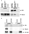
Sip1, Moesin and Slik proteins interact in vivo. (A) A MYC-tagged Sip1 transgene was expressed under the apterous Gal4 (ap Gal4>MYC-Sip1, lane 1) or MS1096 Gal4 (MS1096 Gal4>MYC-Sip1, lane 2) driver in developing pupal-staged animals. Expressed Sip1 was immunoprecipitated using MYC antibodies. Immunoprecipitates were blotted to detect the presence of co-immunoprecipitated endogenous Moesin protein. The control lane contains a w1118>MYC-Sip1 lysate mock immunoprecipitated without anti-MYC antibody. Moesin co-immunoprecipitated with Sip1, suggesting that these proteins form a complex in vivo. (B) An UAS HA-tagged Sip1 transgene was ubiquitously expressed with UAS MYC-Moe and UAS Slik in S2 cells. Expressed Sip1 was immunoprecipitated using anti-HA antibody. Immunoprecipitates were immunoblotted to specifically detect both endogenous and transiently expressed Moesin and Slik kinase protein. Co-precipitating Moesin and Slik are observed in the Sip1 immunoprecipitates but not in the mock immunoprecipitate (no HA antibody control) lane.
Fig. 8.
Fig. 8.
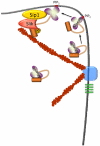
Possible model for Sip1 in Slik-dependent activation of Moesin. Inactive, folded Moesin in the cell cortex might associate with PIP2 in the plasma membrane, inducing a conformational change that results in partial unfolding of Moesin. This event, or other modifications such as phosphorylation of residues in the FERM domain (Krieg and Hunter, 1992), allow Moesin, Sip1 and Slik to form a complex that results in phosphorylation of the C-terminal Thr residue and full activation of Moesin.

Similar articles

Cited by

References

    1. Altschul S. F., Gish W., Miller W., Myers E. W., Lipman D. J. (1990). Basic local alignment search tool. J. Mol. Biol. 215, 403-410 - PubMed
    1. Amieva M. R., Wilgenbus K. K., Furthmayr H. (1994). Radixin is a component of hepatocyte microvilli in situ. Exp. Cell Res. 210, 140-144 - PubMed
    1. Bateman J., Reddy R. S., Saito H., Van Vactor D. (2001). The receptor tyrosine phosphatase Dlar and integrins organize actin filaments in the Drosophila follicular epithelium. Curr. Biol. 11, 1317-1327 - PubMed
    1. Belkina N. V., Liu Y., Hao J. J., Karasuyama H., Shaw S. (2009). LOK is a major ERM kinase in resting lymphocytes and regulates cytoskeletal rearrangement through ERM phosphorylation. Proc. Natl. Acad. Sci. USA 106, 4707-4712 - PMC - PubMed
    1. Berryman M., Franck Z., Bretscher A. (1993). Ezrin is concentrated in the apical microvilli of a wide variety of epithelial cells whereas moesin is found primarily in endothelial cells. J. Cell Sci. 105, 1025-1043 - PubMed

Publication types

MeSH terms