. 2010 Mar 23;107(12):5483-8.
doi: 10.1073/pnas.1000599107. Epub 2010 Mar 8.

Characterization of somatic cell nuclear reprogramming by oocytes in which a linker histone is required for pluripotency gene reactivation

Affiliations

Characterization of somatic cell nuclear reprogramming by oocytes in which a linker histone is required for pluripotency gene reactivation

Jerome Jullien et al. Proc Natl Acad Sci U S A. .

Abstract

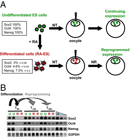
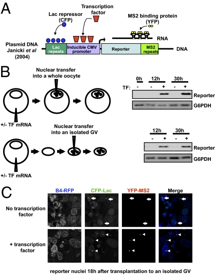
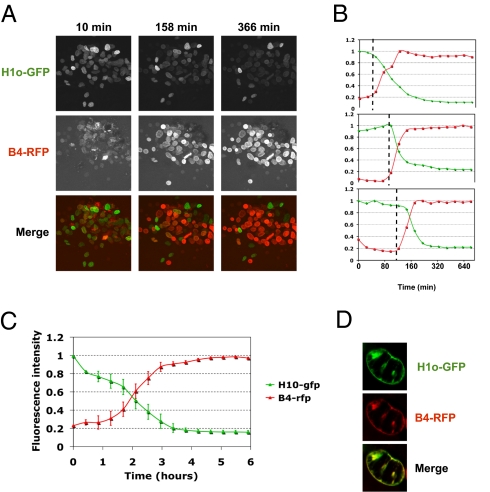
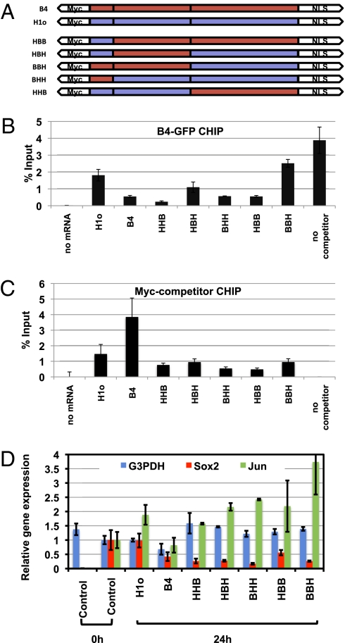
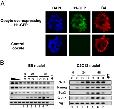
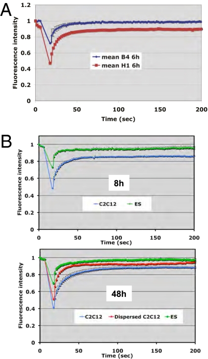
When transplanted into Xenopus oocytes, the nuclei of mammalian somatic cells are reprogrammed to express stem cell genes such as Oct4, Nanog, and Sox2. We now describe an experimental system in which the pluripotency genes Sox2 and Oct4 are repressed in retinoic acid-treated ES cells but are reprogrammed up to 100% within 24 h by injection of nuclei into the germinal vesicle (GV) of growing Xenopus oocytes. The isolation of GVs in nonaqueous medium allows the reprogramming of individual injected nuclei to be seen in real time. Analysis using fluorescence recovery after photobleaching shows that nuclear transfer is associated with an increase in linker histone mobility. A simultaneous loss of somatic H1 linker histone and incorporation of the oocyte-specific linker histone B4 precede transcriptional reprogramming. The loss of H1 is not required for gene reprogramming. We demonstrate both by antibody injection experiments and by dominant negative interference that the incorporation of B4 linker histone is required for pluripotency gene reactivation during nuclear reprogramming. We suggest that the binding of oocyte-specific B4 linker histone to chromatin is a key primary event in the reprogramming of somatic nuclei transplanted to amphibian oocytes.


Conflict of interest statement

The authors declare no conflict of interest.

Figures

Fig. 1.
Fig. 1.
Transplantation of nuclei from growing and differentiated ES cells allows quantification of gene reactivation. (A) Experimental design. NT, nuclear transfer; NR, nuclear reprogramming; RA, retinoic acid. Percentages in boxes show the level of gene expression as measured by real-time PCR on cultured cells before nuclear transfer (three experiments). (B) Example of RT-PCR analysis of the experiment depicted in A. U, undifferentiated ES nuclei; R, RA-differentiated ES nuclei.
Fig. 2.
Fig. 2.
Real-time monitoring of gene activation in nuclei transplanted to isolated Xenopus oocyte GVs. (A) The diagram shows an inducible CMV promoter (24) flanked by lacR binding site repeats thereby allowing visualization of the reporter gene in cells (by binding of CFP-LacR chimeric protein). The promoter drives the expression of a reporter mRNA containing MS2 binding site repeats, so that the transcribed mRNA at the transcription site can be visualized using a MS2-YFP chimeric protein. Nuclei containing this reporter gene for real-time imaging of transcription (24) were used for transplantation into isolated GVs. (B) Genes are reactivated in nuclei transplanted into a GV that already has been isolated from a Xenopus oocyte. Nuclei containing a reporter gene (diagram in A) were transplanted into the GV of a whole oocyte or into an isolated GV. Transplantation was carried out into GVs expressing or not expressing a transcription factor (TF) that is required to activate the reporter gene. RT-PCR analysis shows transcription factor-dependent activation of the reporter gene from both sets of transplanted nuclei. (C) Expression of the B4-RFP protein in the GV allows visualization of transplanted nuclei (Fig. 3), whereas expression of CFP-LacR shows the position of the reporter genes in these nuclei (dots in CFP-Lac channel). The MS2-YFP protein also expressed in the GV shows newly synthesized mRNA at the gene loci, but only when the GV contains the transcription factor specific to the reporter gene (Compare arrows in Upper with arrowheads on Lower Merge image).
Fig. 3.
Fig. 3.
Real-time monitoring of linker histone exchange during nuclear reprogramming, (A) Xenopus oocyte linker histone (B4) replaces somatic linker histone (H1) following NT. Real-time monitoring of H1o-GFP (present in NIH 3T3 nuclei before transplantation) (27) and B4-RFP (expressed in the oocyte by mRNA injection) was carried out during the first 6 h of reprogramming. (A complete series of images is shown in Video S1). (B) Three individual nuclei from the sequence depicted in A were quantified for fluorescence intensity of H1 (green line) and B4 (red line). (C) Average change in fluorescence intensity with time in the experiment shown in A. Errors bars indicate ± SEM (n = 11 nuclei). (D) Confocal images of a nucleus at a time point following transplantation into an oocyte when B4 starts to be incorporated into chromatin and H1 is not yet completely lost from the nucleus.
Fig. 4.
Fig. 4.
Reactivation of a pluripotent gene in transplanted nuclei is prevented by expression of chimeric B4/H1 linker histone. (A) Structure of chimeric linker histones. Amino-terminal, globular, and carboxyl-terminal domains of oocyte- and somatic-linker histones were swapped to generate chimeric linker histones. (B) Chimeric linker histones interfere with B4 binding to chromatin. ChIP analysis of B4-GFP binding to chromatin was performed in the presence of chimeric linker histone as in Fig S4C (B4-GFP, 0.2-fold endogenous B4; Myc-tagged chimeric linker histone, 6- to 9-fold endogenous B4). Error bars indicate the mean ± SEM of triplicate samples. (C) All chimeric linker histones are found associated with chromatin. ChIP analyses with an Myc antibody were performed in same experimental conditions as in B. Error bars represent the mean ± SEM of triplicate samples. (D) Pluripotency gene reactivation is inhibited by chimeric linker histones. RA-ES cells were transplanted into oocytes overexpressing the chimeric linker histones described in A, and gene expression was analyzed by RT-PCR 24 h after NT. The gene reactivation level from RA-ES nuclei 24 h after transplantation into control oocytes is set to 1. Gene expression was averaged from triplicate samples and normalized to the G3PDH level. Error bars indicate the mean ± SEM.
Fig. 5.
Fig. 5.
Loss of somatic H1 from transplanted nuclei is not required for gene reactivation. (A) H1 overexpression in the oocyte maintains H1 presence in transplanted nuclei but does not inhibit B4 incorporation. C2C12 nuclei were transplanted into control or H1-GFP-expressing oocytes. B4-RFP was detected by immunolabeling 24 h after NT. Loss of H1 from transplanted nuclei (Fig. 3A) can be prevented by expressing H1-GFP in the oocyte before NT (Upper Row, Center). B4 is loaded onto chromatin (Right), whereas H1-GFP is maintained (Upper Row, Center) or not (Bottom Row, Center). (B) Pluripotency genes are reactivated even when H1 is maintained in transplanted ES or C2C12 nuclei. RT-PCR analysis was carried out under conditions depicted in A. Pluripotency genes are reactivated whether H1 is maintained or not in transplanted nuclei (compare columns 5 and 7). Data are from three experiments. B, oocyte injected with B4 mRNA; C, control oocyte; H, oocyte injected with H1 mRNA.
Fig. 6.
Fig. 6.
Nuclear reprogramming is associated with an increase in linker histone mobility. (A) B4 is more mobile than H1 in transplanted nuclei. HeLa cell nuclei were transplanted into oocytes overexpressing B4-GFP or H1-GFP (mRNA injection). Linker histone mobility then was measured by FRAP 6 h after nuclear transplantation. The differences in bleach depth (B4 ∼30%, H1 ∼50%) as well as the extent of recovery (B4 ∼100%, H1 ∼90%) reflect the difference in mobility between B4 and H1. Error bars represent the mean ± SEM (n = 10 nuclei). (B) H1 mobility increases during nuclear reprogramming. Using oocytes overexpressing H1-GFP, we measured the mobility of H1 during nuclear reprogramming by FRAP analysis. ES cells (green lines) show high mobility 8 h and 48 h after NT. Committed cell nuclei (C2C12) show a fraction of H1 with low mobility (blue line) 8 h after NT. At 48 h after NR, H1 mobility in C2C12 nuclei (blue line) increases toward that observed in ES cell nuclei (red line). Error bars indicate mean ± SEM (n = 10 nuclei).
Fig. 7.
Fig. 7.
Incorporation of B4 into transplanted nuclei is necessary for pluripotency gene expression in oocytes. (A) ChIP analysis of B4 binding to pluripotency genes in transplanted nuclei. B4 binding to pluripotency gene promoters and to the major satellite region of ES or RA-ES nuclei was analyzed by ChIP 0.5 h, 6 h, and 25 h following transplantation. Error bars represent mean ± SEM. Data are from two experiments. No Ab, control chip performed in the absence of antibody. (B) Incorporation of B4 in transplanted nuclei (control Ab) can be prevented by injection of anti-B4 antibody into the oocyte GV (Lower Row, Center). C2C12 nuclei were transplanted with or without anti-B4 antibody into oocytes expressing B4-GFP. Then B4-GFP labeling of transplanted nuclei was detected by confocal microscopy. The graph on the right shows quantification of the B4-GFP signal. Error bars indicate mean ± SEM (n = 15 nuclei). (C) Pluripotency gene reactivation requires incorporation of B4 into transplanted nuclei. RT-PCR analysis was carried out in conditions depicted in B. Gene reactivation is inhibited in anti-B4 samples at 24 h in both ES and RA-ES transplanted nuclei. Data are from three experiments.

Comment in

Similar articles

Cited by

References

    1. Briggs R, King TJ. Transplantation of living nuclei from blastula cells into enucleated frogs’ eggs. Proc Natl Acad Sci USA. 1952;38:455–463. - PMC - PubMed
    1. Gurdon JB. The developmental capacity of nuclei taken from differentiating endoderm cells of Xenopus laevis. J Embryol Exp Morphol. 1960;8:505–526. - PubMed
    1. Campbell KH, McWhir J, Ritchie WA, Wilmut I. Sheep cloned by nuclear transfer from a cultured cell line. Nature. 1996;380:64–66. - PubMed
    1. Pereira CF, et al. Heterokaryon-based reprogramming of human B lymphocytes for pluripotency requires Oct4 but not Sox2. PLoS Genet. 2008;4:e1000170. - PMC - PubMed
    1. Takahashi K, Yamanaka S. Induction of pluripotent stem cells from mouse embryonic and adult fibroblast cultures by defined factors. Cell. 2006;126:663–676. - PubMed

Publication types

MeSH terms