. 2010:2010:814609.
doi: 10.1155/2010/814609. Epub 2010 Feb 28.

Anticancer Role of PPARgamma Agonists in Hematological Malignancies Found in the Vasculature, Marrow, and Eyes

Affiliations

Anticancer Role of PPARgamma Agonists in Hematological Malignancies Found in the Vasculature, Marrow, and Eyes

P J Simpson-Haidaris et al. PPAR Res. 2010.

Abstract

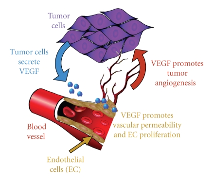
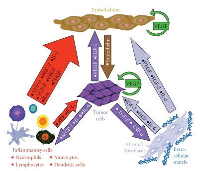
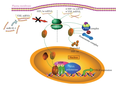
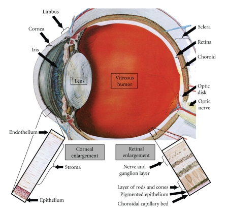
The use of targeted cancer therapies in combination with conventional chemotherapeutic agents and/or radiation treatment has increased overall survival of cancer patients. However, longer survival is accompanied by increased incidence of comorbidities due, in part, to drug side effects and toxicities. It is well accepted that inflammation and tumorigenesis are linked. Because peroxisome proliferator-activated receptor (PPAR)-gamma agonists are potent mediators of anti-inflammatory responses, it was a logical extension to examine the role of PPARgamma agonists in the treatment and prevention of cancer. This paper has two objectives: first to highlight the potential uses for PPARgamma agonists in anticancer therapy with special emphasis on their role when used as adjuvant or combined therapy in the treatment of hematological malignancies found in the vasculature, marrow, and eyes, and second, to review the potential role PPARgamma and/or its ligands may have in modulating cancer-associated angiogenesis and tumor-stromal microenvironment crosstalk in bone marrow.


Figures

Figure 1
Figure 1
Molecular mechanisms of tumor-associated angiogenesis. Angiogenesis is essential for the persistence of solid tumor growth and, only recently, has it been appreciated that angiogenesis plays a role in progression of hematological malignancies as well. Cancer-associated angiogenesis in solid tumors begins once the tumor mass reaches a critical size such that the hypoxic environment inside the tumor leads to cancer cell-specific expression of proangiogenic factors including VEGF to shift the balance from endogenous antiangiogenic factors to tumor supplied proangiogenic factors—the angiogenic switch. Once proangiogenic factors overwhelm antiangiogenic factors, new blood vessels form in response to VEGF-induced endothelial permeability by EC sprouting, migration into the tumor mass, and proliferation from existing blood vessels—molecular mechanisms also induced by VEGF [–67]. The tumor integrity of the vasculature is compromised in that it remains leaky with poor cell-to-cell adhesion, is abnormally branched and not well supported by pericytes (mural cells), the vascular smooth muscle cells that stabilize normal blood vessels [67, 68]. The chronic immaturity of tumor vessels has led Dvorak to characterize a tumor as a “wound that never heals” [69]. Notwithstanding, these features make tumor vessels viable targets for antitumor therapies. Benjamin et al. [70] demonstrated that removal of growth factors leads not only to the cessation of new vessel growth, but also to regression of the immature tumor vasculature [71].
Figure 2
Figure 2
Tumor-associated angiogenesis is sustained through stromal microenvironment crosstalk. Most tumors are associated with the activation of tumor-promoting innate immune responses involving neutrophils, macrophages, and NK cells. Specific (adaptive) antitumor immune responses involving T- or B-lymphocytes are less efficient in suppressing tumor growth. Increased formation of blood and lymphatic vessels in bone marrow and lymph nodes provide oxygen and nutrients to malignant cells. Stromal cells, including ECs, inflammatory cells, and fibroblasts/myofibroblasts, produce cytokines and growth factors that act in a paracrine fashion to promote malignant cell proliferation or survival. In turn, malignant cells produce angiogenic factors and express their cognate receptors establishing functional autocrine loops to perpetuate their survival including signaling through the VEGF pathway [–87, 107]. The secreted factors produced by and in response to those secreted by stromal and tumor cells include, but are not limited to VEGF, FGF-2, PDGF, IGF-1, HSF, TGF-α, TGF-β, TNF-α, IL-8, MCP-1/CCL2, MIF, IL-6, and IL-1 [95]. The potent vasoconstrictor peptide endothelin-1 has been implicated in the pathophysiology of atherosclerosis and its complications [108], as well as tumor angiogenesis and lymphangiogenesis [109, 110]. Proteases important for invasion thorough the basement membrane and remodeling of the ECM, such as plasminogen [96] and MMPs, including MMP-2 and MMP-9 [97], and their inhibitors, PAI-1/2 and TIMPs, respectively, are produced by stromal and tumor cells. Downregulation of endogenous inhibitors of angiogenesis such as thrombospondin (TSP)-1 occurs in the stromal compartment as well to favor angiogenesis, cancer cell growth, and metastasis [98]. In recent years, it has been recognized that a better understanding of the tumor-stromal microenvironment crosstalk may lead to elucidation of new therapeutic strategies for cancer therapy [–102].
Figure 3
Figure 3
PPARγ agonists inhibit Stat3-mediated IL-6 gene expression in myeloma cells. Inactivation of IL-6-activated Stat3 by PPARγ agonists occurs in a PPARγ-dependent manner; however, the molecular mechanisms by which two distinct PPARγ agonists (15d-PGJ2 and troglitazone) suppress IL-6-activated Stat3 in MM cells differ as shown in (a) [211]. Direct complex formation between phosphorylated Stat3 and PPARγ activated by 15d-PGJ2 prevents Stat3 binding to its cognate response element (SBE) on the promoters of target genes ((a), left). This mode of transcriptional inactivation does not require binding of the activated PPARγ transcription factor to DNA in the promoter region and, thus, can occur in the absence of a PPRE. However troglitazone activated PPARγ promotes redistribution of the corepressor SMRT from PPARγ to phosphorylated Stat3 so that Stat3 can no longer recruit the transcriptional machinery necessary for gene expression ((a), right) [211]. High levels of IL-6 are found in MM and promote myeloma cell proliferation and survival and indirectly promote tumor-associated angiogenesis. The PPARγ agonists troglitazone and 15d-PGJ2 have been shown to inhibit transcription of the IL-6 promoter driven by C/EBPβ and NF-κB [212]. Troglitazone-activated PPARγ binds to C/EBPβ preventing binding to its cognate response element on the IL-6 promoter, which is the major mechanistic pathway of troglitazone-mediated downregulation of IL-6 expression. In addition activated PPARγ competes with NF-κB for the PGC-1 coactivator, which leads to decreased NF-κB binding to the κB response element on the IL-6 promoter contributing to inhibition of IL-6 gene expression, albeit to a lesser extent than inhibition of C/EBPβ ((b), left). A slightly different mechanistic emphasis on PPARγ-mediated inhibition of IL-6 gene expression occurs in response to 15d-PGJ2. Although 15d-PGJ2-activated PPARγ inhibits C/EBPβ-mediated transactivation of the IL-6 promoter similarly to troglitazone-activated PPARγ, the predominant mode of inhibition is through 15d-PGJ2-activated PPARγ using the coactivator PGC-1 as a bridging protein to interact with NF-κB to prevent transactivation of the IL-6 promoter. Furthermore, 15d-PGJ2 inactivates NF-κB by inhibiting phosphorylation of IKK and IκB independently of PPARγ activation ((b), right). The schematics in this figure were adapted from [211, 212].
Figure 4
Figure 4
Autocrine production of VEGF in CLL B cells is regulated by miRNA-92-1 inhibition of pVHL production. Expression of high levels of VEGF by tumor cells is critical to promote and sustain the angiogenesis needed for cancer progression. Under normal oxygen tension, the HIF-1α subunit of the transcription factor, HIF-1, is constitutively produced and rapidly degraded by pVHL-induced proteasomal degradation, which prevents transcription of the VEGF gene. In solid tumors, HIF-1-induced VEGF expression occurs when tumor growth exceeds the dimensions where existing blood vessels can feed the tumor and carry away waste products. The resulting hypoxia leads to stabilization of HIF-1α and activation of the HIF-1 heterodimer resulting in high VEGF production by tumor cells. Although solid tumors do not develop in hematological malignancies, angiogenesis is an important process of disease progression. CLL B cells constitutively express high levels of VEGF and VEGFRs leading to autocrine signaling and increased resistance to apoptosis. Recently, Ghosh et al. [273] discovered that HIF-1 is stabilized in CLL B cells due to low levels of pVHL as a result of miR-92-1 overexpression and subsequent repression of translation of the VHL transcript. Therefore, HIF-1 accumulates and translocates to the nucleus where it forms an active complex with the transcriptional coactivator p300 and phosphorylated Stat3 and, together with the basal transcription machinery, transactivates the VEGF promoter. PPARγ agonists could potentially inhibit overexpression of VEGF by inhibiting Stat3 signaling in CLL B cells. The schematic in this figure was adapted from [273].
Figure 5
Figure 5
PPARγ is broadly expressed in the eye providing a pharmacological target for treating ocular angiogenesis. PPARγ expression is found in the retina including RPE cells, REC, pericytes [287], and ganglion cells. In the cornea, PPARγ is most prominently localized in the epithelial and endothelial layers. Excessive angiogenesis is a pathological hallmark of a number of eye diseases, and anti-VEGF/VEGFR strategies are used therapeutically to treat ocular neovascularization. Manifestations of hematological malignancies in the eye have been documented for leukemia, lymphoma, and multiple myeloma. The potential benefits of PPARγ agonist therapy to inhibit tumor-associated angiogenesis could also be applied to treatment of neovascular eye diseases.
Figure 6
Figure 6
Direct and indirect effects of PPARγ agonists on tumor and stromal cells. “Off-target” (PPARγ-independent) effects of PPARγ agonists frequently occur when the agonists are used at high concentrations (much higher than needed to active PPARγ by ligand binding) and in response to electrophilic PPARγ agonists such as 15d-PGJ2 and CDDO, which can promote covalent bond formation with cellular proteins in a redox-sensitive manner to modulate signal transduction pathways. PPARγ agonists have been shown to affect almost every stage of tumor progression from inhibition of uncontrolled tumor growth, induction of apoptosis, inhibition of tumor cell adhesion and invasion through stromal compartments into or out of the blood stream, and inhibition of tumor-associated angiogenesis. PPARγ agonists induce expression of tumor-inhibiting molecules such as CD36, the EC receptor for TSP-1, as well as promote the differentiation of tumor cells, which tends to reduce their invasive and metastatic capabilities. The schematic in this figure was adapted from [181].

Similar articles

Cited by

References

    1. Mangelsdorf DJ, Thummel C, Beato M, et al. The nuclear receptor super-family: the second decade. Cell. 1995;83(6):835–839. - PMC - PubMed
    1. Lazar MA. PPARγ, 10 years later. Biochimie. 2005;87(1):9–13. - PubMed
    1. Garcia-Bates TM, Lehmann GM, Simpson-Haidaris PJ, Bernstein SH, Sime PJ, Phipps RP. Role of peroxisome proliferator-activated receptor gamma and its ligands in the treatment of hematological malignancies. PPAR Research. 2008;2008:18 pages. Article ID 834612. - PMC - PubMed
    1. Goldenberg I, Benderly M, Goldbourt U. Update on the use of fibrates: focus on bezafibrate. Vascular Health and Risk Management. 2008;4(1):131–141. - PMC - PubMed
    1. Sharma AM, Staels B. Review: peroxisome proliferator-activated receptor γ and adipose tissue—understanding obesity-related changes in regulation of lipid and glucose metabolism. Journal of Clinical Endocrinology and Metabolism. 2007;92(2):386–395. - PubMed

LinkOut - more resources