. 2010 Apr 1;123(Pt 7):1039-49.
doi: 10.1242/jcs.059824. Epub 2010 Mar 2.

The Puf-family RNA-binding protein PfPuf2 regulates sexual development and sex differentiation in the malaria parasite Plasmodium falciparum

Affiliations

The Puf-family RNA-binding protein PfPuf2 regulates sexual development and sex differentiation in the malaria parasite Plasmodium falciparum

Jun Miao et al. J Cell Sci. .

Abstract

Translation regulation plays an important role during gametocytogenesis in the malaria parasite, a process that is obligatory for the transmission of the parasite through mosquito vectors. In this study we determined the function of PfPuf2, a member of the Puf family of translational repressors, in gametocytogenesis of Plasmodium falciparum. Tagging of the endogenous PfPuf2 protein with green fluorescent protein showed that PfPuf2 was expressed in both male and female gametocytes, and the protein was localized in the cytoplasm of the parasite. Targeted disruption of the PfPuf2 gene did not affect asexual growth of the parasite, but promoted the formation of gametocytes and differentiation of male gametocytes. Complementation studies were performed to confirm that the resultant phenotypic changes were due to disruption of the PfPuf2 gene. Episomal expression of PfPuf2 under its cognate promoter almost restored the gametocytogenesis rate in a PfPuf2 disruptant to the level of the wild-type parasite. It also partially restored the effect of PfPuf2 disruption on male-female sex ratio. In addition, episomal overexpression of PfPuf2 under its cognate promoter but with a higher concentration of the selection drug or under the constitutive hsp86 promoter in both the PfPuf2-disruptant and wild-type 3D7 lines, further dramatically reduced gametocytogenesis rates and sex ratios. These findings suggest that in this early branch of eukaryotes the function of PfPuf2 is consistent with the ancestral function of suppressing differentiation proposed for Puf-family proteins.


Figures

Fig. 1.
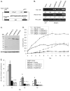
Fig. 1.
GFP-tagging of PfPuf2 and subcellular localization. (A) Predicted integration event of GFP fusion at the endogenous PfPuf2 locus. Top: PfPuf2 locus on chromosome 4. Solid lines indicate introns or intergenic regions, filled boxes represent the exons, and hatched boxes the PfPuf2 region used for homologous recombination in the transfection plasmid. Middle: the plasmid pHD22Y/Puf2-GFP. Bottom: The resultant single-crossover event at the PfPuf2 locus with integration of one copy of the plasmid. The SpeI restriction sites are shown. (B) Confirmation of integration by Southern blot. Genomic DNA from 3D7 and four clones (1-4) of PfPuf2-GFP was digested with SpeI and separated on a 0.6% agarose gel. The blot was probed with the labeled GFP gene. The predicted 12.3 kb DNA band and the 8.2 kb plasmid are marked. (C) Confirmation of PfPuf2-GFP expression by western blotting. Gametocyte lysates from 3D7 control and PfPuf2-GFP clone 2 were separated by 10% SDS-PAGE and probed with anti-GFP antibodies. (D) GFP fluorescence and IFA of gametocytes from the PfPuf2-GFP clone 2. Representative GFP fluorescent images of stage II-IV gametocytes and a female gamete (Gm) showing the expression and localization of PfPuf2-GFP. Gametocytes and gametes were confirmed by IFA with the anti-Pf230 antibodies. Nuclei were counterstained with Hoechst 33342.
Fig. 2.
Fig. 2.
Disruption of the PfPuf2 gene. (A) Predicted PfPuf2 disruption from a single-crossover event. The positions and orientations of the primers on chromosome 4 and the plasmid are shown. Restriction enzyme XbaI (Xb) sites and the expected sizes of DNA fragments after XbaI digestion are also shown. Top: PfPuf2 locus on chromosome 4. Solid lines represent introns or intergenic regions, filled boxes the exons, and hatched boxes the RNA-binding domain. Middle: the transfection plasmid pDT.Tg23/Puf2Δrep8 showing the PfPuf2 genomic fragment and the drug selection cassette TgDHFR. Bottom: the predicted single-crossover event at the PfPuf2 locus showing the integration of one copy of the plasmid. The primers F2d and MR13 were used for identification of this integration event. (B) Confirmation of PfPuf2 disruption by Southern blot. Genomic DNA from wt 3D7, four disruption clones (ΔRep8 1-4) and 100 ng of the plasmid (P) were digested with XbaI and separated in a 0.6% agarose gel. The blot was hybridized to 32P-labeled PCR product using primers F2 × R2 (shown in A). (C) Confirmation of PfPuf2 disruption by RT-PCR. The primers for amplifying the 5′ end (F2d and DR1) and the 3′ end (PB3A and Puf2C) of PfPuf2 are shown in A. The constitutively expressed gene PF13_0272 was used as a control. The sizes of the RT-PCR products are shown on the right. Two representative PfPuf2-disruption clones ΔRep8-1 and ΔRep8-3, wt 3D7, and a transfection control (Control) were tested for PfPuf2 expression in mixed gametocytes.
Fig. 3.
Fig. 3.
Asexual growth and gametocytogenesis. Four clones, wt 3D7, transfection control and two PfPuf2 disruptants (ΔRep8-1 and ΔRep8-3) were compared. (A) Asexual growth. Synchronized parasite cultures were initiated at 0.5% parasitemia on day 1. Parasitemia was determined daily from Giemsa-stained thin smears. Each point represents the mean and standard deviation from three experiments. Statistical analysis by two-way ANOVA followed by Tukey's pairwise comparison showed that daily parasitemias were not significantly different among the parasite lines (P>0.05). (B) Daily gametocytemias after induction of gametocytogenesis. Values are means ± standard deviations of triplicate samples. Statistical analysis showed that the gametocytemias were significantly different in the PfPuf2 disruptants and controls (two-way ANOVA and Tukey's t-test; P<0.0001), whereas gametocytemias were not significantly different in the two disruptants or in the two controls (P>0.05). Symbols are the same as in A. (C) Gametocyte development and maturation dynamics. The dynamics of different gametocyte stages (except stage 1) was determined for wt 3D7 (upper panel) and ΔRep8-1 (lower panel) lines. Gametocyte stages were differentiated on the basis of morphology of the gametocytes. Values are the means and standard deviations of stage-specific gametocytemias determined from three replicates. (D) Real-time RT-PCR of representative genes expressed during early gametocyte stages in the PfPuf2 disruptant clone ΔRep8-1 and wt 3D7. The data were log-transformed and the two samples from the same day were compared by t-test. Asterisks indicate significant differences between the two samples: *P=0.05 and **P=0.01.
Fig. 4.
Fig. 4.
Sex differentiation and male gametocyte exflagellation. (A) Mature male:female ratios in ΔRep8-1, ΔRep8-3, transfection control and wt 3D7 clones on day 8, 10 and 12. The means and standard deviations are shown from three gametocyte induction experiments. Daily sex ratios in the clones were compared by one-way ANOVA with Bonferroni corrections. For each time point, different letters indicate significant differences between the clones (P<0.05). (B) Sex ratios (male:female) of all gametocytes were compared between the ΔRep8-1 clone and transfection control. From day 6 to 12, IFA was performed with anti-Pfs230 antibodies to detect all gametocytes, and anti-α-tubulin II antibodies to detect male gametocytes. Data were analyzed by t-tests with Bonferroni corrections. For each time point, asterisks indicate significant differences between the two parasite clones: *P=0.05, **P=0.01. (C) The ratios of relative mRNA levels, determined by real-time RT-PCR, of the male-specific α-tubulin II gene (α-tub II) and female-specific gene Pfs47. Asterisks indicate significant differences between the two samples: *P=0.05 and **P=0.01 (t-test with Bonferroni corrections). (D) Comparison of male gametocyte exflagellation centers. Exflagellation centers were counted and normalized to mature male gametocytemia of each clone. One-way ANOVA did not detect significant differences among these clones (P>0.05).
Fig. 5.
Fig. 5.
Complementation of PfPuf2 disruption. (A) Diagram of the pCC4 expression plasmid used for the episomal expression of PfPuf2-GFP. PfPuf2-GFP was directed by either the PfPuf2 or hsp86 promoter, and GFP expression in the pCC4-GFP control (Vector) was directed by the PfPuf2 promoter. (B) Episomal expression of PfPuf2 in the ΔRep8-1 clone. RT-PCR was performed to detect PfPuf2 expression at N- and C-termini using the primers (F2d × DR1) and primers (PB3A × Puf2C), respectively (same as in Fig. 2A). PF13_0272 was used as an internal control. Parasites were transfected with either pCC4-GFP control vector or PfPuf2 coding sequence driven by the hsp86 promoter (Hsp86/Puf2) or PfPuf2 promoter (Puf2/Puf2). Puf2/Puf2(1) and Puf2/Puf2(2) were selected with 2.5 and 10 μg/ml of BSD, respectively. The numbers below the DNA bands indicate average relative expression levels of PfPuf2 in each parasite line from real-time RT-PCR analysis determined using the ΔCt method with PF13_0272 as the control. (C) Expression of the recombinant PfPuf2-GFP in transfected PfPuf2 disruptant. The GPF-tagged PfPuf2 was detected by western blotting using anti-GFP antibodies (upper panel) and protein loading was monitored with anti-HSP70 antibodies (lower panel). (D) Gametocytogenesis in the parasite lines described in B. Two-way ANOVA showed significant difference between each line (d.f.=3, 88; F=1166; P<0.001) except between Hsp86/Puf2 and Puf2/PfPuf2(2). Bonferroni simultaneous tests indicate that gametocytemias between 3D7+vector and ΔRep8-1+Puf2/Puf2(1) were significantly different from day 2 to 6 (P<0.05), but not significantly different at subsequent time points (P>0.05). (E) Sex ratios (male:female) in the parasite lines described in B. One-way ANOVA and Bonferroni simultaneous pairwise comparisons were performed for each time point. Different letters indicate significant differences between the parasite lines (P<0.05).
Fig. 6.
Fig. 6.
Overexpression of PfPuf2 in wt 3D7. (A) Expression of PfPuf2-GFP. The wt 3D7 was transfected with the plasmids shown in Fig. 5A. The GFP-tagged PfPuf2 protein expression was determined by western blotting with anti-GFP antibodies (upper panel), and protein loading was monitored with anti-HSP70 antibodies (lower panel). 3D7 parasites were transfected with either pCC4-GFP control vector (Vector) or PfPuf2-GFP driven by the hsp86 (Hsp86/Puf2) or PfPuf2 promoter (Puf2/Puf2). Puf2/Puf2(1) and Puf2/Puf2(2) were selected with 2.5 and 10 μg/ml of BSD, respectively. (B) Gametocytogenesis in the parasite lines described in A. Two-way ANOVA showed significant difference between pCC4-GFP control (Vector) and Puf2/Puf2(1) from day 7 to day 12 (P<0.05). No significant difference was found between Hsp86/Puf2 and Puf2/Puf2(2) at all time points (P>0.05), whereas these two parasite lines showed significant difference in daily gametocytemias from the control (Vector) and Puf2/Puf2(1) lines (P<0.001). (C) Sex ratios (male:female) in the parasite lines described in A. One way ANOVA and Bonferroni simultaneous pairwise comparison were performed for each time point. Different letters indicate significant difference between the parasite lines (P<0.05).

Similar articles

Cited by

References

    1. Alano P. (2007). Plasmodium falciparum gametocytes: still many secrets of a hidden life. Mol. Microbiol. 66, 291-302 - PubMed
    1. Alano P., Premawansa S., Bruce M. C., Carter R. (1991). A stage specific gene expressed at the onset of gametocytogenesis in Plasmodium falciparum. Mol. Biochem. Parasitol. 46, 81-88 - PubMed
    1. Asaoka-Taguchi M., Yamada M., Nakamura A., Hanyu K., Kobayashi S. (1999). Maternal Pumilio acts together with Nanos in germline development in Drosophila embryos. Nat. Cell Biol. 1, 431-437 - PubMed
    1. Bachorik J. L., Kimble J. (2005). Redundant control of the Caenorhabditis elegans sperm/oocyte switch by PUF-8 and FBF-1, two distinct PUF RNA-binding proteins. Proc. Natl. Acad. Sci. USA 102, 10893-10897 - PMC - PubMed
    1. Bhattacharyya M. K., Kumar N. (2001). Effect of xanthurenic acid on infectivity of Plasmodium falciparum to Anopheles stephensi. Int. J. Parasitol. 31, 1129-1133 - PubMed

Publication types

MeSH terms