. 2010 Apr 30;285(18):14052-61.
doi: 10.1074/jbc.M109.068262. Epub 2010 Feb 26.

Cdk5 nuclear localization is p27-dependent in nerve cells: implications for cell cycle suppression and caspase-3 activation

Affiliations

Cdk5 nuclear localization is p27-dependent in nerve cells: implications for cell cycle suppression and caspase-3 activation

Jie Zhang et al. J Biol Chem. .

Abstract

Initiation of a cell cycle in an adult neuron leads to cell death, placing great importance on the mechanisms that normally suppress the neuronal cell cycle. We have previously shown that the cyclin-dependent kinase Cdk5 is an important part of this process, but only when it is present in the nucleus. We report here that Cdk5 nuclear localization relies on its binding to the cyclin-dependent kinase inhibitor p27. Cdk5 has no intrinsic nuclear localization signal; in the absence of p27, two weak nuclear export signals that bind CRM1 cause it to shuttle to the cytoplasm. When a neuron is subjected to stress, such as exposure to beta-amyloid, the Cdk5-p27 interaction is lost, reducing Cdk5 levels in the nucleus and depriving the neuron of a major cell cycle suppression mechanism. Caspase-3 is activated within hours, but death is not immediate; elevated levels of cytoplasmic Cdk5 appear to retard neuronal death by a mechanism that may involve Bcl2. These data suggest a model in which Cdk5 exerts a double protective function in neurons: chronically suppressing the cell cycle when located in the nucleus and transiently delaying cell death in the cytoplasm.


Figures

FIGURE 1.
FIGURE 1.
A, N2a cells were treated with nocodazole for 48 h and then returned to drug free medium. The cells were harvested at the indicted times. Nuclear fractions were run on Western blots and probed with antibodies against Cdk5, p27, and Cdk4. The RNA binding protein hnRNP was used as loading control. B, the bands in A were quantified by National Institutes of Health ImageJ. Western blots of nuclear and cytoplasm fractions of lysates from embryonic day 16.5 Cdk5+/+, Cdk5+/−, and Cdk5−/− are shown. C and D, embryonic cortex (C) and mouse embryonic fibroblasts (D, MEFs) of the same genotypes. Actin and HSP90 were used as cytoplasmic loading controls. Lamin B1 and Histone H3 were used as nuclear loading controls. *, p < 0.05 by analysis of variance. The error bars represent S.E., n = 3.
FIGURE 2.
FIGURE 2.
A, serial Cdk5 truncations with an in-frame NLS were co-expressed with HA-tagged p27. The cell lysates were immunoprecipitated (IP) with HA and blotted (IB) with GFP. Western blots of the lysates show the equal expression levels both within and between runs. B, fine mapping of the p27 binding site on Cdk5. One-by-one deletion of amino acid residues from 12 to 17 identifies the threonine residue at position 17 as crucial for Cdk5 binding to p27. C, N-terminal truncations from N13 to N31 were transfected into N2a cells. The percentage of cells with GFP excluded from the nucleus is shown. D, Western blots of the truncated Cdk5. Note that even the single amino acid differences between two mutations are detectable as a small shift in the migration of the band. E, representative micrographs of the behavior of truncated Cdk5 N13, N17, N18, and N26 in primary mouse cortical neuron.
FIGURE 3.
FIGURE 3.
A, location of the Cdk5 nuclear export signals determined by truncation mutation of Cdk5. The upper panel shows the position of the mutation. The dashed lines indicate the amino acid fragments with NES activity. B, the percentage of cells with GFP excluded from the nucleus in A. C, each panel shows the behavior of representative truncation mutations after expression in N2a cells. D, top panel is a sequence comparison of the well known NES signal in the MAPKKK protein compared with various fragments in the Cdk5 sequence. The bottom panels illustrate the behavior NES activity of the fragments indicated toward the GFP protein. The middle panels illustrate the 4′,6′-diamino-2-phenylindole counterstain only. E, GFP-Cdk5 was transfected into N2a cells without (left panel, LMB−) or with (right panel, LMB+) leptomycin-B treatment. F, Cdk5, Cdk5(1–64), Cdk5(170–292), and Cdk5(L78A/L142A) were expressed as GFP fusion proteins. A GFP-only vector was expressed as a control. All were co-transfected with HA-CRM-1 into N2a cells. Their interaction was evaluated by immunoprecipitation (IP) with CRM-1 and immunoblotting (IB) with GFP.
FIGURE 4.
FIGURE 4.
A, diagram of the behavior of the caspase-3-sensitive dsRed construct during cell death stimulation. B, a GFP-only control vector was co-transfected with caspase-3-sensitive vector into Cdk5−/− neurons, and dsRed localization was monitored at varying times after the administration of 10 μm β-amyloid. Caspase-3 cleavage time is determined by noting the time after Aβ addition when the dsRed marker is completely nuclear. C, decreasing β-amyloid concentrations increase the latency between administration and nuclear import. D and E, GFP-Cdk5-NLS (D) and GFP-Cdk5-NES (E) were co-transfected with caspase-3 vector into Cdk5−/− neurons. After 10 μm β-amyloid administration, caspase-3 cleavage time was monitored. F, the quantification of the time needed for the constructs indicated to lead to nuclear import of dsRed (activate caspase-3). *, p < 0.05 by analysis of variance. The error bars represent S.E., n = 4.
FIGURE 5.
FIGURE 5.
Primary mouse cortical neurons were transfected by Amaxa mouse nucleofector transfection kit. A, cleaved caspase-3 and Bcl-2 levels were detected by Western blotting after 10 μm Aβ1–42 administration for 3 h. B, statistics of the bands in A.
FIGURE 6.
FIGURE 6.
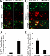
A, BrdUrd staining was performed in NIH 3T3 or NIH 3T3 p27(D51) cells with transfection by GFP-Cdk5N13-NLS and GFP-Cdk5N26-NLS. B and C, the percentages of double-labeled cells percentage in NIH 3T3 cells (B) or NIH 3T3 p27(D51) cells (C). *, p < 0.05 by analysis of variance. The error bars represent S.E., n = 4.
FIGURE 7.
FIGURE 7.
A, caspase-3 sensitive vector was co-transfected into Cdk5−/− neurons with cyclin D1-GFP reporter construct. After 1 μm β-amyloid administration, the dsRed and GFP signals were monitored. B and C, wild type primary neurons were treated with 3 μm β-amyloid 24 h. B, double immunostaining for endogenous Cdk5 (green) and BrdUrd (red). C, double immunostaining for endogenous p27 (red) and BrdUrd (Brdu, green). Nuclear Cdk5 and p27 both reveal in inverse relationship with the appearance of BrdUrd incorporation. DAPI, 4′,6′-diamino-2-phenylindole. DIC, differential interference contrast.
FIGURE 8.
FIGURE 8.
GFP-Cdk5-NLS and GFP-Cdk5-NES were transfected into Cdk5−/− neurons. A and C, after treatment with 3 μm β-amyloid, BrdUrd incorporation (A) and cleaved caspase-3 (C) were detected by immunocytochemistry (red). Note that in the nucleus Cdk5 prevents BrdUrd incorporation (no double-labeling) but permits caspase activation, whereas in the cytoplasm it prevents caspase activation but permits BrdUrd incorporation. B and D, the percentage of double-labeled cells was quantified for BrdUrd (B) and cleaved caspase-3 (D). E, *, p < 0.05 by analysis of variance. The error bars represent S.E.
FIGURE 9.
FIGURE 9.
When a postmitotic neuron is subjected to cell cycle or cell death stimulation (such as in Alzheimer disease), the interaction between Cdk5 and p27 is lost. When this happens, p27 is transported into the cytoplasm. At the same time, the two atypical NES in the Cdk5 sequence allow its transport to the cytoplasm by the CRM-1 pathway. The reduction in nuclear Cdk5 and p27 deprives the postmitotic neuron of its cell cycle suppression activity. The stressed neurons re-enter the cell cycle, but with the enhanced levels of cytoplasmic Cdk5, the neurons are temporarily protected from cell death. As yet, it is unknown what event causes the ultimate failure of this temporary protection. Driving expression of exogenous nuclear Cdk5 blocks the cell cycle; driving exogenous cytoplasmic Cdk5 attenuates cell death.

Similar articles

Cited by

References

    1. Dhavan R., Tsai L. H. (2001) Nat. Rev. Mol. Cell Biol. 2, 749–759 - PubMed
    1. van den Heuvel S., Harlow E. (1993) Science 262, 2050–2054 - PubMed
    1. Cruz J. C., Tsai L. H. (2004) Trends Mol. Med. 10, 452–458 - PubMed
    1. Cicero S., Herrup K. (2005) J. Neurosci. 25, 9658–9668 - PMC - PubMed
    1. Zhang J., Cicero S. A., Wang L., Romito-Digiacomo R. R., Yang Y., Herrup K. (2008) Proc. Natl. Acad. Sci. U.S.A. 105, 8772–8777 - PMC - PubMed

Publication types

MeSH terms