. 2010 May;149(2):263-272.
doi: 10.1016/j.pain.2010.02.010. Epub 2010 Mar 1.

Serine proteases and protease-activated receptor 2-dependent allodynia: a novel cancer pain pathway

Affiliations

Serine proteases and protease-activated receptor 2-dependent allodynia: a novel cancer pain pathway

D K Lam et al. Pain. 2010 May.

Abstract

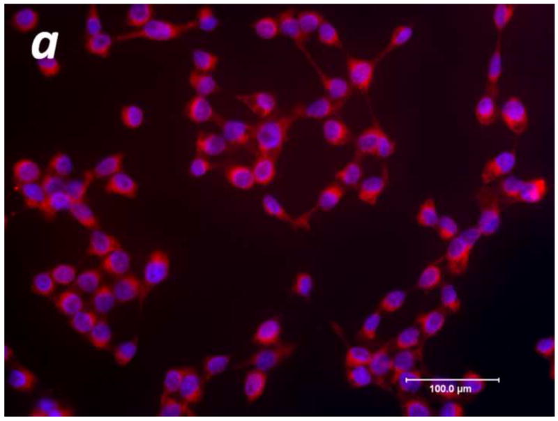
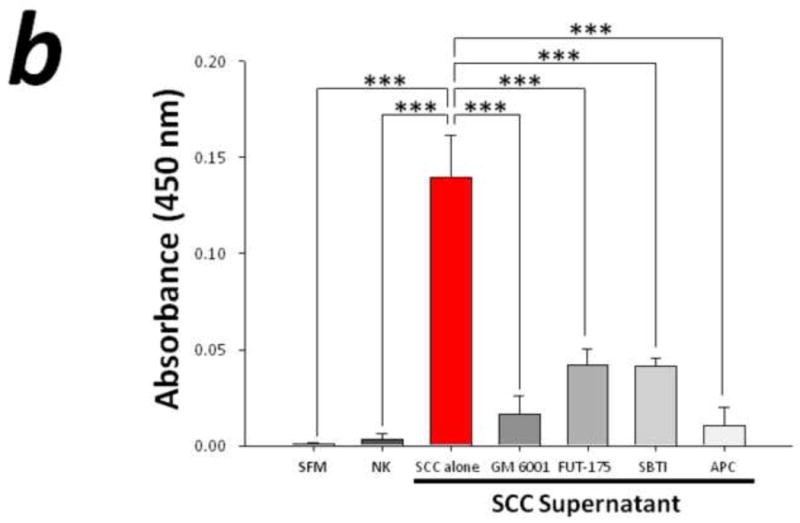
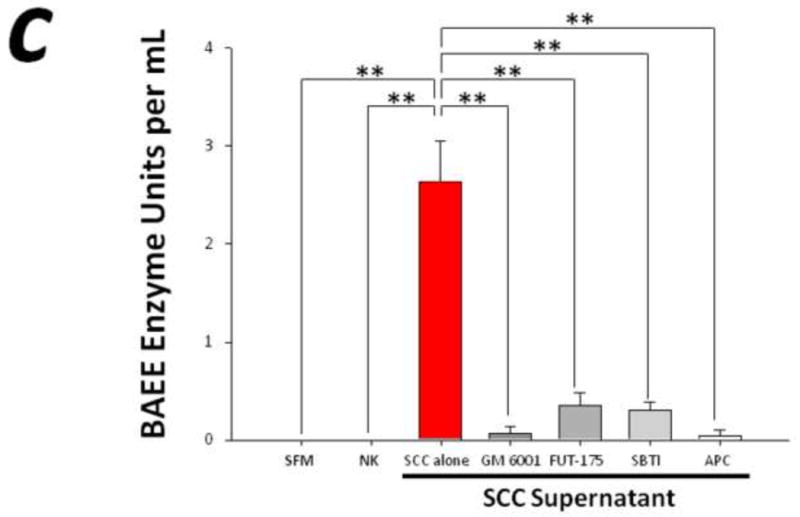
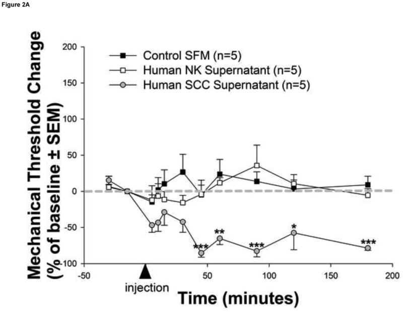
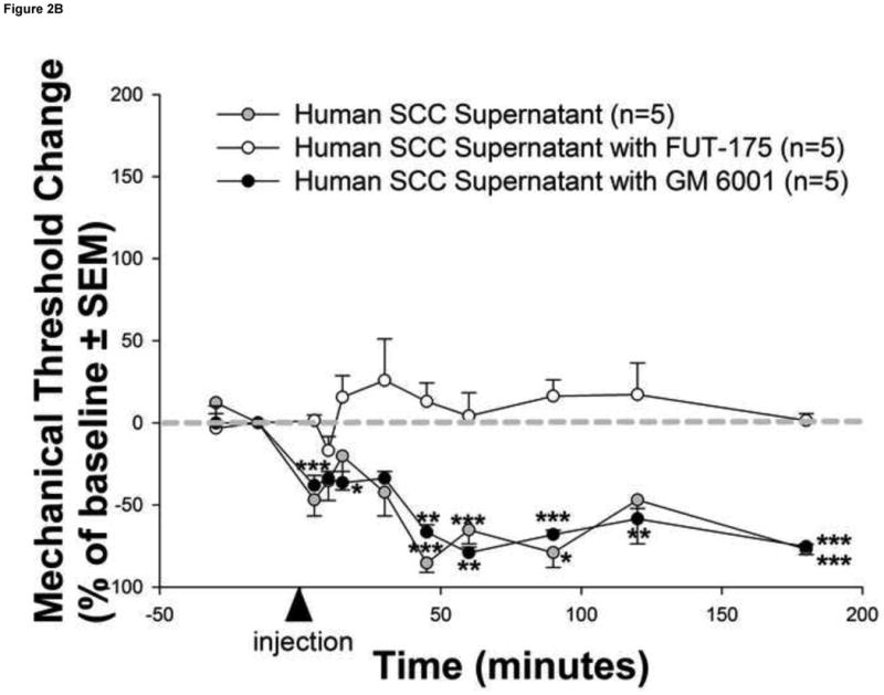
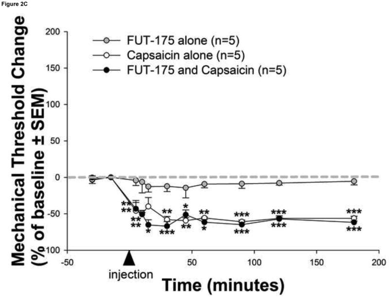
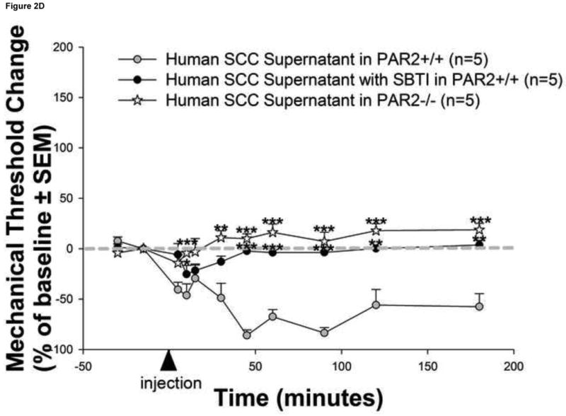
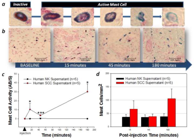
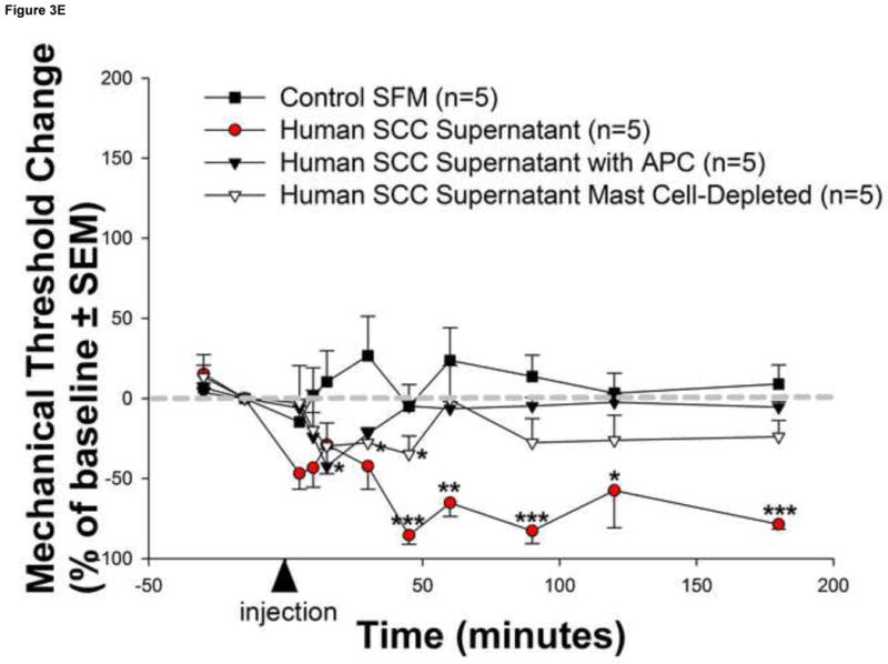
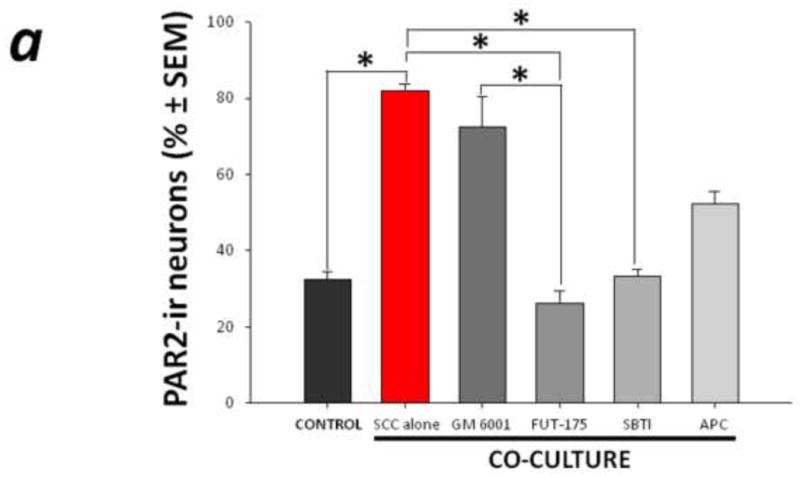
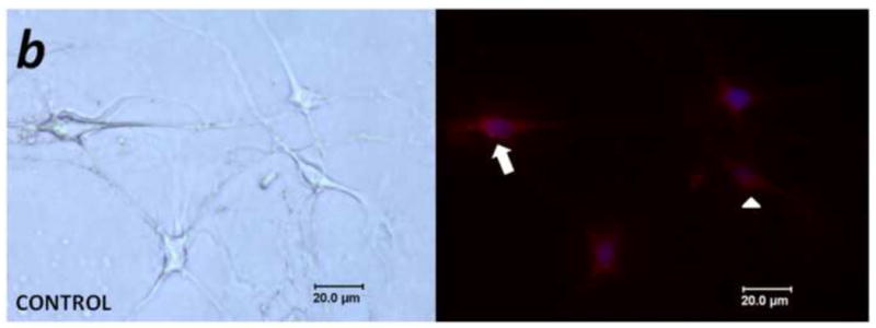
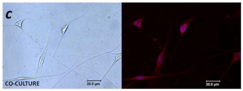
Mediators involved in the generation of pain in patients with cancer are poorly understood. Using a combined molecular, pharmacologic, behavioral, and genetic approach, we have identified a novel mechanism of cancer-dependent allodynia induced by protease-activated receptor 2 (PAR2). Here we show that human head and neck carcinoma cells have increased levels of proteolytic activity compared to normal human cell controls. Supernatant from human carcinoma cells, but not controls, caused marked and prolonged mechanical allodynia in mice, when administered into the hindpaw. This nociceptive effect was abolished by serine protease inhibition, diminished by mast cell depletion and absent in PAR2-deficient mice. In addition, non-contact co-culture of trigeminal ganglion neurons with human head and neck carcinoma cells increased the proportion of neurons that exhibited PAR2-immunoreactivity. Our results point to a direct role for serine proteases and their receptor in the pathogenesis of cancer pain. This previously unrecognized cancer pain pathway has important therapeutic implications wherein serine protease inhibitors and PAR2 antagonists may be useful for the treatment of cancer pain.


Conflict of interest statement

The authors declare no conflicts of interest for this study.

Figures

Figure 1
Figure 1. Elevated levels of proteolytic activity in human cancer
(a) Immunofluorescence of human squamous cell carcinoma (SCC) cells revealed homogeneous cytoplasmic trypsin staining, (b) and (c) Proteolytic activity of human SCC supernatant was markedly elevated compared to control serum free media (SFM) and human normal keratinocyte (NK) supernatant. Matrix metalloproteases and serine proteases contribute to this increased proteolytic activity as suggested by the attenuation in SCC-induced proteolysis by pre-incubation with GM 6001 and FUT-175, respectively. In particular, both trypsin and tryptase serine proteases significantly modulate SCC-induced proteolysis as demonstrated by pre-incubation with trypsin (SBTI) and tryptase (APC) inhibitors (**P<0.01, ***P<0.001, RM ANOVA, Holm-Sidak).
Figure 2
Figure 2. Human cancer cells induce serine protease and PAR2-dependent cancer pain
(a) Injection of human squamous cell carcinoma (SCC) supernatant into the hindpaw of mice produced marked and prolonged reductions in mechanical withdrawal-thresholds (grey circles). These nociceptive responses are mediated by serine proteases since (b) pre-incubation of human SCC supernatant with the serine protease inhibitor, FUT-175, abolished human SCC supernatant-induced reductions in withdrawal-thresholds (white circles). In contrast, pre-incubation with the matrix metalloprotease inhibitor, GM 6001, had no effect on SCC supernatant-induced reductions in withdrawal-thresholds (black circles). The anti-nociceptive effect of FUT-175 is specific to SCC-associated serine protease inhibition since (c) injection of FUT-175 alone into the hindpaw of mice did not affect baseline mechanical withdrawal-thresholds (grey circles) and, in contrast to its attenuation of SCC supernatant-induced mechanical allodynia, co-injection of FUT-175 with 0.1% capsaicin (black circles) did not affect capsaicin-induced reductions in mechanical withdrawal-thresholds (white circles). This cancer-induced nociceptive mechanism is trypsin and PAR2-dependent since (d) human SCC supernatant-induced mechanical withdrawal-threshold reductions were markedly attenuated when pre-incubated with trypsin inhibitor (SBTI) and injected in wild-type (PAR2+/+) littermate mice (black circles) and abolished when injected alone in PAR2-/- mice (white stars) (*P<0.05, **P<0.01, ***P<0.001, RM ANOVA, Holm-Sidak, Bonferroni or Dunn's as appropriate).
Figure 3
Figure 3. Mast cells mediate human carcinoma-induced cancer pain
Human squamous cell carcinoma (SCC) supernatant-induced changes in mast cell number and activity in the mouse hindpaw were assessed with a differential Alcian Blue/Safranin staining method. Mast cells stained uniformly red with safranin but not with alcian blue are inactive, while those that stained with alcian blue alone or mixed are active (a). Mast cells were persistently activated by human SCC supernatant as depicted in (b) images (200× magnification) of representative sections from 5 distinct animals at 15, 45 and 180 minutes post-injection and (c) graphically as the ratio of active/non-active (AB/S) mast cells over time. However, there was no change in the total mast cell count in the dermis (P>0.05, RM ANOVA) (d). The magnitude and duration of human SCC supernatant–induced reduction in withdrawal-thresholds was attenuated in (e) mast cell-depleted mice (white triangles) as well as in naive mice injected with human SCC supernatant pre-incubated with the tryptase inhibitor APC (black triangles) (*P<0.05, **P<0.01, ***P<0.001, RM ANOVA, Bonferroni or Dunn's as appropriate).
Figure 4
Figure 4. PAR2-immunoreactivity increased in trigeminal neurons co-cultured with human cancer cells
(a) Non-contact co-culture of trigeminal ganglion neurons with head and neck carcinoma cells (SCC) for 48 h increased the proportion of neurons that exhibited PAR2-immunoreactivity. The increased neuronal PAR2-immunoreactivity was attenuated in the presence of the serine protease inhibitor FUT-175 and trypsin inhibitor SBTI (*P<0.05, RM ANOVA-on-ranks, Tukey test). (b) and (c) Representative bright field (left panel) and corresponding PAR2 immunofluorescence (right panel) images of trigeminal ganglion neurons maintained in vitro for 48 h in control (b) and co-culture (c) conditions. In (b), arrow designates a PAR2-immunoreactive neuron, and the arrowhead indicates a non-labeled neuron.

Similar articles

Cited by

References

    1. Amadesi S, Cottrell GS, Divino L, Chapman K, Grady EF, Bautista F, Karanjia R, Barajas-Lopez C, Vanner S, Vergnolle N, Bunnett NW. Protease-activated receptor 2 sensitizes TRPV1 by protein kinase Cε- and A-dependent mechanisms in rats and mice. J Physiol. 2006;575:555–571. - PMC - PubMed
    1. Aromando RF, Pérez MA, Heber EM, Trivillin VA, Tomasi VH, Schwint AE, Itoiz ME. Potential role of mast cells in hamster cheek pouch carcinogenesis. Oral Oncol. 2008;44:1080–1087. - PubMed
    1. Bjordal K, Ahlner-Elmqvist M, Hammerlid E, Boysen M, Evensen JF, Biörklund A, Jannert M, Westin T, Kaasa S. A prospective study of quality of life in head and neck cancer patients. Part II: Longitudinal data. Laryngoscope. 2001;111:1440–1452. - PubMed
    1. Brasseur L. Review of current pharmacologic treatment of pain. Drugs. 1997;53:10–17. - PubMed
    1. Cenac N, Andrews CN, Holzhausen M, Chapman K, Cottrell G, Andrade-Gordon P, Steinhoff M, Barbara G, Beck P, Bunnett NW, Sharkey KA, Ferraz JG, Shaffer E, Vergnolle N. Role for protease activity in visceral pain in irritable bowel syndrome. J Clin Invest. 2007;117:636–647. - PMC - PubMed

Publication types

MeSH terms