. 2010 Apr 23;285(17):13022-31.
doi: 10.1074/jbc.M109.079426. Epub 2010 Feb 22.

DHHC5 interacts with PDZ domain 3 of post-synaptic density-95 (PSD-95) protein and plays a role in learning and memory

Affiliations

DHHC5 interacts with PDZ domain 3 of post-synaptic density-95 (PSD-95) protein and plays a role in learning and memory

Yi Li et al. J Biol Chem. .

Abstract

A family of integral membrane proteins containing a signature DHHC motif has been shown to display protein S-acyltransferase activity, modifying cysteine residues in proteins with fatty acids. The physiological roles of these proteins have largely been unexplored. Here we report that mice homozygous for a hypomorphic allele of a previously uncharacterized member, DHHC5, are born at half the expected rate, and survivors show a marked deficit in contextual fear conditioning, an indicator of defective hippocampal-dependent learning. DHHC5 is highly enriched in a post-synaptic density preparation and co-immunoprecipitates with post-synaptic density protein-95 (PSD-95), an interaction that is mediated through binding of the carboxyl terminus of DHHC5 and the PDZ3 domain of PSD-95. Immunohistochemistry demonstrated that DHHC5 is expressed in the CA3 and dentate gyrus in the hippocampus. These findings point to a previously unsuspected role for DHHC5 in post-synaptic function affecting learning and memory.


Figures
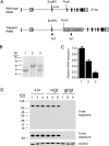
FIGURE 1.
FIGURE 1.
Insertional mutagenesis of the mouse DHHC5 gene. A, shown is a schematic of the insertional mutation in DHHC5. The β-geo cassette was found to be inserted into intron 3 of the DHHC5 gene. Binding sites for primers fp1 and rp1 are also shown. B, a Southern blot of EcoRv/PvuII digest of genomic mouse tail DNA using an exon 3 probe is shown. The wild-type allele yielded a 6.0-kb band, and the mutant allele yielded a 4.7-kb band. Lane 1, homozygous gt/gt; lane 2, homozygous +/+; lane 3, heterozygous +/gt. C, DHHC5 mRNA levels were determined by quantitative real-time PCR performed on total RNA extracted from DHHC5 +/+, +/gtv and gt/gt mouse brains. Expression levels were normalized to glyceraldehyde-3-phosphate dehydrogenase, n = 5, in each group. D, DHHC5 immunoblotting of membrane extracts (25 μg of protein) prepared from DHHC5 +/+, +/gt, and gt/gt mouse brains is shown. The rabbit polyclonal anti-DHHC5 antibody was used for detection (1:1000, Sigma). Three mice are shown for each genotype. COXIV was immunostained to serve as a loading control.
FIGURE 2.
FIGURE 2.
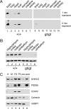
DHHC5 tissue distribution and subcellular fractionation. A, tissue distribution of DHHC5 in wild-type and DHHC5 gene-targeted mice is shown. Immunoblotting of whole tissue extracts (25 μg of protein/lane) was performed using an anti-DHHC5 polyclonal antibody (1:1000, Sigma). B, regional distribution of DHHC5 is shown. Brain tissue was dissected, and immunoblotting was performed on whole tissue homogenates. COXIV was used as a loading control. C, DHHC5 in pre- and post-synaptic membrane fractions is shown. Twenty micrograms of protein were loaded in each lane. H, homogenate; S1, post-nuclear supernatant; P2, mitochondrial pellet; P3, second mitochondrial pellet; pre, pre-synaptic membrane fractions; post, post-synaptic membrane fraction. Markers shown were used to detect post-synaptic membranes (PSD95), mitochondria (COXIV), endoplasmic reticulum (calnexin), and pre-synaptic membranes (VAMP1). Results shown were from one of two independent experiments giving similar results.
FIGURE 3.
FIGURE 3.
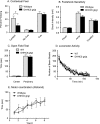
Impaired contextual fear learning in DHHC5 gt/gt mice. A, contextual and cued fear conditioning is shown. Significant deficits in contextual (p < 0.025, unpaired t test, two-tailed), but not cued freezing, were observed in the DHHC5 gt/gt mice (n = 10), compared with their +/+ littermate controls (n = 12). B, foot-shock sensitivity is shown. The same mice used in A were tested for foot-shock sensitivity. No significant difference was observed between DHHC5 +/+ and gt/gt animals. C, an open field test is shown. The average amount of time spent in the center, non-periphery, and periphery of an open field by DHHC5 +/+ and gt/gt mice was calculated. The means (±S.E.) are presented, and n = 12 for wild-type (WT) and n = 11 for DHHC gt/gt mice. D, locomotor activity is shown. DHHC5 +/+ (n = 12) and DHHC5 gt/gt mice (n = 11) were observed in a clean, plastic mouse cage located inside a dark Plexiglas box. Data are expressed as the average number of beam breaks at each 5-min bin per 2-h test period ± S.E. E, motor coordination (Rotarod test) is shown. The time to fall from a rotating rod that was accelerated from 5 to 45 rpm over 5 min was assessed. Each mouse was tested four times (15–30-min intertrial interval) each day for two consecutive days. n = 12 for wild-type and n = 10 for DHHC5 gt/gt mice.
FIGURE 4.
FIGURE 4.
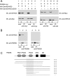
Coimmunoprecipitation of DHHC5 and PSD-95 in transfected cells and brain and determination of the interaction site. A, co-immunoprecipitation of PSD-95 and DHHC5 carboxyl-terminal cytoplasmic domain in transfected cells is shown. HEK-293 cells were transfected with PSD-95-myc and HA-CtermDHHC5, a construct that consisted of an HA epitope tag fused to the carboxyl-terminal cytoplasmic domain of DHHC5. A construct in which the final four amino acids were deleted (ΔEISV) was also tested. In the left panel, immunoprecipitation was performed using an anti-Myc rabbit polyclonal antibody to precipitate PSD-95, and in the right panel, an anti-HA rabbit polyclonal antibody was used to precipitate DHHC5. Whole cell extracts (wce) consisting of 5% of the total mixture (lanes 1, 4, 7, and 10) or entire immunoprecipitate (IP) (other lanes) were loaded onto SDS-PAGE gels and immunoblotted (IB) using an anti-DHHC5 chicken IgY antibody or anti-Myc mouse monoclonal antibody as indicated. B, DHHC5 was detected in an immunoprecipitate of PSD-95 from mouse brain. Mouse brain lysates were prepared (lanes 1 and 4) and incubated with anti-PSD-95 monoclonal antibody (lanes 2 and 5) or control mouse IgG (lanes 4 and 6) and subjected to SDS-PAGE and immunoblotting with the anti-DHHC5 polyclonal antibody (upper panel) or anti-PSD-95 rabbit polyclonal antibody (lower panel). (upper panel, 1% of input; lower panel, 2% of input). Lanes 1–3, wild-type brain, lanes 4–6, DHHC gt/gt brain. The experiment was repeated three times with similar results. C, deletion analysis of PSD-95 clones in a yeast two-hybrid assay with the carboxyl-terminal cytoplasmic domain of DHHC5 is shown. L40 yeast strains harboring the bait vector (pLexN) expressing the amino acids 220–715 of mouse DHHC5 protein and prey vectors (pVP16-3) expressing different fragments of PDZ domain-containing proteins were grown on CSM-WLH (−Trp/−Leu/−His) plates. Only those that contain DHHC5-interacting partners survive the −His selection. GK, guanylate kinase-like domain.
FIGURE 5.
FIGURE 5.
Altered DHHC5 immunohistochemistry in DHHC5 gt/gt mouse brain. Immunohistochemical staining revealed widespread expression of DHHC5 in the hippocampal formation (A, C, E, F) and cortex (I, K) of wild-type mice, with DHHC5 immunoreactivity present homogeneously in the neuropil, dentate granule neurons, and pyramidal neurons in the CA1–3 of the hippocampal formation (A) and laminae III and V of the cortex (I). In contrast, DHHC5 immunoreactivity was markedly reduced in the neuropil of DHHC5 gt/gt mice, with DHHC5 expression retained in subsets of neurons. The changes in distribution of DHHC5 immunoreactivity were most pronounced in the hippocampal formation, with staining virtually abolished in dentate granule neurons (D and H) and relatively reduced in CA1 (B and G) compared with the retained DHHC5 immunoreactivity evident in the ventrolateral portion of CA3 in these mutant mice (as indicated by arrows in B). Within the primary somatosensory cortex-barrel field (S1BF) of DHHC5 gt/gt mice, staining for DHHC5 was effectively absent from the neuropil (J), and although generally reduced DHHC5 immunoreactivity was present, this was more pronounced in subpopulations of neurons in lamina V (L, LamV). Hp, hippocampus; DG, dentate gyrus; CA1 and CA3, hippocampal subfields. Bars, A and B, 1200 μm; C and D, 500 μm; E–H, 75 μm; I and J, 100 μm, K and L, 50 μm.

Similar articles

Cited by

References

    1. Bartels D. J., Mitchell D. A., Dong X., Deschenes R. J. (1999) Mol. Cell Biol. 19, 6775–6787 - PMC - PubMed
    1. Lobo S., Greentree W. K., Linder M. E., Deschenes R. J. (2002) J. Biol. Chem. 277, 41268–41273 - PubMed
    1. Roth A. F., Feng Y., Chen L., Davis N. G. (2002) J. Cell Biol. 159, 23–28 - PMC - PubMed
    1. Putilina T., Wong P., Gentleman S. (1999) Mol. Cell Biochem. 195, 219–226 - PubMed
    1. Hines R. M., Kang R., Goytain A., Quamme G. A. (2010) J. Biol. Chem. 285, 4621–4628 - PMC - PubMed

Publication types

MeSH terms