. 2010 Feb 16;5(2):e9226.
doi: 10.1371/journal.pone.0009226.

Delayed onset of positive feedback activation of Rab5 by Rabex-5 and Rabaptin-5 in endocytosis

Affiliations

Delayed onset of positive feedback activation of Rab5 by Rabex-5 and Rabaptin-5 in endocytosis

Huaiping Zhu et al. PLoS One. .

Abstract

Background: Rabex-5 is a guanine nucleotide exchange factor (GEF) that specifically activates Rab5, i.e., converting Rab5-GDP to Rab5-GTP, through two distinct pathways to promote endosome fusion and endocytosis. The direct pathway involves a pool of membrane-associated Rabex-5 that targets to the membrane via an early endosomal targeting (EET) domain. The indirect pathway, on the other hand, involves a cytosolic pool of Rabex-5/Rabaptin-5 complex. The complex is recruited to the membrane via Rabaptin-5 binding to Rab5-GTP, suggesting a positive feedback mechanism. The relationship of these two pathways for Rab5 activation in the cell is unclear.

Methodology/principal findings: We dissect the relative contribution of each pathway to Rab5 activation via mathematical modeling and kinetic analysis in the cell. These studies show that the indirect pathway constitutes a positive feedback loop for converting Rab5-GDP to Rab5-GTP on the endosomal membrane and allows sensitive regulation of endosome fusion activity by the levels of Rab5 and Rabex-5 in the cell. The onset of this positive feedback effect, however, contains a threshold, which requires above endogenous levels of Rab5 or Rabex-5 in the cell. We term this novel phenomenon "delayed response". The presence of the direct pathway reduces the delay by increasing the basal level of Rab5-GTP, thus facilitates the function of the Rabex-5/Rabaptin-5-mediated positive feedback loop.

Conclusion: Our data support the mathematical model. With the model's guidance, the data reveal the affinity of Rabex-5/Rabaptin-5/Rab5-GTP interaction in the cell, which is quantitatively related to the Rabex-5 concentration for the onset of the indirect positive feedback pathway. The presence of the direct pathway and increased Rab5 concentration can reduce the Rabex-5 concentration required for the onset of the positive feedback loop. Thus the direct and indirect pathways cooperate in the regulation of early endosome fusion.


Conflict of interest statement

Competing Interests: The authors have declared that no competing interests exist.

Figures

Figure 1
Figure 1. Direct and indirect pathways in Rabex-5-mediated Rab5 activation.
Rabex-5 targets to the early endosomal membrane in two parallel pathways: direct targeting via the EET domain and indirect targeting via forming complex with Rabaptin-5 that binds to Rab5-GTP. The reaction scheme of the direct pathway contains two steps: newly synthesized cytosolic Rabex-5 binds to the membrane, and the membrane-bound Rabex-5 can then act on the membrane-bound substrate Rab5-GDP. The reaction scheme of the indirect pathway contains three steps: cytosolic Rabex-5 binds to Rabaptin-5 in the cytoplasm, the Rabex-5/Rabaptin-5 complex remains soluble in the cytoplasm until its level and/or Rab5-GTP level reach a threshold and the complex is recruited to the membrane by binding to Rab5-GTP, and the membrane-bound Rabex-5 in turn converts more Rab5-GDP to Rab5-GTP. The superscripts “s” and “m” denote soluble and membrane-bound forms of Rabex-5 in the cell.
Figure 2
Figure 2. Mathematical model of Rab5 activation via direct and indirect pathways.
Rab5 activity is plotted against Rabex-5 concentration in the cell, when there is only direct or indirect pathway as indicated. The response curves are plotted according to the mathematical model (eq. 6 and eq. 7) described in the text, with rate constants arbitrarily assigned as: α = 1, β = 1, γ = 1, λ = 100, and σ = 0.1.
Figure 3
Figure 3. Inducible expression of Rabex-5 constructs and Rabaptin-5 in BHK cells.
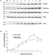
A. BHK cells were transfected with pBI/myc-Rabex-5(135–480)/Rabaptin-5 or pBI/myc-Rabex-5(1–399), pTet-Off, and pcDNA3/GFP-Rab5 (3∶3∶1) and incubated at 37°C for 15 hours in the presence of Dox. Upon removal of Dox, myc-Rabex-5(135–480)/Rabaptin-5 or myc-Rabex-(1–399) expression was induced for the indicated times. Shown are immunoblots of the cell lysates with anti-myc, anti-Rabaptin-5, and anti-Rab5 antibodies. Control cells (Con) were transfected with the empty pBI vector. Endogenous Rab5 serves a loading control. Molecular mass standards (in kDa) are indicated on the left side of the panel. B. Shown is the quantification of myc-Rabex-5(135–480) expression from the immunoblot in A by densitometry. The graph shows inducible myc-Rabex-5(135–480) or myc-Rabex-(1–399) expression over the indicated time course, with intracellular protein concentrations calculated based on the standard curve described in the Materials and Methods and error bars indicating SEM from three independent immunoblot experiments.
Figure 4
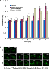
Figure 4. Kinetics of Rabex-5(135–480)/Rabaptin-5 and Rabex-5(1–399)-mediated Rab5 activation in BHK cells with ectopic expression of GFP-Rab5.
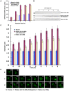
A. GST pull-down assay showing increased levels of GTP-bound GFP-Rab5 over the time course of inducible expression of the indicated Rabex-5 proteins (see Figure 3). GTP-bound GFP-Rab5 in each cell lysate was detected by its binding to GST-R5BD, followed by immunoblot analysis with an anti-Rab5 mAb and quantification by densitometry. Endogenous Rab5-GTP level was too low to be detected with the same amount of lysates. The graph shows the quantification of GTP-bound GFP-Rab5 in each cell lysate, and error bars represent SEM of three independent experiments. Representative immunoblots from one of the experiments are shown in B. Molecular mass standards (in kDa) are indicated on the left side of each panel. C. Confocal fluorescence microscopy analysis of the size increase of GFP-Rab5-labeled early endosomes over the time course of inducible expression of the indicated Rabex-5 proteins. The graph quantifies the maximal size of early endosomes in cells expressing Rabex-5(1–399) or Rabex-5(135–480) with Rabaptin-5, as indicated. In control cells transfected with the empty vector, the size of endogenous endosomes did not change over time and was slightly smaller than that in cells expressing Rabex-5(1–399) at 0 h (see panel D). The 0 h value of control cells is shown in the graph to serve as a background control. The diameters of 90 largest GFP-Rab5-labeled endosomes in 30 cells were measured in each case and the graph shows the mean and calculated SEM. Representative confocal fluorescence microscopy images of the GFP-Rab5-labeled early endosomes used in the quantification are shown in D. X indicates control cells transfected with the empty pBI vector; Y indicates cells expressing Rabex-5(135–480) and Rabaptin-5; Z indicates cells expressing Rabex-5(1–399). Bar = 16 µm.
Figure 5
Figure 5. Only the direct pathway allows Rabex-5 to target to early endosomes and activate Rab5 in BHK cells with endogenous level of Rab5.
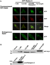
A. Confocal fluorescence microscopy images of BHK cells expressing the indicated GFP-Rabex-5 proteins with or without co-expression with RFP-Rab5. The cells were transfected with the indicated constructs and processed for microscopy 15 hours after the transfection. Bar = 16 µm. B. Immunoblots showing the expression of the indicated GFP-Rabex-5 constructs and Rabaptin-5. The GFP-Rabex-5 constructs contain a myc-epitope downstream of GFP and was probed with the anti-myc antibody (top panel), while Rabaptin-5 expression was identified with the anti-Rabaptin-5 antibody (bottom panel). Molecular mass standards (in kDa) are indicated on the left side of each panel.
Figure 6
Figure 6. Kinetics of GFP-Rabex-5-mediated Rab5 activation in BHK cells with endogenous level of Rab5.
A. Immunoblot showing inducible expression of GFP-Rabex-5. BHK cell monolayers were transfected either with the empty pBI vector (control) or with pBI/GFP-Rabex-5 and pTet-Off, then incubated at 37°C for 15 hours in the presence of Dox. Upon Dox removal to induce GFP-Rabex-5 expression, cell lysates were prepared at the indicated times for immunoblot analysis with the anti-Rabex-5 antibody. Endogenous Rabex-5 in the same lysates serves an internal loading control, as indicated. Molecular mass standards (in kDa) are indicated on the left side of the panel. B. Confocal fluorescence microscopy showing the size increase of GFP-Rabex-5-labeled early endosomes over the time course of inducible expression of the protein as indicated. Bar = 16 µm. C. The graph quantifies the maximum size of GFP-Rabex-5-labeled early endosomes shown in B. The diameters of 90 largest GFP-Rabex5-labeled endosomes in 30 cells were measured in each case and the graph shows the mean and calculated SEM.
Figure 7
Figure 7. Inducible expression of Rabex-5 constructs and Rabaptin-5 in Rabex-5-deficient mouse embryo fibroblasts.
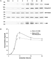
A. Cells were transfected with pBI/myc-Rabex-5(135–480)/Rabaptin-5 or pBI/myc-Rabex-5(1–399), pTet-Off, and pcDNA3/GFP-Rab5 (3∶3∶1) and incubated at 37°C for 15 hours in the presence of Dox. Upon removal of Dox, myc-Rabex-5(135–480)/Rabaptin-5 or myc-Rabex-5(1–399) expression was induced for the indicated times. Shown are immunoblots of the cell lysates with anti-myc, anti-Rabaptin-5, and anti-Rab5 antibodies. Control cells were transfected with the empty pBI vector. Endogenous Rab5 in the same lysates serves an internal loading control. Molecular mass standards (in kDa) are indicated on the left side of the panel. B. Shown is the quantification of myc-Rabex-5(1–399) or myc-Rabex-5(135–480) expression from the immunoblot in A by densitometry. The graph shows inducible myc-Rabex-5(135–480) or myc-Rabex-(1–399) expression over the indicated time course, with intracellular protein concentrations calculated based on the standard curve described in the Materials and Methods and error bars indicating SEM from three independent immunoblot experiments.
Figure 8
Figure 8. Kinetics of Rabex-5(135–480)/Rabaptin-5 and Rabex-5(1–399)-mediated Rab5 activation in Rabex-5-deficient cells with ectopic expression of GFP-Rab5.
A. Confocal fluorescence microscopy analysis of the size increase of GFP-Rab5-labeled early endosomes over the time course of inducible expression of the indicated Rabex-5 proteins (see Figure 7). The graph quantifies the maximal size of early endosomes in cells expressing Rabex-5(1–399) or Rabex-5(135–480) with Rabaptin-5, as indicated. In control cells transfected with the empty vector, the size of endogenous endosomes did not change over time and was similar to that in cells expressing Rabex-5(1–399) or Rabex-5(135–480) at 0 h (see panel B). The 0 h value of control cells is shown in the graph to serve as a background control. The diameters of 90 largest GFP-Rab5-labeled endosomes in 30 cells were measured in each case and the graph shows the mean and calculated SEM. Representative confocal fluorescence microscopy images of the GFP-Rab5-labeled early endosomes used in the quantification are shown in B. X indicates control cells transfected with the empty pBI vector; Y indicates cells expressing Rabex-5(135–480) and Rabaptin-5; Z indicates cells expressing Rabex-5(1–399). Bar = 16 µm.

Similar articles

Cited by

References

    1. Horiuchi H, Lippe R, McBride HM, Rubino M, Woodman P, et al. A novel Rab5 GDP/GTP exchange factor complexed to Rabaptin-5 links nucleotide exchange to effector recruitment and function. Cell. 1997;90:1149–1159. - PubMed
    1. Bucci C, Parton RG, Mather IM, Stunnenberg H, Simons K, et al. The small GTPase Rab5 functions as a regulatory factor in the early endocytic pathway. Cell. 1992;70:715–728. - PubMed
    1. Gorvel J-P, Chavrier P, Zerial M, Gruenberg J. rab5 controls early endosome fusion in vitro. Cell. 1991;64:915–925. - PubMed
    1. Li G, Barbieri MA, Colombo MI, Stahl PD. Structural features of the GTP-binding defective Rab5 mutants required for their inhibitory activity on endocytosis. J Biol Chem. 1994;269:14631–14635. - PubMed
    1. Li G, Liang Z. Phosphate-binding loop and Rab GTPase function: mutations at Ser29 and Ala30 of Rab5 lead to loss-of-function as well as gain-of-function phenotype. Biochem J. 2001;355:681–689. - PMC - PubMed

Publication types

MeSH terms