

Unconventional ubiquitin recognition by the ubiquitin-binding motif within the Y family DNA polymerases iota and Rev1
- PMID: 20159559
- PMCID: PMC2841503
- DOI: 10.1016/j.molcel.2009.12.038
Unconventional ubiquitin recognition by the ubiquitin-binding motif within the Y family DNA polymerases iota and Rev1
Abstract
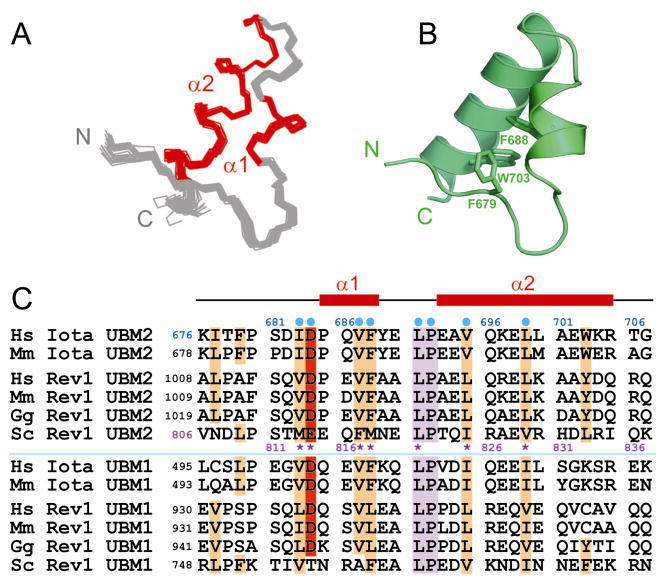
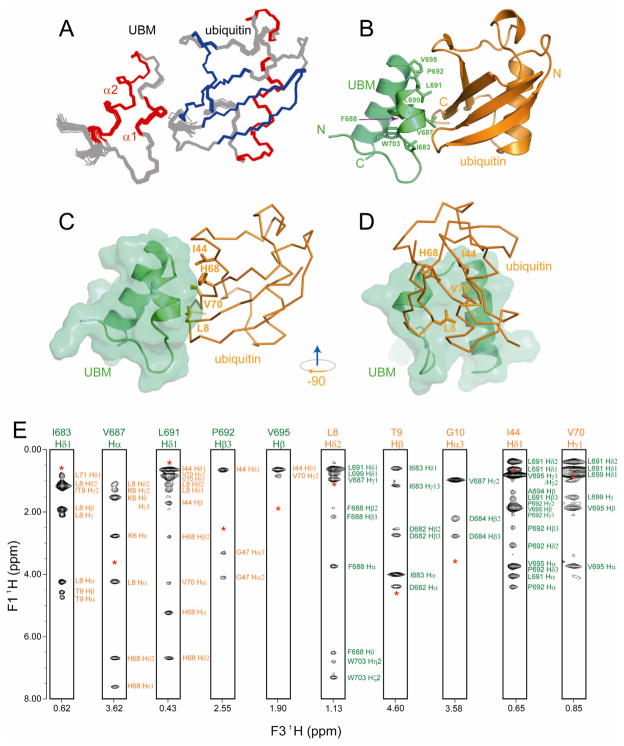
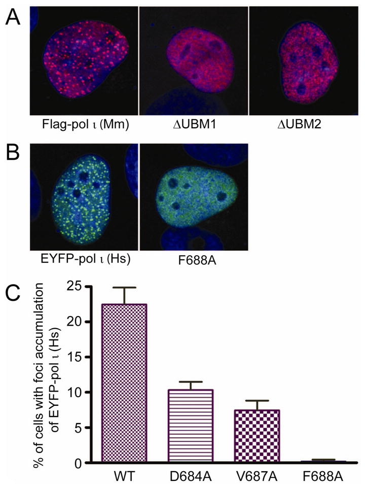
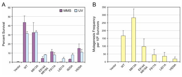
Translesion synthesis is an essential cell survival strategy to promote replication after DNA damage. The accumulation of Y family polymerases (pol) iota and Rev1 at the stalled replication machinery is mediated by the ubiquitin-binding motifs (UBMs) of the polymerases and enhanced by PCNA monoubiquitination. We report the solution structures of the C-terminal UBM of human pol iota and its complex with ubiquitin. Distinct from other ubiquitin-binding domains, the UBM binds to the hydrophobic surface of ubiquitin centered at L8. Accordingly, mutation of L8A, but not I44A, of ubiquitin abolishes UBM binding. Human pol iota contains two functional UBMs, both contributing to replication foci formation. In contrast, only the second UBM of Saccharomyces cerevisiae Rev1 binds to ubiquitin and is essential for Rev1-dependent cell survival and mutagenesis. Point mutations disrupting the UBM-ubiquitin interaction also impair the accumulation of pol iota in replication foci and Rev1-mediated DNA damage tolerance in vivo.
Figures
Similar articles
-
Structural analysis of the conserved ubiquitin-binding motifs (UBMs) of the translesion polymerase iota in complex with ubiquitin.J Biol Chem. 2011 Jan 14;286(2):1364-73. doi: 10.1074/jbc.M110.135038. Epub 2010 Oct 6. J Biol Chem. 2011. PMID: 20929865 Free PMC article.
-
NMR mapping of PCNA interaction with translesion synthesis DNA polymerase Rev1 mediated by Rev1-BRCT domain.J Mol Biol. 2013 Sep 9;425(17):3091-105. doi: 10.1016/j.jmb.2013.05.029. Epub 2013 Jun 7. J Mol Biol. 2013. PMID: 23747975
-
The Proliferating Cell Nuclear Antigen (PCNA)-interacting Protein (PIP) Motif of DNA Polymerase η Mediates Its Interaction with the C-terminal Domain of Rev1.J Biol Chem. 2016 Apr 15;291(16):8735-44. doi: 10.1074/jbc.M115.697938. Epub 2016 Feb 22. J Biol Chem. 2016. PMID: 26903512 Free PMC article.
-
Eukaryotic mutagenesis and translesion replication dependent on DNA polymerase zeta and Rev1 protein.Biochem Soc Trans. 2001 May;29(Pt 2):187-91. doi: 10.1042/0300-5127:0290187. Biochem Soc Trans. 2001. PMID: 11356151 Review.
-
Eukaryotic translesion synthesis DNA polymerases: specificity of structure and function.Annu Rev Biochem. 2005;74:317-53. doi: 10.1146/annurev.biochem.74.082803.133250. Annu Rev Biochem. 2005. PMID: 15952890 Review.
Cited by
-
Protein Assemblies in Translesion Synthesis.Genes (Basel). 2024 Jun 24;15(7):832. doi: 10.3390/genes15070832. Genes (Basel). 2024. PMID: 39062611 Free PMC article. Review.
-
Ubiquitin mediates the physical and functional interaction between human DNA polymerases η and ι.Nucleic Acids Res. 2013 Feb 1;41(3):1649-60. doi: 10.1093/nar/gks1277. Epub 2012 Dec 16. Nucleic Acids Res. 2013. PMID: 23248005 Free PMC article.
-
An overview of Y-Family DNA polymerases and a case study of human DNA polymerase η.Biochemistry. 2014 May 6;53(17):2793-803. doi: 10.1021/bi500019s. Epub 2014 Apr 23. Biochemistry. 2014. PMID: 24716551 Free PMC article. Review.
-
REV7 has a dynamic adaptor region to accommodate small GTPase RAN/Shigella IpaB ligands, and its activity is regulated by the RanGTP/GDP switch.J Biol Chem. 2019 Oct 25;294(43):15733-15742. doi: 10.1074/jbc.RA119.010123. Epub 2019 Sep 4. J Biol Chem. 2019. PMID: 31484720 Free PMC article.
-
Structural analysis of the conserved ubiquitin-binding motifs (UBMs) of the translesion polymerase iota in complex with ubiquitin.J Biol Chem. 2011 Jan 14;286(2):1364-73. doi: 10.1074/jbc.M110.135038. Epub 2010 Oct 6. J Biol Chem. 2011. PMID: 20929865 Free PMC article.
References
-
- Akutsu M, Kawasaki M, Katoh Y, Shiba T, Yamaguchi Y, Kato R, Kato K, Nakayama K, Wakatsuki S. Structural basis for recognition of ubiquitinated cargo by Tom1-GAT domain. FEBS Lett. 2005;579:5385–5391. - PubMed
-
- Ball G, Meenan N, Bromek K, Smith BO, Bella J, Uhrin D. Measurement of one-bond 13Calpha-1Halpha residual dipolar coupling constants in proteins by selective manipulation of CalphaHalpha spins. J Magn Reson. 2006;180:127–136. - PubMed
-
- Bartels C, Xia T, Billeter M, Güntert P, Wüthrich K. The program XEASY for computer-supported NMR spectral analysis of biological macromolecules. J Biol NMR. 1995;6:1–10. - PubMed
-
- Bienko M, Green CM, Crosetto N, Rudolf F, Zapart G, Coull B, Kannouche P, Wider G, Peter M, Lehmann AR, et al. Ubiquitin-binding domains in Y-family polymerases regulate translesion synthesis. Science. 2005;310:1821–1824. - PubMed
Publication types
MeSH terms
Substances
Grants and funding
LinkOut - more resources
Full Text Sources
Molecular Biology Databases
Miscellaneous
