. 2009 May 21;1(7):637-51.
doi: 10.18632/aging.100054.

The role of p38b MAPK in age-related modulation of intestinal stem cell proliferation and differentiation in Drosophila

Affiliations

The role of p38b MAPK in age-related modulation of intestinal stem cell proliferation and differentiation in Drosophila

Joung-Sun Park et al. Aging (Albany NY). .

Abstract

It is important to understand how age-related changes in intestinal stem cells (ISCs) may contribute to age-associated intestinal diseases, including cancer. Drosophila midgut is an excellent model system for the study of ISC proliferation and differentiation. Recently, age-related changes in the Drosophila midgut have been shown to include an increase in ISC proliferation and accumulation of mis-differentiated ISC daughter cells. Here, we show that the p38b MAPK pathway contributes to the age-related changes in ISC and progenitor cells in Drosophila. D-p38b MAPK is required for an age-related increase of ISC proliferation. In addition, this pathway is involved in age and oxidative stress-associated mis-differentiation of enterocytes and upregulation of Delta, a Notch receptor ligand. Furthermore, we also show that D-p38b acts downstream of PVF2/PVR signaling in these age-related changes. Taken together, our findings suggest that p38 MAPK plays a crucial role in the balance between ISC proliferation and proper differentiation in the adult Drosophila midgut.

Keywords: Delta/Notch pathway; Drosophila; PVR signaling; differentiation; gut; intestinal stem cell; oxidative stress; p38b MAPK; aging; proliferation.


Conflict of interest statement

The authors in this manuscript have no conflict of interests to declare.

Figures

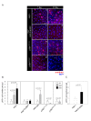
Figure 1.
Figure 1.. Increased expression of D-p38b in ISCs and EBs within aged gut.
(A) Increased expression of D-p38b-lacZ reporter construct in the adult posterior midgut with age. The midguts of 5- and 30-day-old flies, including those with two copies of the D-p38b-lacZ, were examined by X-gal staining. Squared boxes are enlarged images. Arrow head indicates β-gal-positive cells. Original magnification is 400x. (B) Increased expression of D-p38b-lacZ reporter construct in the Delta-positive and neighboring cells in aged gut. The midguts of 30-day-old flies were examined with anti-β-gal and anti-Delta. Anti-β-gal, green; anti-Delta, red; DAPI, blue. Scale bar, 1 μM.
Figure 2.
Figure 2.. Effect of D-p38b MAPK signaling on DNA synthesis of intestinal cells and ISC division.
(A) Effects of D-p38b MAPK modulation on BrdU incorporation levels in the adult midgut. Twenty-five day-old flies expressing esg>+ (a and b), esg>UAS-D-p38bas(c and b), p38bEY11174 (e and f) or p38bKG01337 (g and h) were fed on 0.2 mg/ml BrdU media for 4 days, and stained with anti-BrdU. Overlay (DAPI, blue; anti-BrdU, red). Asterisk indicates enlarged EC nuclei. Arrow indicates small ISC, EB or EE cell nuclei. Scale bar, 5 μM. Original magnification is 400x. (B) Effect of D-p38b MAPK activity on the number of PH3-positive cells within the adult gut. Number of PH3-positive cells detected per midgut of 5-, 30- and 60-day-old esg>+ or esg>UAS-D-p38bas flies and 3- and 30-day-old control flies, p38bEY11174 or p38bKG01337. The number of PH3-positive cells detected per midgut of 5-day-old flies was set as 1. White bar, 5-day-old flies; gray bar, 30-day-old flies; black bar, 60-day-old flies. P-values were calculated using Student's t-test. (C) Effect of D-p38b MAPK activation on the number of PH3-positive cells. Number of PH3-positive cells in the midguts of 5-day-old flies carrying esg>+ or esg>UAS-D-p38b+ were analyzed. White bar, esg>+; gray bar, esg>UAS-D-p38b+. The number of PH3-positive cells detected per midgut of 5-day-old flies was set as 1. P-values were calculated using Student's t-test.
Figure 3.
Figure 3.. Effect of D-p38b activity on colony size in heat-shock FLP-catalysed site-specific recombination.
(A) Lineage-marking random dividing cells by Flp-out cassette. Genotype: a-a'', y,w,hs-FLP122/+;actin>y+>gal4,UAS-GFP; b-b'', y,w,hs-FLP122/UAS-D-p38bas;actin>y+>gal4,UAS-GFP and y,w,hs-FLP122/UAS-D-p38b+;actin>y+>gal4,UAS-GFP. All clones were marked with GFP (green) and Prospero (red). An asterisk represents non-enteroendocrine small cells. Arrow head represents a transient enterocyte colony. Arrow indicates large EC cell nuclei. Original magnification is 400x. (B) Histograms showing colony analysis in each genotype. Genotype: closed circular, y,w,hs-FLP122/+;actin>y+>gal4,UAS-GFP; open triangle, y,w,hs-FLP122/UAS-D-p38bas;actin>y+>gal4,UAS-GFP, open circular, y,w,hs-FLP122/UAS-D-p38b+;actin>y+>gal4,UAS-GFP.
Figure 4.
Figure 4.. D-p38b MAPK plays a role in age-related defects in the differentiation of ISCs and progenitor cells.
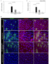
(A) Graph showing the ratio of Su(H)GBE-positive to total cells. Effects of D-p38b activity on age-related changes in the number of Su(H)GBE-positive cells in the posterior midgut. Midguts of esg>+;Su(H)GBE-lacZ or esg>UAS-D-p38bas;Su(H)GBE-lacZ flies were stained with DAPI, anti-β-gal and anti-GFP. The numbers of each cell type were counted in a 0.06 x 0.04 cm area of the posterior midgut. The ratio of Su(H)GBE-positive to total cells counted in the posterior midgut of 5-day-old flies was set as 1. White square, 5-day-old flies; black square, 30-day-old flies. P-values were determined using Student's t-test. (B) Graph showing the ratio of EE to total cells. Midguts of esg>+;Su(H)GBE-lacZ or esg>UAS-D-p38bas;Su(H)GBE-lacZ flies were stained with DAPI, anti-Prospero and anti-GFP. Numbers of each cell type were counted in a 0.06 x 0.04 cm area of posterior midgut. The ratio of EE to total cells counted in the posterior midgut of 5-day-old flies was set as 1. White square, 5-day-old flies; black square, 30-day-old flies. P-values were determined using Student's t-test. (C) Effects of D-p38bas expression on ISC and EB cell morphology of esg-positive cells and differentiation of EEs. Midguts of esg>+ (a-b) or esg>UAS-D-p38bas (c-d) flies were stained with anti-Prospero (red), anti-GFP (green) and DAPI (blue). Enlarged images, panels b and d. Scale bar, 5 μM. Arrow heads indicate esg-positive cells. Asterisks indicate EEs. (D) Graph showing the ratio of EC to Su(H)GBE-positive cells. Midguts of esg>+; Su(H)GBE-lacZ or esg>UAS-D-p38bas; Su(H)GBE-lacZ flies were stained with DAPI, anti-β-gal and anti-GFP. Numbers of each cell type were counted in a 0.06 x 0.04 cm area of posterior midgut. The ratio of EC to Su(H)GBE-positive cells counted in the posterior midgut of 5-day-old flies was set as 1. White square, 5-day-old flies; black square, 30-day-old flies. P-values were determined using Student's t-test. (E) Effect of D-p38bas expression in ISCs and EBs on age-related accumulation of EC-like large esg- and Su(H)GBE-positive cells. The guts of 5- and 30-day-old flies were labeled with anti-β-gal and anti-GFP. (a-f) esg>+;Su(H)GBE-lacZ, (g-l) esg>UAS-D-p38bas;Su(H)GBE-lacZ, (m-r) esg>UAS-D-p38b+; Su(H)GBE-lacZ. (a, d, g, j, m, and p - green) anti-GFP; (b, e, h, k, n, and q - red) anti-β-gal; (c, f, I, l, o, and r) merged image. (DAPI, blue). Arrow indicates EC-like large esg-GAL4. Asterisk indicates large Su(H)GBE-positive cell. Scale bar, 5 μM. (F) Effect of D-p38b activity on the expression levels of Delta mRNA ISCs and EBs in adult guts. Delta mRNA was measured by quantitative RT-PCR in cDNA prepared from dissected guts from 5- and 30-day-old esg>+, esg>UAS-D-p38bas, wild-type, p38bEY11174, or p38bKG01337 flies. Expression was normalized to the expression of rp49. Expression level of Delta mRNA in the midgut of 5-day-old flies was set as 1. White bar, 5-day-old flies; black bar, 30-day-old flies. P-values were determined using Student's t-test. (G) Effect of D-p38b+ on the expression of Delta in ISCs and EBs in adult gut. The level of Delta mRNA was measured by real-time RT-PCR in cDNA prepared from dissected gut from 5-day-old esg>+ or esg>UAS-D-p38b+ flies. Expression was normalized to the expression of rp49. Expression level of Delta mRNA in midgut of 5-day-old flies was set as 1. White bar, esg>+; black bar, esg>UAS-D-p38b+. P-value was determined using Student's t-test.
Figure 5.
Figure 5.. D-p38b MAPK plays a role in oxidative stress-induced aberrant Delta/ Notch signaling within the adult midgut.
(A) Effect of D-p38b knockdown in ISCs and EBs on oxidative stress-induced accumulation of abnormal large esg- and Su(H)GBE-positive cells within the adult midgut. The midguts of 5-day-old esg>+;Su(H)GBE-lacZ (a-f) or esg>UAS-D-p38bas;Su(H)-GEB-lacZ (g-l) flies exposed to 10 mM PQ in 1% sucrose (d-f and j-l) or 1% sucrose media control (a-c and g-i) were labeled for 16 h with anti-GFP (a, d, g, and j) and anti-?-gal (b, e, h, and k - red). (c, f, i, and l) merged image. (DAPI, blue) Arrow indicates EC-like large esg-GAL4. Asterisk indicates large Su(H)GBE-positive cell. Scale bar, 5 µM. (B) Effect of D-p38b knockdown on oxidative stress-induced increase of Delta expression. The level of Delta mRNA was measured by quantitative RT-PCR of dissected guts from 5-day-old esg>+, esg>UAS-D-p38bas flies exposed to 10 mM PQ in 1% sucrose or 1% sucrose media control for 16 h. Expression was normalized to the expression of rp49. The level of Delta mRNA in midgut of 5-day-old flies was set as 1. White bar, 1% sucrose media; black bar, 10 mM PQ in 1% sucrose media. P-values were determined using Student′s t-test.
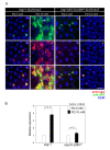
Figure 6.
Figure 6.. Effects of p38b MAPK knockdown in ISCs and EBs on PVR-induced phenotypes.
(A) Effect of D-p38b knockdown in ISCs/EBs on ectopic PVR-induced ISC proliferation. The PH3-positive cells in the midgut of the 3-day-old flies were counted. White bar, esg>+; black bar, esg>UAS-PVR; dark gray bar, esg>UAS-D-p38bas;UAS-PVR; gray bar, esg>UAS-D-p38bas. The number of PH3-positive cells detected per midgut of esg>+ flies was set as 1. P-values were calculated using Student's t-test. (B) Effect of D-p38b knockdown in ISCs and EBs on PVR-induced accumulation of large esg- and Delta-positive cells. The guts of 3-day-old flies were labeled with anti-Delta and anti-GFP. (a-a''), esg>+; (b-b''), esg>UAS-PVR; (c-c''), esg>UAS-D-p38bas;UAS-PVR; (d-d''), esg>UAS-D-p38bas. Overlay (DAPI, blue; anti-Delta, red; anti-GFP, green). Asterisk indicates EC-like large esg-positive cell. Arrow indicates Delta-positive cells. Scale bar, 5 μM. C. Effect of D-p38b knockdown in ISCs and EBs on PVR-induced Delta mRNA expression. Expression of Delta was measured by quantitative RT-PCR of dissected gut from 3-day-old flies. White bar, esg>+; black bar, esg>UAS-PVR; dark gray bar, esg>UAS-D-p38bas;UAS-PVR; gray bar, esg>UAS-D-p38bas. Expression was normalized to the expression of rp49. The level of Delta mRNA in the midgut of esg>+ flies was set as 1. P-values were determined using Student's t-test.

Similar articles

Cited by

References

    1. Casali A, Batlle E. Intestinal stem cells in mammals and Drosophila. Cell Stem Cell. 2009;4:124–127. - PubMed
    1. Finkel T, Holbrook NJ. Oxidants, oxidative stress and the biology of ageing. Nature. 2000;408:239–247. - PubMed
    1. Beckman KB, Ames BN. The free radical theory of aging matures. Physiol Rev. 1998;78:547–581. - PubMed
    1. Ohlstein B, Spradling A. The adult Drosophila posterior midgut is maintained by pluripotent stem cells. Nature. 2006;439:470–474. - PubMed
    1. Micchelli CA, Perrimon N. Evidence that stem cells reside in the adult Drosophila midgut epithelium. Nature. 2006;439:475–479. - PubMed

Publication types

MeSH terms