Haematopoietic stem cells derive directly from aortic endothelium during development
- PMID: 20154733
- PMCID: PMC2858358
- DOI: 10.1038/nature08738
Haematopoietic stem cells derive directly from aortic endothelium during development
Abstract
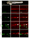
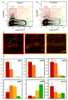
A major goal of regenerative medicine is to instruct formation of multipotent, tissue-specific stem cells from induced pluripotent stem cells (iPSCs) for cell replacement therapies. Generation of haematopoietic stem cells (HSCs) from iPSCs or embryonic stem cells (ESCs) is not currently possible, however, necessitating a better understanding of how HSCs normally arise during embryonic development. We previously showed that haematopoiesis occurs through four distinct waves during zebrafish development, with HSCs arising in the final wave in close association with the dorsal aorta. Recent reports have suggested that murine HSCs derive from haemogenic endothelial cells (ECs) lining the aortic floor. Additional in vitro studies have similarly indicated that the haematopoietic progeny of ESCs arise through intermediates with endothelial potential. Here we have used the unique strengths of the zebrafish embryo to image directly the generation of HSCs from the ventral wall of the dorsal aorta. Using combinations of fluorescent reporter transgenes, confocal time-lapse microscopy and flow cytometry, we have identified and isolated the stepwise intermediates as aortic haemogenic endothelium transitions to nascent HSCs. Finally, using a permanent lineage tracing strategy, we demonstrate that the HSCs generated from haemogenic endothelium are the lineal founders of the adult haematopoietic system.
Figures


Similar articles
-
Blood stem cells emerge from aortic endothelium by a novel type of cell transition.Nature. 2010 Mar 4;464(7285):112-5. doi: 10.1038/nature08761. Epub 2010 Feb 14. Nature. 2010. PMID: 20154732
-
In vivo imaging of haematopoietic cells emerging from the mouse aortic endothelium.Nature. 2010 Mar 4;464(7285):116-20. doi: 10.1038/nature08764. Epub 2010 Feb 14. Nature. 2010. PMID: 20154729
-
The first wave of T lymphopoiesis in zebrafish arises from aorta endothelium independent of hematopoietic stem cells.J Exp Med. 2017 Nov 6;214(11):3347-3360. doi: 10.1084/jem.20170488. Epub 2017 Sep 20. J Exp Med. 2017. PMID: 28931624 Free PMC article.
-
[The aortic endothelium in the embryo: genesis and role in hematopoiesis].J Soc Biol. 2009;203(2):155-60. doi: 10.1051/jbio/2009018. Epub 2009 Jun 16. J Soc Biol. 2009. PMID: 19527628 Review. French.
-
[From the primitive to the definitive aorta: angioblasts and hemangioblasts during aorta-associated haematopoiesis].J Soc Biol. 2005;199(2):85-91. doi: 10.1051/jbio:2005009. J Soc Biol. 2005. PMID: 16485595 Review. French.
Cited by
-
A single-cell resolution developmental atlas of hematopoietic stem and progenitor cell expansion in zebrafish.Proc Natl Acad Sci U S A. 2021 Apr 6;118(14):e2015748118. doi: 10.1073/pnas.2015748118. Proc Natl Acad Sci U S A. 2021. PMID: 33785593 Free PMC article.
-
Myelopoiesis and myeloid leukaemogenesis in the zebrafish.Adv Hematol. 2012;2012:358518. doi: 10.1155/2012/358518. Epub 2012 Jul 19. Adv Hematol. 2012. PMID: 22851971 Free PMC article.
-
Retinoic acid signaling plays a restrictive role in zebrafish primitive myelopoiesis.PLoS One. 2012;7(2):e30865. doi: 10.1371/journal.pone.0030865. Epub 2012 Feb 17. PLoS One. 2012. PMID: 22363502 Free PMC article.
-
Glucose metabolism impacts the spatiotemporal onset and magnitude of HSC induction in vivo.Blood. 2013 Mar 28;121(13):2483-93. doi: 10.1182/blood-2012-12-471201. Epub 2013 Jan 22. Blood. 2013. PMID: 23341543 Free PMC article.
-
Ginger stimulates hematopoiesis via Bmp pathway in zebrafish.PLoS One. 2012;7(6):e39327. doi: 10.1371/journal.pone.0039327. Epub 2012 Jun 25. PLoS One. 2012. PMID: 22761764 Free PMC article.
References
Publication types
MeSH terms
Grants and funding
LinkOut - more resources
Full Text Sources
Other Literature Sources
Medical
Molecular Biology Databases
Research Materials

