. 2010 Feb 5;140(3):372-83.
doi: 10.1016/j.cell.2009.12.054.

Chromatin-bound nuclear pore components regulate gene expression in higher eukaryotes

Affiliations

Chromatin-bound nuclear pore components regulate gene expression in higher eukaryotes

Maya Capelson et al. Cell. .

Abstract

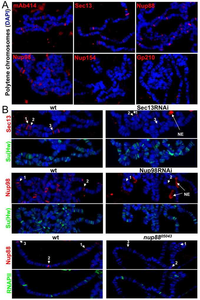
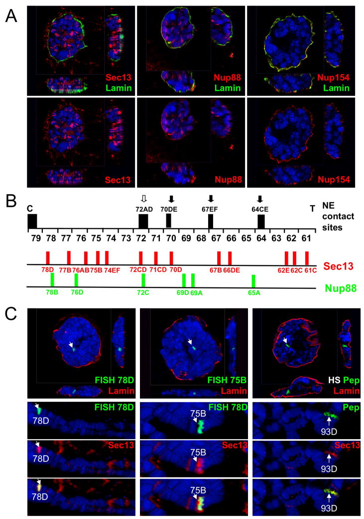
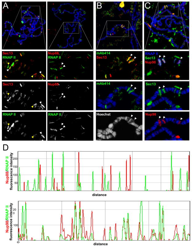
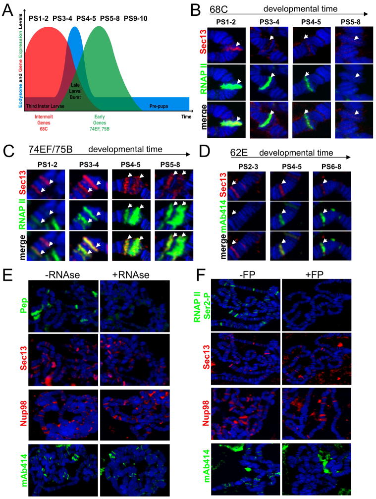
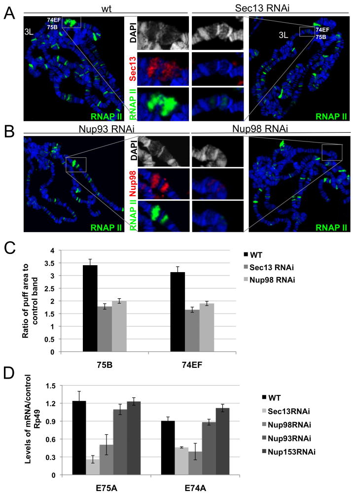
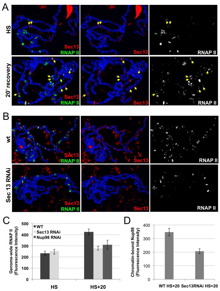
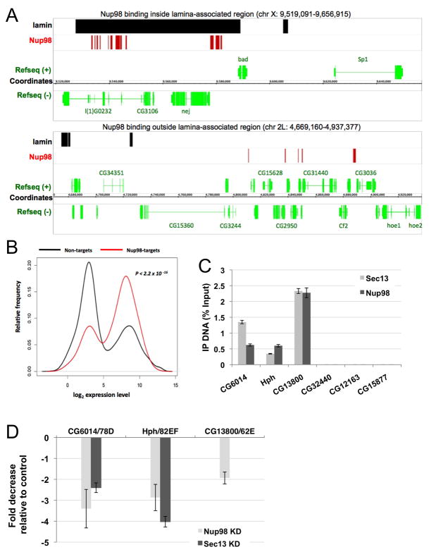
Nuclear pore complexes have recently been shown to play roles in gene activation; however their potential involvement in metazoan transcription remains unclear. Here we show that the nucleoporins Sec13, Nup98, and Nup88, as well as a group of FG-repeat nucleoporins, bind to the Drosophila genome at functionally distinct loci that often do not represent nuclear envelope contact sites. Whereas Nup88 localizes to silent loci, Sec13, Nup98, and a subset of FG-repeat nucleoporins bind to developmentally regulated genes undergoing transcription induction. Strikingly, RNAi-mediated knockdown of intranuclear Sec13 and Nup98 specifically inhibits transcription of their target genes and prevents efficient reactivation of transcription after heat shock, suggesting an essential role of NPC components in regulating complex gene expression programs of multicellular organisms.


Figures

Figure 1
Figure 1. A subset of nuclear pore components binds specific sites in the Drosophila genome
(A) Polytene chromosome spreads were stained with mAb414, anti-Sec13, anti-Nup88, anti-Nup98, anti-Nup154 and anti-gp210 as indicated; chromosomes are stained with DAPI (blue) here and throughout the paper, unless otherwise indicated. (B) Polytene chromosomes of third instar larvae of wandering LB stage from wt and Sec13 RNAi (top), Nup98 RNAi (middle) or nup8805043 homozygotes (bottom) were stained with anti-Sec13 and anti-Su(Hw), anti-Nup98 and a nti-Su(Hw), anti-Nup88 and anti-RNAP II, respectively. White arrows with numbers denote the same genomic locations for a particular Nup between chromosomes of wt and mutant larvae in a given panel. See also Figure S1.
Figure 2
Figure 2. Nup-chromatin interactions occur independently of the NE
(A) Fixed intact polytene nuclei were stained with anti-Lamin and anti-Sec13, anti-Nup88 or anti-Nup154 antibodies and analyzed by confocal microscopy. 3D reconstructions were generated from multiple z-stacks. (B) NE contact sites, which correspond to regions of high probability of contacting NE, derived from (Hochstrasser et al., 1986), were plotted against mapped binding sites of Sec13 and Nup88 along chromosome 3L. 61–79 correspond to 23 cytological divisions of 3L, each of which is further subdivided into 6 subdivisions A–F; C and T mark the centromere and telomere, respectively. NE contacts sites are marked by black (intercalary heterochromatin) or un-filled (not intercalary heterochromatin) arrows and approximate cytological locations. For Sec13, all observed binding sites on 3L during wandering third instar larval development are shown. (C) FISH probes generated from BAC clones spanning 78D and 75B were hybridized to either fixed intact polytene nuclei, followed by an anti-Lamin staining, or to chromosomes from the sister salivary glands, followed by anti-Sec13 staining; alternatively (right panel), fixed intact nuclei and chromosomes from the sister glands of heat-shocked (HS) larvae were stained with anti-Pep, which predominantly stains 93D after HS, and anti-Lamin or anti-Sec13 antibodies. Top panels show 3D reconstructions from z-stacks.
Figure 3
Figure 3. Nucleoporins Sec13, Nup98 and FG-repeat Nups are recruited to sites of active transcription
(A) Chromosome spreads were co-stained with either anti-Sec13 (left) or anti-Nup88 (right) and RNAP II (MARA3 antibody that recognizes phosphorylated RNAP II). Proportional or non-proportional binding behavior of Sec13 is indicated with white and yellow arrows, respectively. Nup88 is found at sites devoid of RNAP II. (B) Chromosomes were co-stained with anti-Sec13 and mAb414, which shows recruitment of FG Nups to a subset of Sec13-positive sites, particularly at highly transcribing puffs. (C) Chromosomes were triple-stained with anti-Nup98, anti-Sec13 and anti-RNAP II (farred, color-coded as blue), showing co-localization of all 3 at multiple sites. (D) Co-localization analysis of a typical chromosomal arm, co-stained with either Nup88 and RNAP II (top plot) or with Nup98 and RNAP II (bottom plot), demonstrating lack of co-localization or co-localization, respectively. See also Figure S2.
Figure 4
Figure 4. Recruitment of Sec13 and mAb414-reactive Nups to transcribing genes during development, which can depend on RNAP II CTD Ser2 phosphorylation
(A) Schematic illustration of the gene expression program of third instar larval stage, showing the expression profiles of Intermolt genes (red), Early genes (green), and the relative levels of ecdysone hormone (blue), with representative Puff Stages (PS) shown above. (B–C) Polytene chromosomes from larvae of different developmental stages (PS) were stained with anti-Sec13 and RNAP II; (B) during down-regulation of an Intermolt locus 68C, Sec13 levels are lost as RNAP II levels are decreased; (C) Developmental binding of Sec13 during up-regulation of Early loci 74EF and 75B, demonstrating presence of Sec13 either preceding or coinciding with appearance of phosphorylated RNAP II. (D) Polytene chromosomes from larvae of different developmental stages (PS) were stained with mAb414 and Sec13. Locus 62E is shown. (E) Isolated chromosomes were treated with buffer +/−RNase A and stained with anti-Pep, anti-Sec13, anti-Nup98 or mAb414. (F) Salivary glands were treated with buffer −/+FP and stained with control H5 antibody (anti-phosphoSer2 CTD of RNAP II), anti-Sec13, anti-Nup98 or mAb414. See also Figure S3.
Figure 5
Figure 5. Sec13 and Nup98 play a functional role in transcriptional regulation
(A) Levels of phosphorylated RNAP II and of chromatin decondensation (puffing) of Early genes at 74EF and 75B (inset) are reduced in Sec13 RNAi knock-down larvae as compared to wt; and (B) in Nup98 RNAi knock-down larvae relative to Nup93 RNAi knock-down larvae. (C) Measured puff areas at 75B and 74EF were significantly reduced in Sec13 RNAi animals relative to wt, when normalized to the control non-puffing band. (D) RT-PCR analysis using primers for the Early gene transcripts E75A (75B) and E74A (74EF) in Sec13 RNAi and Nup98 RNAi relative to wt, Nup93 RNAi and Nup153 RNAi larvae. Ratios of mRNA levels of Early gene transcripts to control gene rp49 are shown. See also Figure S4.
Figure 6
Figure 6. Nucleoporins Sec13 and Nup98 are involved in re-activation of transcription after heat shock-induced repression
(A) Chromosomes were stained with RNAPII and Sec13 right after heat shock (HS) or after 20 min recovery (HS + recovery). Yellow arrows point to sites of bound Sec13 that do not correspond to the 9 known heat shock loci (marked by RNAPII at the HS time point). (B) Chromosomes of wt or Sec13 RNAi larvae were stained with RNAP II and Sec13 at the HS + recovery time point. RNAP II accumulation at the recovering transcription sites, observed in wt, is not seen in the Sec13 RNAi larvae. (C) Genome-wide levels of active RNAP II were compared between HS and HS + recovery time points for wt, Sec13 RNAi and Nup98 RNAi larvae. (D) Genome-wide levels of Nup98 recruited during heat shock recovery are shown to be reduced in Sec13 RNAi larvae relative to wt. See also Figure S5.
Figure 7
Figure 7. Nucleoporins bind and play a functional role in expression of target genes in tissue culture cells
(A) A representative example of Nup98 target sites obtained from ChIP-on-chip analysis, plotted relative to Lamin binding sites and annotated genes. (B) Distribution of Nup98 target and non-target genes relative to expression levels. (C) ChIP analysis of candidate target genes of Sec13 and Nup98 (CG6014/78D, Hph/82EF, CG13800/62E) and of control neighboring genes in Drosophila S2 cells, showing enrichment of target genes in fractions immunoprecipitated by Nup antisera (blue and red bars, respectively) as percentage of input DNA, with levels immunoprecipitated by the normal rabbit IgG subtracted. (D) Fold decrease in mRNA levels of Nup target genes in S2 cells treated with dsRNA against Sec13 or Nup98, relative to S2 cells treated with control dsRNA, as detected by microarray analysis. See also Figures S6 and S7.

Similar articles

Cited by

References

    1. Agudo D, Gomez-Esquer F, Martinez-Arribas F, Nunez-Villar MJ, Pollan M, Schneider J. Nup88 mRNA overexpression is associated with high aggressiveness of breast cancer. Int J Cancer. 2004;109:717–720. - PubMed
    1. Akhtar A, Gasser SM. The nuclear envelope and transcriptional control. Nat Rev Genet. 2007;8:507–517. - PubMed
    1. Alber F, Dokudovskaya S, Veenhoff LM, Zhang W, Kipper J, Devos D, Suprapto A, Karni-Schmidt O, Williams R, Chait BT, et al. Determining the architectures of macromolecular assemblies. Nature. 2007a;450:683–694. - PubMed
    1. Alber F, Dokudovskaya S, Veenhoff LM, Zhang W, Kipper J, Devos D, Suprapto A, Karni-Schmidt O, Williams R, Chait BT, et al. The molecular architecture of the nuclear pore complex. Nature. 2007b;450:695–701. - PubMed
    1. Andres AJ, Thummel CS. Methods for quantitative analysis of transcription in larvae and prepupae. Methods Cell Biol. 1994;44:565–573. - PubMed

Publication types

MeSH terms