Comparative Study
. 2010 Feb;133(Pt 2):389-405.
doi: 10.1093/brain/awp341. Epub 2010 Feb 3.

Novel forms of neurofascin 155 in the central nervous system: alterations in paranodal disruption models and multiple sclerosis

Affiliations
Comparative Study

Novel forms of neurofascin 155 in the central nervous system: alterations in paranodal disruption models and multiple sclerosis

Anthony D Pomicter et al. Brain. 2010 Feb.

Erratum in

  • Brain. 2011 Apr;134(Pt 4):1250

Abstract

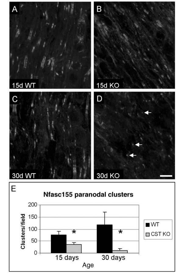
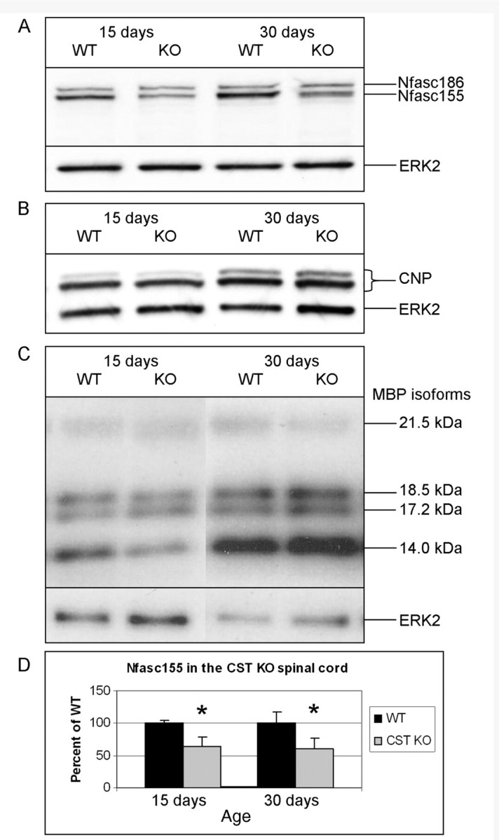
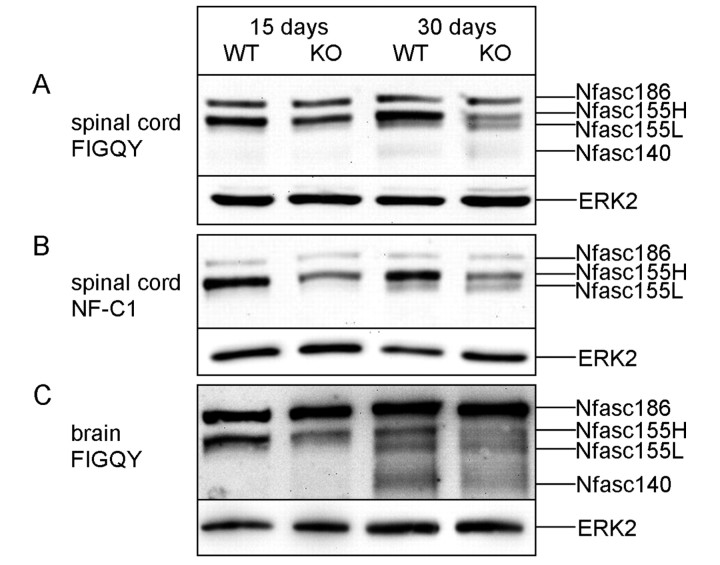
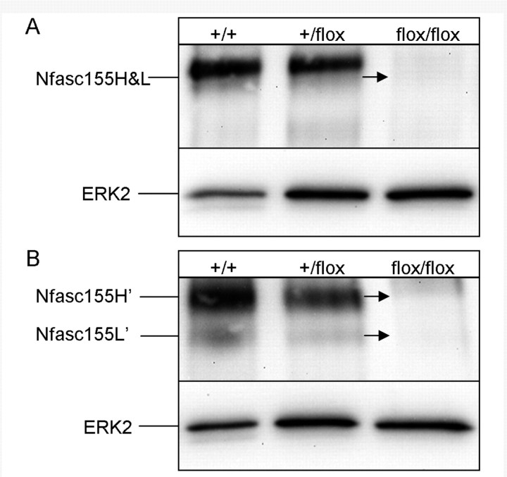
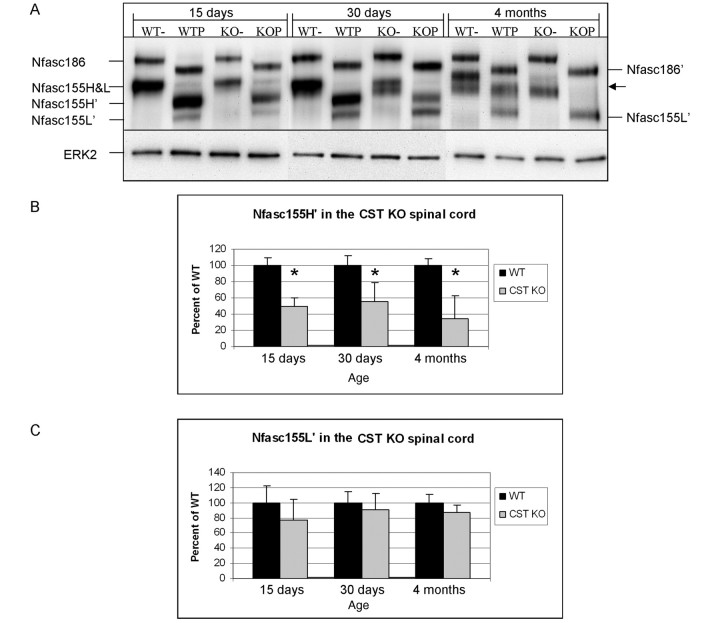
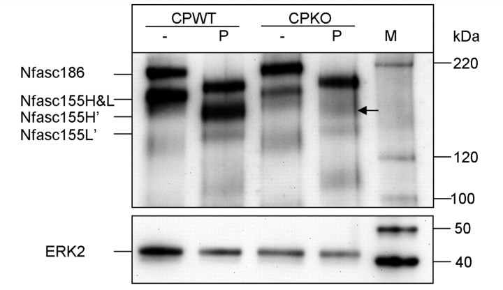
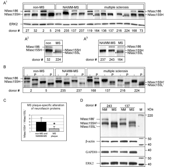
Stability of the myelin-axon unit is achieved, at least in part, by specialized paranodal junctions comprised of the neuronal heterocomplex of contactin and contactin-associated protein and the myelin protein neurofascin 155. In multiple sclerosis, normal distribution of these proteins is altered, resulting in the loss of the insulating myelin and consequently causing axonal dysfunction. Previously, this laboratory reported that mice lacking the myelin-enriched lipid sulphatide are characterized by a progressive deterioration of the paranodal structure. Here, it is shown that this deterioration is preceded by significant loss of neurofascin 155 clustering at the myelin paranode. Interestingly, prolonged electrophoretic separation revealed the existence of two neurofascin 155 bands, neurofascin 155 high and neurofascin 155 low, which are readily observed following N-linked deglycosylation. Neurofascin 155 high is observed at 7 days of age and reaches peak expression at one month of age, while neurofascin 155 low is first observed at 14 days of age and constantly increases until 5 months of age. Studies using conditional neurofascin knockout mice indicated that neurofascin 155 high and neurofascin 155 low are products of the neurofascin gene and are exclusively expressed by oligodendrocytes within the central nervous system. Neurofascin 155 high is a myelin paranodal protein while the distribution of neurofascin 155 low remains to be determined. While neurofascin 155 high levels are significantly reduced in the sulphatide null mice at 15 days, 30 days and 4 months of age, neurofascin 155 low levels remain unaltered. Although maintained at normal levels, neurofascin 155 low is incapable of preserving paranodal structure, thus indicating that neurofascin 155 high is required for paranodal stability. Additionally, comparisons between neurofascin 155 high and neurofascin 155 low in human samples revealed a significant alteration, specifically in multiple sclerosis plaques.


Figures

Figure 1
Figure 1
Paranodal clustering of neurofascin 155 is significantly reduced in the absence of sulphatide. Cervical spinal cord sections immunolabelled with the NF-C1 pan-neurofascin antibody revealed abundant paired clusters of neurofascin (Nfasc) 155 in the myelin paranode of wild-type (WT) animals at 15 (A) and 30 (C) days of age. Note that the labelling pattern in the 15-day-old animals was frequently rectangular in shape representing immunolabelling of both neurofascin 155 in the myelin paranode and neurofascin 186 in the (axonal) node. In contrast, by 30 days of age, nodal labelling was less apparent while paranodal labelling remained prevalent. In the CST knockout (KO) spinal cord sections, the number of paranodal clusters of neurofascin 155 was reduced by ∼50 and 90% at 15 (B) and 30 (D) days of age, respectively, compared to littermate wild-type mice. Also, note that by 30 days of age, the prevalence of small, single clusters (arrows) was increased in the CST knockout. (E) Quantitative analyses revealed that the reduction of paranodal clustering of neurofascin 155 was significantly reduced at both 15 and 30 days of age. Scale bar = 10 µm; see Table 2. *P<0.05.
Figure 2
Figure 2
CST knockout spinal cords contain reduced amounts of neurofascin 155 at 15 and 30 days of age. (A) Western blot analysis showed that CST knockout (KO) mice contain ∼40% less (Table 2) neurofascin (Nfasc) 155 at 15 and 30 days of age than wild-type (WT) littermates. However, no change in the levels of either the CNP monomers (B) or myelin basic proteins (MBP) (C) was observed in the absence of sulphatide, demonstrating that the reduction in neurofascin 155 is not due to a global decrease in myelin proteins. (D) Graphical representation of the reduction in neurofascin 155. ERK2 = extracellular signal-regulated kinase 2. *P<0.05.
Figure 3
Figure 3
Extended electrophoresis reveals two forms of neurofascin 155. At 15 days of age, the FIGQY (A) and NF-C1 (B) pan-neurofascin antibodies labelled one band representing neurofascin (Nfasc) 155 in both the wild-type (WT) and the CST knockout (KO) spinal cord samples. By 30 days of age, wild-type samples displayed the protein now called neurofascin 155H with a shadow-like band immediately below, while the CST knockout samples showed both neurofascin 155H and neurofascin 155L. (C) Mouse brain samples incubated with FIGQY show neurofascin 155H and neurofascin 155L in both wild-type and CST knockout at 30 days of age and, when compared to 15-day-old samples, suggest a developmental pattern for neurofascin 155L in wild-type as well as CST knockout mice. ERK2 = extracellular signal-regulated kinase 2.
Figure 4
Figure 4
Neurofascin 155H and neurofascin 155L are developmentally regulated glycoproteins in rat spinal cord. (A) Rat spinal cord homogenates revealed low levels of neurofascin 155H at 7 days of age. Neurofascin 155H steadily increased with age, reaching a peak at 27 days of age followed by a decrease which is maintained through 5 months of age. Neurofascin 155L was first seen as a clearly delineated band at 3 months of age and remained prevalent at 5 months. Interestingly, neurofascin 155L appeared to form gradually from the shadow-like band below neurofascin 155H. (B) Following PNGase F treatment neurofascin 155L’ was observed in 3 and 5 month samples (as expected from Fig. 4A) as well as at 14, 21 and 27 days of age, with increasing abundance with increasing age. Neurofascin 155H’ was barely visible at 7 days of age, became increasingly intense through 27 days of age, then decreased in abundance at 3 and 5 months of age consistent with neurofascin 155H in Fig. 4A. (C) At 7 days of age, when neurofascin 155L’ was not present, the pan-neurofascin antibody FIGQY labelled neurofascin 186 at the node of Ranvier and neurofascin 155H at the paranode in the rat spinal cord. Voltage gated sodium channels are indicated in dark blue (a and e), Caspr is indicated in red (b and f), and FIGQY labelling is shown in green (c and g). The panels on the left (a–d) show a fully formed node:paranode region and in the merged image (d) light blue represents voltage gated sodium channels and neurofascin 186 at the node and the flame colour represents Caspr and neurofascin 155H at the paranode. The panels on the right (e–h) show an immature node:paranode region with a pair of voltage gated sodium channel/neurofascin 186-labelled clusters flanking an unlabelled region that is presumed to develop into the node. Notice that the flame colour is observed in the merged image (h) indicating that Caspr and neurofascin 155H are present in the developing paranode. d = days; m = months; P = PNGase F treated; ERK2 = extracellular signal-regulated kinase 2; Nfasc = neurofascin; Nav = voltage gated sodium. Scale bar = 5 µm.
Figure 5
Figure 5
Neurofascin 155H and neurofascin 155L are products of the Nfasc gene. Spinal cords from 16-day-old CNP-Cre;Nfascflox/flox mice contained no ‘neurofascin 155H&L’ and no neurofascin 155H’ or neurofascin 155L’ (arrows) when observed without (A) or with (B) PNGase F treatment. ERK2 = extracellular signal-regulated kinase 2.
Figure 6
Figure 6
Neurofascin 155H’ is reduced in CST knockout spinal cords. (A) Following PNGase F treatment, spinal cord homogenates showed an ∼50% decrease in neurofascin 155H’ at 15 and 30 days of age. Surprisingly, at 4 months of age no definitive neurofascin 155H’ (or neurofascin 155H) band was observed in the CST knockout samples (arrow). Quantitative analyses revealed that neurofascin 155H’ (B) was significantly reduced in the CST knockout mice at all ages examined while the levels of neurofascin 155L’ (C) were not altered; see Table 3. − = untreated; P = PNGase F treated; KO = knockout; WT = wild-type; Nfasc = neurofascin; ERK2 = extracellular signal-regulated kinase 2. *P<0.05.
Figure 7
Figure 7
Spinal cords from Caspr knockout mice, similar to CST knockout animals, contain reduced amounts of neurofascin 155H and neurofascin 155H’. Comparable to 15-day-old CST knockout mice, spinal cords samples from 19-day-old Caspr knockout mice treated with PNGase F showed dramatically reduced neurofascin 155H and no band representing neurofascin 155H’ was observed following PNGase F treatment (arrow). Neurofascin 155L’ appears modestly reduced. The Magic Mark Western Protein standard (M) revealed the shift in molecular mass following treatment with PNGase F. CP = Caspr; − = untreated; P = PNGase F treated; KO = knockout; WT = wild-type; Nfasc = neurofascin; ERK2 = extracellular signal-regulated kinase 2.
Figure 8
Figure 8
The pattern of neurofascin 155H’ and neurofascin 155L’ is altered in multiple sclerosis plaques. (A) Following the extended electrophoresis protocol, clearly delineated neurofascin 155L was not observed in non-multiple sclerosis (A1 and A2) or normal-appearing white matter-multiple sclerosis (A1 and A3) samples but was observed in multiple sclerosis plaque samples (black stars). (B) Following PNGase F treatment, no clearly delineated neurofascin 155L’ band was observed in either the non-multiple sclerosis or normal-appearing white matter-multiple sclerosis samples. In contrast, neurofascin 155L’ was clearly observed in the majority of multiple sclerosis brains analysed (white stars). (C) The ratio of neurofascin 155H’/neurofascin 155L’ was significantly reduced in multiple sclerosis plaque samples (n = 8; P < 0.006) compared to non-multiple sclerosis and normal-appearing white matter-multiple sclerosis samples (n = 7). (D) Side-by-side analysis of PNGase F treated samples of normal-appearing white matter-multiple sclerosis and multiple sclerosis plaque samples from the same donors shows neurofascin 155L’ only in the plaque samples. It is noteworthy that the pattern of neurofascin 155H’ and neurofascin 155L’ is similar between inactive (#243) and active (#137) plaque samples. Additionally, neurofascin 186’ was frequently not observed in multiple sclerosis plaque samples. The commonly used loading control proteins β-actin, glyceraldehyde 3-phosphate dehydrogenase and extracellular signal-regulated kinase 2 are shown and demonstrate a lack of autolysis within the samples. − = untreated; P = PNGase F treated; NM or NAWM = normal appearing white matter from multiple sclerosis donor; M = Magic Mark XP Protein Standard; MS = multiple sclerosis; Nfasc = neurofascin; ERK2 = extracellular signal-regulated kinase 2; GAPDH = glyceraldehyde 3-phosphate dehydrogenase. *P<0.05.

Similar articles

Cited by

References

    1. Abad-Rodriguez J, Ledesma MD, Craessaerts K, Perga S, Medina M, Delacourte A, et al. Neuronal membrane cholesterol loss enhances amyloid peptide generation. J Cell Biol. 2004;167:953–60. - PMC - PubMed
    1. Arvanitis DN, Min W, Gong Y, Heng YM, Boggs JM. Two types of detergent-insoluble, glycosphingolipid/cholesterol-rich membrane domains from isolated myelin. J Neurochem. 2005;94:1696–710. - PubMed
    1. Bae JS, Yang L, Rezaie AR. Lipid raft localization regulates the cleavage specificity of protease activated receptor 1 in endothelial cells. J Thromb Haemost. 2008;6:954–61. - PubMed
    1. Basak S, Raju K, Babiarz J, Kane-Goldsmith N, Koticha D, Grumet M. Differential expression and functions of neuronal and glial neurofascin isoforms and splice variants during PNS development. Dev Biol. 2007;311:408–22. - PubMed
    1. Bhat MA, Rios JC, Lu Y, Garcia-Fresco GP, Ching W, St Martin M, et al. Axon-glia interactions and the domain organization of myelinated axons requires neurexin IV/Caspr/Paranodin. Neuron. 2001;30:369–83. - PubMed

Publication types

MeSH terms