. 2010 Apr 23;285(17):12935-47.
doi: 10.1074/jbc.M109.071696. Epub 2010 Jan 29.

Wild-type p53-induced phosphatase 1 dephosphorylates histone variant gamma-H2AX and suppresses DNA double strand break repair

Affiliations

Wild-type p53-induced phosphatase 1 dephosphorylates histone variant gamma-H2AX and suppresses DNA double strand break repair

Sung-Hwan Moon et al. J Biol Chem. .

Abstract

In response to DNA double strand breaks, the histone variant H2AX at the break site is phosphorylated at serine 139 by DNA damage sensor kinases such as ataxia telangiectasia-mutated, forming gamma-H2AX. This phosphorylation event is critical for sustained recruitment of other proteins to repair the break. After repair, restoration of the cell to a prestress state is associated with gamma-H2AX dephosphorylation and dissolution of gamma-H2AX-associated damage foci. The phosphatases PP2A and PP4 have previously been shown to dephosphorylate gamma-H2AX. Here, we demonstrate that the wild-type p53-induced phosphatase 1 (WIP1) also dephosphorylates gamma-H2AX at serine 139 in vitro and in vivo. Overexpression of WIP1 reduces formation of gamma-H2AX foci in response to ionizing and ultraviolet radiation and blocks recruitment of MDC1 (mediator of DNA damage checkpoint 1) and 53BP1 (p53 binding protein 1) to DNA damage foci. Finally, these inhibitory effects of WIP1 on gamma-H2AX are accompanied by WIP1 suppression of DNA double strand break repair. Thus, WIP1 has a homeostatic role in reversing the effects of ataxia telangiectasia-mutated phosphorylation of H2AX.


Figures

FIGURE 1.
FIGURE 1.
γ-H2AX is dephosphorylated by WIP1 in vitro. A, H2AX pS139 phosphopeptide was incubated with human recombinant WIP1 protein. p38 pT180 and UNG2 pT31 phosphopeptides were used as a positive and negative control, respectively. Free phosphate released from the phosphopeptide was measured by malachite green phosphate assay to determine relative phosphatase activities on each phosphopeptide. Error bars correspond to standard error (n = 3). O.A., okadaic acid. WIP1 PD is a phosphatase-dead point mutant used as a control. B, 293T cells were transfected with myc-H2AX expression plasmid and treated with bleomycin for 1 h to induce γ-H2AX phosphorylation. Myc-tagged full-length H2AX protein was immunopurified using anti-Myc antibody and incubated with human recombinant WIP1 protein, WIP1 phosphatase-dead protein, or PP2C alpha in vitro. γ-H2AX phosphorylation levels were determined by immunoblotting using a γ-H2AX-specific antibody. The key components of each assay are indicated below each lane.
FIGURE 2.
FIGURE 2.
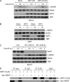
WIP1 impairs γ-H2AX phosphorylation in cells. A, overexpression of WIP1 suppresses γ-H2AX in IR-treated HeLa cells. HeLa cells were either mock transfected or transfected with WIP1-FLAG expression plasmid. At 24 h after transfection, cells were irradiated with 5-Gy IR and harvested. Nuclear lysates were immunoblotted to assess γ-H2AX and total H2AX protein, and WIP1 protein. TBP (TATA-binding protein) served as a loading control. B, overexpression of WIP1 suppresses γ-H2AX in IR and UV-treated 293T cells. 293T cells were transfected as in A. After 24 h, cells were irradiated with either 5-Gy IR or 50 J/m2 UV and harvested 30 min after IR or 2 h after UV treatment. Nuclear lysates were immunoblotted as indicated. C, WIP1 knockdown increased γ-H2AX levels in IR-treated MCF7 cells. Cells were transduced with lentivirus expressing WIP1 shRNA (#1 and #2). After 48 h, cells were irradiated with 5-Gy IR, harvested, and then analyzed by immunoblot with the indicated antibodies. D, WIP1 and H2AX physically interact in 293T cells. 293T cells were co-transfected with myc-H2AX and WIP1-FLAG expression plasmids. Interaction between H2AX and WIP1 was examined by immunoprecipitation with anti-FLAG antibody followed by immunoblotting with anti-Myc antibody and vice versa.
FIGURE 3.
FIGURE 3.
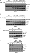
WIP1 reverses ATR-mediated γ-H2AX phosphorylation. A and B, WIP1 suppresses γ-H2AX phosphorylation after UV (A) and hydroxyurea treatment (B). HeLa cells were transfected with empty vector or WIP1-FLAG expression plasmid. At 24 h after transfection, cells were irradiated with 50 J/m2 of UV (A) or treated with 10 mm hydroxyurea (HU) (B) and harvested at the indicated time points. Nuclear lysates were immunoblotted as indicated. C, WIP1 suppresses γ-H2AX phosphorylation in the absence of ATM. GM09607 (ATM null) fibroblasts were transfected with either empty vector or WIP1-FLAG expression plasmid. After 24 h, cells were irradiated with 50 J/m2 of UV and harvested. Nuclear lysates were immunoblotted as indicated. D, suppression of WIP1 in MCF-7 cells results in enhanced γ-H2AX phosphorylation following UV irradiation. MCF-7 cells were transduced with one of two different shRNA lentiviral vectors prior to 10 J/m2 UV irradiation. Nuclear lysates were harvested 1 h post-UV treatment along with un-irradiated control cells and mock transduced controls. Immunoblot analysis for WIP1 protein levels, H2AX protein levels, and γ-H2AX levels was performed.
FIGURE 4.
FIGURE 4.
Wip1 null mice exhibit increased γ-H2AX phosphorylation after IR treatment. A, absence of Wip1 in irradiated mouse tissue increases γ-H2AX phosphorylation. Wild-type, Wip1−/−, Atm−/−, and Atm−/− Wip1−/− double knock-out mice were exposed to 5 Gy of whole body ionizing irradiation. Six hours after irradiation, spleens were harvested from each mouse and tissue lysates were immunoblotted with the indicated antibodies. B, quantitative analysis of γ-H2AX phosphorylation levels in A. Immunoblot bands were quantitated with NIH ImageJ software. γ-H2AX bands were normalized to the total H2AX bands. The γ-H2AX level in irradiated wild-type tissue (lane 3 in A) was set to 1.0.
FIGURE 5.
FIGURE 5.
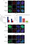
WIP1 inhibits damage-induced focus formation. A, overexpression of WIP1 inhibits IR-induced damage foci in HeLa cells. HeLa cells were mock-transfected or transfected with either wild-type or D314A mutant WIP1-FLAG expression plasmid. At 24 h after transfection, cells were irradiated with 5-Gy IR. After 30 min, cells were fixed and immunostained with anti-γ-H2AX (red fluorescence) and anti-FLAG antibodies (green fluorescence) to assess damage-induced foci formation. 4′,6-Diamidino-2-phenylindole staining (DAPI, blue fluorescence) was used to identify nuclei. B, quantitative analysis of γ-H2AX-asociated damage foci in IR-treated HeLa cells with or without overexpressed WIP1. The fluorescence intensity values of γ-H2AX from >1300 independent cells from the mock-transfected and WIP1-transfected cells were measured by high throughput microscopic imaging and plotted. Error bars represent the standard error. C, fluorescence intensity of γ-H2AX-associated damage foci is inversely correlated with WIP1 fluorescence intensity. At 30-min post-IR, individual WIP1-transfected HeLa cells were subdivided into low (n = 138), medium (n = 406), or high (n = 181) WIP1-expressing cells based on high throughput quantitative fluorescence imaging. The fluorescence intensity values for γ-H2AX for each cell were also measured, and mean γ-H2AX fluorescence for each of the three WIP1 expression categories was plotted. Error bars represent the standard error (Student's t test; p < 10−10 between Low and Med and p < 10−40 between Med and High). D, knockdown of WIP1 in MCF7 cells results in increased γ-H2AX-associated damage foci. MCF7 cells were transfected with negative control siRNA or WIP1 siRNA. At 24 h after transfection, cells were irradiated with 5-Gy IR, fixed at 3 h after irradiation, and immunostained with anti-γ-H2AX (red fluorescence) and anti-WIP1 antibodies (green fluorescence). E, WIP1 inhibition γ-H2AX-associated damage foci is ATM-independent. GM09607 (ATM null) fibroblasts were transfected with WIP1-FLAG expression plasmid. After 24 h, cells were irradiated with 5-Gy IR, fixed 30 min after irradiation and then immunostained with the indicated antibodies.
FIGURE 6.
FIGURE 6.
WIP1 inhibits recruitment of repair complex proteins MDC1 and 53BP1 to DNA damage foci. A, WIP1 inhibits recruitment of MDC1 to IR-induced damage foci. HeLa cells were transfected with WIP1-FLAG expression plasmid. At 24 h after transfection, cells were irradiated with 5-Gy IR. Thirty minutes later, cells were fixed and immunostained with anti-MDC1 (far-red fluorescence), anti-γ-H2AX (green fluorescence), and anti-FLAG (red fluorescence) antibodies. B, Wip1 inhibits recruitment of 53BP1 to IR-induced damage foci. HeLa cells were mock transfected or transfected with WIP1-FLAG expression plasmid, treated with 5-Gy IR, fixed, and immunostained with anti-53BP1 and anti-FLAG antibodies as in A.
FIGURE 7.
FIGURE 7.
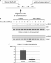
WIP1 inhibits DSB repair and γ-H2AX DSB association. A, strategy employed to measure γ-H2AX binding adjacent to a DSB (B) and the efficiency of DNA DSB repair (C) using the I-PpoI system. Real-time PCR with primers flanking the I-PpoI cut site allow measurement of DSB break repair efficiency. γ-H2AX association was monitored with ChIP followed by PCR with primers 50 and 280 bp downstream of the I-PpoI cleavage site. B, ChIP assay for γ-H2AX association at I-PpoI cleavage site shows increased γ-H2AX when WIP1 levels are decreased. MCF7 cells expressing HA-ER-I-PpoI were infected with either empty or WIP1 shRNA lentivirus. After 4-OHT treatment, cells were fixed at the indicated time points and subjected to the ChIP assay with primers adjacent to the single I-PpoI cut site in chromosome 1. A ChIP assay with primers from the GAPDH region not near an I-PpoI cut site (GAPDH) serves as a negative control. C, DNA DSB repair is enhanced when WIP1 levels are reduced. I-PpoI was activated in MCF7 cells by 4-OHT treatment for 12 h. Genomic DNA was obtained and analyzed with real-time PCR to measure the efficiency of DSB repair. The relative amounts of the trans-DSB PCR fragment were assessed by the ΔΔCT method. Error bars represent the standard error (n = 3).
FIGURE 8.
FIGURE 8.
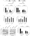
WIP1 inhibits repair of DNA DSBs. A, recombination substrates pLB4 and pTNeo99-7 in stable cell lines, LB4 and TNeo99-7. Each substrate contains a functional hygromycin gene (hyg), used to select for stably transfected cells, and a tk-neo fusion gene that is disrupted by a 22-bp oligonucleotide containing the recognition site for endonuclease I-SceI. Substrate pLB4 contains a 2.5-kb HindIII fragment containing a complete HSV-1 tk gene that serves as a donor for the homologous recombination with the disrupted tk-neo gene. B, WIP1, but not phosphatase-dead WIP1 inhibits DSB repair. I-SceI and wild-type/mutant WIP1 or control expression vector DNA were transfected into cells, and the frequency of recovery of G418R clones was measured. C, WIP1 has dose-dependent inhibitory effects on DSB repair. As indicated, varying amounts of WIP1 vector DNA or control vector DNA were introduced into cells, and the frequency of recovery of G418R clones was measured. D, suppression of WIP1 activity by WIP1 shRNA and the WIP1 inhibitors arsenic trioxide (ATO) and CCT007093 enhances DSB repair. LB4 and TNeo99-7 cells were transduced with a lentiviral WIP1 shRNA vector or were incubated in 10 μm ATO or 25 μm CCT007093 prior to I-SceI introduction. Formation of G418R clones was then measured. E, the effects of WIP1 on DSB repair are not solely dependent on ATM. LB4 and TNeo99-7 cells were transfected with control or WIP1 expression vector DNA, treated with neocarzinostatin (200 ng/ml) and with or without KU55933 (10 μm). Proteins were detected by their specific antibodies in immunoblotting (left panel). The effects of WIP1 on DSB repair were also examined in LB4 and TNeo99-7 cells in the presence or absence of KU55933 (right panel).

Similar articles

Cited by

References

    1. Abraham R. T. (2001) Genes Dev. 15, 2177–2196 - PubMed
    1. Shiloh Y. (2003) Nat. Rev. Cancer 3, 155–168 - PubMed
    1. Lovejoy C. A., Cortez D. (2009) DNA Repair 8, 1004–1008 - PMC - PubMed
    1. Bakkenist C. J., Kastan M. B. (2004) Trends Cell Biol. 14, 339–341 - PubMed
    1. Fiscella M., Zhang H., Fan S., Sakaguchi K., Shen S., Mercer W. E., Vande Woude G. F., O'Connor P. M., Appella E. (1997) Proc. Natl. Acad. Sci. U.S.A. 94, 6048–6053 - PMC - PubMed

Publication types

MeSH terms

Substances