. 2010 Apr 10;399(2):201-11.
doi: 10.1016/j.virol.2010.01.012. Epub 2010 Feb 8.

The E6 protein from vaccinia virus is required for the formation of immature virions

Affiliations

The E6 protein from vaccinia virus is required for the formation of immature virions

Olga Boyd et al. Virology. .

Abstract

An IPTG-inducible mutant in the E6R gene of vaccinia virus was used to study the role of the E6 virion core protein in viral replication. In the absence of the inducer, the mutant exhibited a normal pattern DNA replication, concatemer resolution and late gene expression, but it showed an inhibition of virion structural protein processing it failed to produce infectious particles. Electron microscopic analysis showed that in the absence of IPTG viral morphogenesis was arrested before IV formation: crescents, aberrant or empty IV-like structures, and large aggregated virosomes were observed throughout the cytoplasm. The addition of IPTG to release a 12-h block showed that virus infectious particles could be formed in the absence of de novo DNA synthesis. Our observations show that in the absence of E6 the association of viroplasm with viral membrane crescents is impaired.


Figures

Fig. 1
Fig. 1
Construction of vE6i inducible mutant. A diagram of the modified regions in vE6i is shown in (A): the upper lines represent the J2R and E5R - E6R regions that were modified. The white rectangles represent the unmodified E5R and E6R ORF. The shaded rectangles represent the DNA insertions encoding for the bacteriophage T7 RNA polymerase (T7RNApol) regulated by the late P11 promoter (P11) and lac operator (lacO); E. coli lac repressor (lacI) regulated by the VACV p7.5 early/late promoter (P7.5); E. coli xanthine-guanine phosphoribosyltransferase (E-gpt) regulated by p7.5. The E6R natural promoter was substituted by a T7 promoter (PT7), lacO, and encephalomyocarditis internal ribosomal entry site (IRES) regulatory sequence. The IPTG requirement for VACV replication and the optimal concentration of the inducer was determined by titering vE6i in the presence of increasing concentration of IPTG. After 72 hours, the cells were stain with crystal violet and the viral plaques counted and graphed (B). To verify is the small plaque phenotype was constant with time the plaque assay was incubated up 7 days before the cells were stained with crystal violet (C).
Fig. 2
Fig. 2
Expression and accumulation of E6. To analyze the accumulation of E6 protein and the expression of VACV early and late genes, BSC40 cells were infected with moi =10 with VACV-WR or vE6i virus in the absence or in the presence of IPTG. At indicated times, cell extracts were prepared and the proteins were separated on 11% SDS-PAGE and transferred to a nitrocellulose membrane. (A) The blot was probed with E6 rabbit polyclonal antibody and developed by chemiluminescence. (B) The blots were probed with E6 and L4 rabbit polyclonal antibodies and A18 mouse monoclonal antibody. The dashed line indicates the separation of two different membranes. The lane (M) represents the mock-infected cells.
Fig. 3
Fig. 3
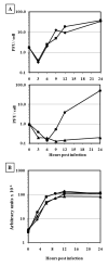
One-step growth and DNA replication. BSC40 cells were infected at moi = 10 pfu/ml with VACV-WR or vE6i virus in the absence or in the presence of IPTG. At indicated hours post infection samples were removed and the virus yield was determined by plaque assay (A) and the amount of DNA measured by slot blot hybridization (B). WR (−) IPTG (◆); WR (+) IPTG (■); vE6i (−) IPTG (▲);vE6i (+) IPTG (●).
Fig. 4
Fig. 4
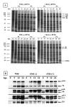
Protein synthesis and proteolytic processing of viral proteins. BSC40 cells were infected at moi = 10 pfu/cell and incubated in the presence or absence of IPTG. (A) Cells were pulse-labeled (PL) with 35S methionine at various times post-infection, indicated in hours at the top of each autoradiogram. The pulse-chase (PC) was performed by incubating the cells with an excess of non-radioactive methionine, for the times indicated at the top of each lane. Proteins were separated on an 11% SDS-PAGE, and gels were dried and exposed to film. The virus and the incubation condition is indicated at the top of each gel. Approximate molecular weights, in kDa, are indicated to the left of each autoradiograms. The major viral core proteins p4a/4a, p4b/4b and 23 kDa are indicated on the right of the autoradiograms. (B) Western blot analysis was performed on infected cell extracts prepared as described above. The cells were harvested, a cell extract was prepared, and the proteins were separated on an 11% SDS-PAGE, transferred to a nitrocellulose membrane, and blotted against specific vaccinia virus proteins, as indicated on the right side of the figure.
Fig. 5
Fig. 5
Electron micrograph of the vE6i infected cells. BSC40 cells were infected with vE6i in the presence of IPTG (A, B) or in the absence of the inducer (C – F). After 24 hpi (A – D) or 48 hpi (E – F) the cells were processed for microscopy as described in Methods. IV= immature virions; MV= mature virions; WV= wrapped virions; C= crescents; AV = aggregated virosome; N=nucleus.
Fig. 6
Fig. 6
Telomere resolution. A diagram showing relevant features of the viral genome concatemer junction and mature DNA hairpin ends is shown in the top of the figure. Shaded areas represent direct repeats within the viral termini. The arrows represent the site of cleavage of the BstE II restriction enzyme and the double arrows represent the size of the fragment that is generated in both resolved and non-resolved condition. The autoradiogram of the Southern blot is shown in the bottom part of the figure. BSC40 cells were infected with WR or mutant virus at moi = 10 pfu/cell and incubated in absence or presence of IPTG. Cts53 virus, used a positive control, was grown at 31 and 40°C. DNA was extracted at various times post-infection, indicated in h at the top of each lane, cleaved with BstE II, the fragments separated in an 0.8% agarose gel, blotted, and probed with a 32P-labeled DNA containing concatemer junction sequences. The 2.6- and 1.3-kb bands, indicated to the right of the autoradiogram, represent unresolved and resolved end fragments, respectively. The slower migrating bands represent internal fragments hybridized to the probe due to the presence of tandem repeat sequences.
Fig. 7
Fig. 7
Release of the IPTG blockage. A diagram on the top shows the scheme for the procedure. The vertical arrows represent the hpi where samples were collected for plaque assay. The horizontal arrows represent the hours of the blockage release. The result of the plaque assay is presented in the bottom part of the figure. BSC40 cells were infected with vE6i at moi = 10 pfu/cell and incubated in absence IPTG. At 12 hpi, IPTG or IPTG + CAR in fresh media were added to the cells. Cells that did not receive the drugs were also supplemented with fresh media. At the indicated hpi, samples were removed and the virus yield was measured by plaque titration.
Fig. 8
Fig. 8
Electron micrograph after the IPTG blockage release. BSC40 cells were infected with vE6i at moi = 10 pfu/cell and incubated in absence IPTG. At 12 hpi, fresh media with no drugs (A, B) or containing IPTG (C, D) or IPTG + CAR (E, F) were added to the cells. After 24h of the blockage release (36 hpi), the cells were processed for microscopy as described in Methods. EIV = empty immature virions; IV= immature virions; MV= mature virions; WV= wrapped virions; C= crescents; RAV = rough aggregated virosome; SAV = smooth aggregated virosome; N=nucleus.
Fig. 9
Fig. 9
Stability of E6 in cells infected with mutants of the seven-protein complex. BSC40 cells were infected with VACV mutants of the seven-protein complex (7PC) at moi = 10 pfu/cell and incubated 31°C (P) or 40°C (N) for 24 hours. After this period cell extracts were prepared, proteins separated in a 11% SDS-PAGE, transferred to a nitrocellulose membrane and blotted using rabbit antiserum against the VACV proteins G7, A30, A10 (p4a/4a) and E6. A 15% SDS-PAGE was used for the analysis of the A30 protein. The mutant viruses indicated in the lanes correspond to following genes: D46 (A30), C11 (G7), C05 (D3), C45 (J1), C54 (F10), E52 (D2), and C16 (I7). vE6i was incubated in the absence (−) or in the presence (+) of IPTG. The arrows in the right of the figure indicate the VACV precursor (p) or the protein. The numbers in the left of the figure represents the molecular weight markers in kDa.

Similar articles

Cited by

References

    1. Alexander WA, Moss B, Fuerst TR. Regulated expression of foreign genes in vaccinia virus under the control of bacteriophage T7 RNA polymerase and the Escherichia coli lac repressor. J.Virol. 1992;66:2934–2942. - PMC - PubMed
    1. Ansarah-Sobrinho C, Moss B. Role of the I7 protein in proteolytic processing of vaccinia virus membrane and core components. J.Virol. 2004;78:6335–6343. - PMC - PubMed
    1. Assarsson E, Greenbaum JA, Sundstrom M, Schaffer L, Hammond JA, Pasquetto V, Oseroff C, Hendrickson RC, Lefkowitz EJ, Tscharke DC, Sidney J, Grey HM, Head SR, Peters B, Sette A. Kinetic analysis of a complete poxvirus transcriptome reveals an immediate-early class of genes. Proc.Natl.Acad.Sci.U.S.A. 2008;105:2140–2145. - PMC - PubMed
    1. Black EP, Moussatche N, Condit RC. Characterization of the interactions among vaccinia virus transcription factors G2R, A18R, and H5R. Virology. 1998;245:313–322. - PubMed
    1. Boyd O, Strahl AL, Rodeffer C, Condit RC, Moussatche N. Temperature-sensitive mutant in the vaccinia virus E6 protein produce virions that are transcriptionally inactive. Virology. 2010 - PMC - PubMed

Publication types