Monitoring drug-induced gammaH2AX as a pharmacodynamic biomarker in individual circulating tumor cells
- PMID: 20103672
- PMCID: PMC2818670
- DOI: 10.1158/1078-0432.CCR-09-2799
Monitoring drug-induced gammaH2AX as a pharmacodynamic biomarker in individual circulating tumor cells
Abstract
Purpose: Circulating tumor cells (CTC) in peripheral blood of patients potentially represent a fraction of solid tumor cells available for more frequent pharmacodynamic assessment of drug action than is possible using tumor biopsy. However, currently available CTC assays are limited to cell membrane antigens. Here, we describe an assay that directly examines changes in levels of the nuclear DNA damage marker gammaH2AX in individual CTCs of patients treated with chemotherapeutic agents.
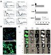
Experimental design: An Alexa Fluor 488-conjugated monoclonal gammaH2AX antibody and epithelial cancer cell lines treated with topotecan and spiked into whole blood were used to measure DNA damage-dependent nuclear gammaH2AX signals in individual CTCs. Time-course changes in both CTC number and gammaH2AX levels in CTCs were also evaluated in blood samples from patients undergoing treatment.
Results: The percentage of gammaH2AX-positive CTCs increased in a concentration-dependent manner in cells treated with therapeutically relevant concentrations of topotecan ex vivo. In samples from five patients, percent gammaH2AX-positive cells increased post-treatment from a mean of 2% at baseline (range, 0-6%) to a mean of 38% (range, 22-64%) after a single day of drug administration; this increase was irrespective of increases or decreases in the total CTC count.
Conclusions: These data show promise for monitoring dynamic changes in nuclear biomarkers in CTCs (in addition to CTC count) for rapidly assessing drug activity in clinical trials of molecularly targeted anticancer therapeutics as well as for translational research.
Figures


Similar articles
-
The prognostic and therapeutic implications of circulating tumor cell phenotype detection based on epithelial-mesenchymal transition markers in the first-line chemotherapy of HER2-negative metastatic breast cancer.Cancer Commun (Lond). 2019 Jan 3;39(1):1. doi: 10.1186/s40880-018-0346-4. Cancer Commun (Lond). 2019. PMID: 30606259 Free PMC article. Clinical Trial.
-
Circulating tumor cells as a predictive biomarker in patients with small cell lung cancer undergoing chemotherapy.Lung Cancer. 2017 Oct;112:118-125. doi: 10.1016/j.lungcan.2017.08.008. Epub 2017 Aug 12. Lung Cancer. 2017. PMID: 29191584
-
Circulating tumor cells: a multifunctional biomarker.Clin Cancer Res. 2014 May 15;20(10):2553-68. doi: 10.1158/1078-0432.CCR-13-2664. Clin Cancer Res. 2014. PMID: 24831278 Review.
-
Circulating tumor cells and survival in abiraterone- and enzalutamide-treated patients with castration-resistant prostate cancer.Prostate. 2018 May;78(6):435-445. doi: 10.1002/pros.23488. Epub 2018 Feb 12. Prostate. 2018. PMID: 29431193
-
Can γH2AX be used to personalise cancer treatment?Curr Mol Med. 2013 Dec;13(10):1591-602. doi: 10.2174/1566524013666131111124531. Curr Mol Med. 2013. PMID: 24206133 Review.
Cited by
-
Dietary phytochemicals, HDAC inhibition, and DNA damage/repair defects in cancer cells.Clin Epigenetics. 2011;3(1):4. doi: 10.1186/1868-7083-3-4. Epub 2011 Oct 26. Clin Epigenetics. 2011. PMID: 22247744 Free PMC article.
-
Circulating Tumor Cells in Breast Cancer Patients: A Balancing Act between Stemness, EMT Features and DNA Damage Responses.Cancers (Basel). 2022 Feb 16;14(4):997. doi: 10.3390/cancers14040997. Cancers (Basel). 2022. PMID: 35205744 Free PMC article. Review.
-
Molecular analysis of circulating tumour cells-biology and biomarkers.Nat Rev Clin Oncol. 2014 Mar;11(3):129-44. doi: 10.1038/nrclinonc.2013.253. Epub 2014 Jan 21. Nat Rev Clin Oncol. 2014. PMID: 24445517 Review.
-
Clinical and pharmacologic evaluation of two dosing schedules of indotecan (LMP400), a novel indenoisoquinoline, in patients with advanced solid tumors.Cancer Chemother Pharmacol. 2016 Jul;78(1):73-81. doi: 10.1007/s00280-016-2998-6. Epub 2016 May 11. Cancer Chemother Pharmacol. 2016. PMID: 27169793 Free PMC article. Clinical Trial.
-
Development and validation of an immunoassay for quantification of topoisomerase I in solid tumor tissues.PLoS One. 2012;7(12):e50494. doi: 10.1371/journal.pone.0050494. Epub 2012 Dec 28. PLoS One. 2012. PMID: 23284638 Free PMC article.
References
-
- Dowlati A, Haaga J, Remick SC, et al. Sequential tumor biopsies in early phase clinical trials of anticancer agents for pharmacodynamic evaluation. Clin Cancer Res. 2001;7:2971–6. - PubMed
-
- Helft PR, Daugherty CK. Are we taking without giving in return? The ethics of research-related biopsies and the benefits of clinical trial participation. J Clin Oncol. 2006;24:4793–5. - PubMed
-
- Cristofanilli M, Hayes DF, Budd GT, et al. Circulating tumor cells: a novel prognostic factor for newly diagnosed metastatic breast cancer. J Clin Oncol. 2005;23:1420–30. - PubMed
-
- Hayes DF, Smerage J. Is there a role for circulating tumor cells in the management of breast cancer? Clin Cancer Res. 2008;14:3646–50. - PubMed
-
- Smerage JB, Hayes DF. The prognostic implications of circulating tumor cells in patients with breast cancer. Cancer Invest. 2008;26:109–14. - PubMed
Publication types
MeSH terms
Substances
Grants and funding
LinkOut - more resources
Full Text Sources
Other Literature Sources

