. 2010 Jan 26:9:15.
doi: 10.1186/1476-4598-9-15.

Retinoic acid protects human breast cancer cells against etoposide-induced apoptosis by NF-kappaB-dependent but cIAP2-independent mechanisms

Affiliations

Retinoic acid protects human breast cancer cells against etoposide-induced apoptosis by NF-kappaB-dependent but cIAP2-independent mechanisms

Ana M Jiménez-Lara et al. Mol Cancer. .

Abstract

Background: Retinoids, through their cognate nuclear receptors, exert potent effects on cell growth, differentiation and apoptosis, and have significant promise for cancer therapy and chemoprevention. These ligands can determine the ultimate fate of target cells by stimulating or repressing gene expression directly, or indirectly through crosstalking with other signal transducers.

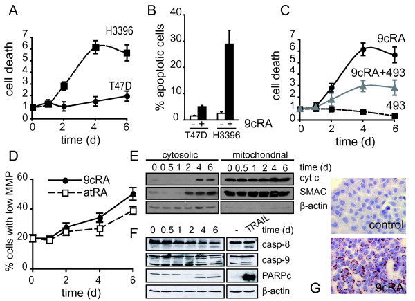
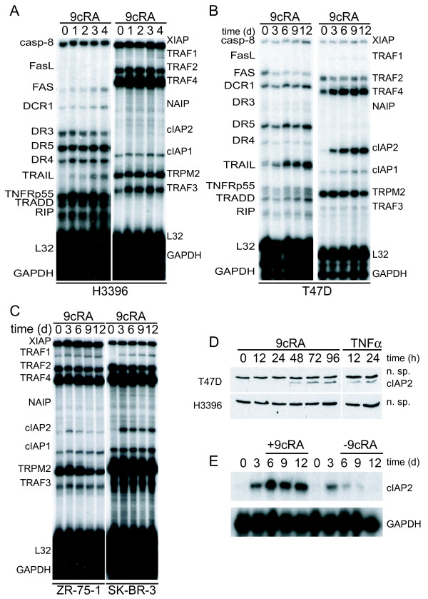
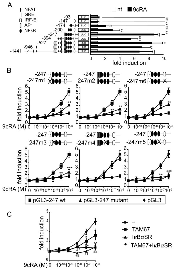
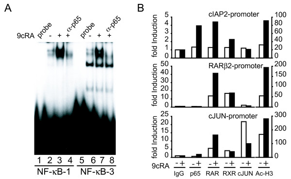
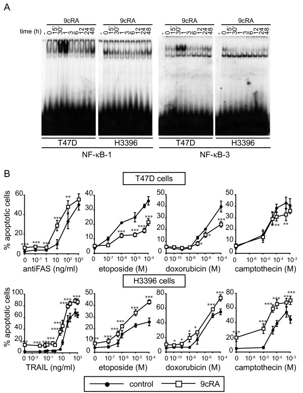
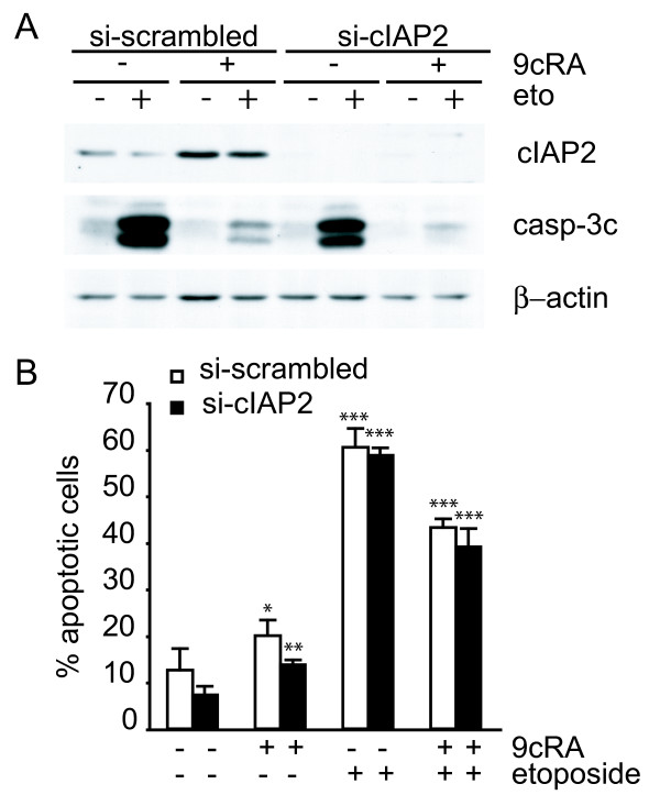
Results: Using different breast cancer cell models, we show here that depending on the cellular context retinoids can signal either towards cell death or cell survival. Indeed, retinoids can induce the expression of pro-apoptotic (i.e. TRAIL, TNF-Related Apoptosis-Inducing Ligand, Apo2L/TNFSF10) and anti-apoptotic (i.e. cIAP2, inhibitor of apoptosis protein-2) genes. Promoter mapping, gel retardation and chromatin immunoprecipitation assays revealed that retinoids induce the expression of this gene mainly through crosstalk with NF-kappaB. Supporting this crosstalk, the activation of NF-kappaB by retinoids in T47D cells antagonizes the apoptosis triggered by the chemotherapeutic drugs etoposide, camptothecin or doxorubicin. Notably apoptosis induced by death ligands (i.e. TRAIL or antiFAS) is not antagonized by retinoids. That knockdown of cIAP2 expression by small interfering RNA does not alter the inhibition of etoposide-induced apoptosis by retinoids in T47D cells reveals that stimulation of cIAP2 expression is not the cause of their anti-apoptotic action. However, ectopic overexpression of a NF-kappaB repressor increases apoptosis by retinoids moderately and abrogates almost completely the retinoid-dependent inhibition of etoposide-induced apoptosis. Our data exclude cIAP2 and suggest that retinoids target other regulator(s) of the NF-kappaB signaling pathway to induce resistance to etoposide on certain breast cancer cells.

Conclusions: This study shows an important role for the NF-kappaB pathway in retinoic acid signaling and retinoic acid-mediated resistance to cancer therapy-mediated apoptosis in breast cancer cells, independently of cIAP2. Our data support the use of NF-kappaB pathway activation as a marker for screening that will help to develop novel retinoids, or retinoid-based combination therapies with improved efficacy.


Figures

Figure 1
Figure 1
Retinoic acid promotes either differentiation or cell death of breast cancer cells in a cell-context dependent manner. (A) T47D and H3396 cells were treated with 9-cis-retinoic acid (9-cis-RA) for the indicated time and analyzed for the presence of DNA fragments in their cytosol as a measurement of cell death. Y-axis refers to the enrichment of histone complexed DNA fragments (mono- and oligonucleosomes) in the cytoplasm of apoptotic cells. The values represent the mean ± SD of three different experiments performed in triplicate. (B) T47D and H3396 cells were treated with 9-cis-RA for 6 days. Cell death was determined by FACS analysis after staining with propidium iodide as described in "Materials and Methods". The values represent the mean ± SD of three different experiments performed in duplicate. (C) H3396 cells were treated with 9-cis-RA and the RAR-antagonist BMS493, as indicated, and analyzed as described in (A). (D) H3396 cells were analyzed by flow cytometry to assess the population of cells with lower mitochondria membrane potential following all-trans retinoic acid (atRA) or 9-cis-RA treatment for the indicated time. The values represent the mean ± SD of three different experiments performed in duplicate. (E) Release of mitochondrial proteins to the cytosol was analyzed by western blotting after subcellular fractionation of H3396 cells treated with 9-cis-RA for the indicated time. (F) Effect of 9-cis-RA treatment on the cleavage of caspase-8 (casp-8), caspase-9 (casp-9) and PARP assessed by western blotting in H3396 cells. (G) Oil-red O staining of T47D cells untreated or treated with 1 μM 9-cis-RA for 72 h. The images shown are from one representative experiment performed three times with similar results.
Figure 2
Figure 2
9-cis-RA induces the expression of cIAP2 in breast cancer cells in a cell context dependent manner. (A, B, C) Multiplex RNase protections assays (RPAs) to monitor the effect of 9-cis-RA on the expression of death receptor, death ligands, IAP and TRAF family members in four different breast cancer cell lines. Breast cancer cells were treated for the indicated time with 9-cis-RA at a concentration of 10-6 M. (D) Western blot of whole cell extracts of 9-cis-RA-treated T47D cells and H3396 cells for 0, 12, 24, 48, 72 and 96 hours with anti-cIAP2. The nonspecific signal (n. sp.) confirms equal loading. As a positive control, breast cancer cells were treated with 50 μg/ml of hTNFα for 24 and 48 hours. (E) Reversibility of 9-cis-RA-induced cIAP2 gene expression. T47D cells were treated either in the absence or presence of 1 μM 9- cis-RA and after 3 days, total RNA was extracted. In parallel flasks, the medium was removed, and cells were washed and treated with either fresh control medium or medium with 10-6 M of 9-cis-RA and grown for additional 3, 6 and 9 days. Media and ligands were renewed every 3 days. RNA was isolated and analyzed by RPA as described in (A). Equal loading was confirmed by GAPDH RNA level. The images shown are from one representative experiment performed twice with similar results.
Figure 3
Figure 3
9-cis-RA activates cIAP2 transcription through NF-κB response elements. (A) Illustration of the reporter constructs containing 5'-deletion fragments of the cIAP2 upstream regulatory region. Luciferase activity in SK-BR-3 cells transiently transfected with the indicated cIAP2 reporter gene in the presence (black bars) and absence (white bars) of 9-cis-RA. The data shown represent the mean ± SD of three independent experiments performed in duplicate. The luciferase levels were normalized with those of β-galactosidase and expressed as the induction over the controls. (B) Transfection experiments as described in (A) but using site-specific mutants derived from -247-cIAP2 promoter as depicted (Black triangle), the wild-type promoter (Black square) and the backbone vector pGL3 (Black diamond). After transfection, SK-BR-3 cells were treated with different doses of 9-cis-RA, as indicated. The data shown represent the mean ± SD of three independent experiments performed in duplicate. The luciferase levels were normalized with those of β-galactosidase and expressed as the induction over the controls. Asterisks denote the existence of statistically significant differences between the wild-type and mutant promoter constructs. (C) SK-BR-3 cells were transiently co-transfected with the -247- cIAP2 reporter gene and pSG5, pSG5-IκBαSR(S32A/S36A) or pcDNA-TAM54 (a dominant-negative version of c-JUN) and either untreated or treated with 9-cis-RA, as described in (B). The luciferase levels were normalized with those of β-galactosidase and expressed as the induction over the controls. The values represent the mean ± SD of three experiments performed in duplicate. Asterisks denote statistically significant differences against cells transfected with an empty vector.
Figure 4
Figure 4
9-cis-RA treatment results in in vivo recruitment of p65 and retinoic-acid receptors, RAR and RXR to the cIAP2 promoter. (A) EMSA performed with probes bearing the NF-κB-1 and NF-κB-3 sites of the cIAP2 promoter and nuclear extracts from T47D cells treated with 1 μM 9-cis-RA for 1 h. When indicated, antibodies directed against p65 were present in the binding assays. The images shown are from one representative experiment performed twice with similar results. B) T47D breast cancer cells were treated as indicated in Material and Methods and ChIPs assays were performed using antibodies against p65, RARs, RXRα, cJUN or acetyl-H3 histone. Immunoprecipitated chromatin was analyzed by real time PCR using primers specific for the cIAP2, RARβ2 and cJUN promoters. Shown are data from a representative experiment, expressed as fold induction relative to the values obtained with immunoglobulin G (IgG) used as a negative control, which were set to 1. This experiment was repeated three times with similar results.
Figure 5
Figure 5
9-cis-RA pretreatment prevents apoptosis induced by cancer chemotherapy insults in T47D cells, but increases chemotherapy-induced apoptosis in H3396 cells: correlation with NF-κB activity induction by 9-cis-RA. (A) EMSA performed with probes bearing the NF-κB-1 and NF-κB-3 sites of the cIAP2 promoter and nuclear extracts from T47D and H3396 cells treated with 1 μM 9-cis-RA for different times, as indicated. The images shown are from one representative experiment performed twice with similar results. (B) T47D and H3396 cells were pretreated with or without 9-cis-RA for 30 h, followed by treatment with different doses of the indicated chemotherapeutic agents for 72 h and death ligands for 48 h. Apoptotic cells were determined by FACS analysis after staining with propidium iodide as described in "Materials and Methods". The values represent the mean ± SD of three independent experiments performed in duplicate. Asterisks denote statistically significant differences between untreated cells and cells treated with 9-cis-RA.
Figure 6
Figure 6
Suppression of cIAP2 expression is not sufficient to abrogate 9-cis-RA inhibition of etoposide-induced apoptosis in T47D cells. (A) T47D cells were transfected with either scrambled-siRNA or cIAP2-siRNA and pretreated with or without 9-cis-RA for 30 h, followed by treatment with etoposide 100 μM for 24 h. Cell lysates were analyzed by western blot for the expression of cleaved caspase-3, cIAP2 and β-actin using specific antibodies. The images shown are from one representative experiment performed three times with similar results. (B) T47D cells were transfected with either scrambled-siRNA (white bars) or cIAP2-siRNA (black bars). After 24 h, lipid-siRNA complexes were removed from media and cells were pretreated with or without 1 μM 9-cis-RA for 30 h, followed by treatment with etoposide 100 μM for 72 h. The percentage of apoptotic cells was determined by FACS analysis after staining with propidium iodide. The values represent the mean ± SD of three experiments performed in duplicate. Asterisks denote statistically significant differences against the corresponding untreated cells.
Figure 7
Figure 7
Over-expression of the super-repressor of NF-κB activation, IκBα-SR(S32A/S36A), leads to significant abrogation of retinoic acid-mediated inhibition of etoposide-induced apoptosis. (A) T47D-vector and T47D-IκBαSR cells were untreated or incubated for the indicated time with hTNFα (50 ng/ml). Extracts were analyzed by western blotting with an IκBα antibody. Equal loading was confirmed with an anti-β-actin antibody. The images shown are from one representative experiment performed three times with similar results. (B) T47D-vector or T47D-IκBαSR cells were untreated or treated with 9-cis-RA for 48 h and expression of cIAP2 and β-actin were analysed by Reverse-Transcriptase Polymerase Reaction and real time PCR. The values represent the mean ± SD of three different experiments performed in duplicate. (C) T47D-vector or T47D-IκBαSR cells were pretreated with or without 9-cis-RA for 30 h, followed by treatment with etoposide 100 μM as previously indicated. At the indicated times, cell lysates were analyzed by western blot for the expression of cleaved caspase-3, cIAP2 and β-actin using specific antibodies. The images shown are from one representative experiment performed three times with similar results. (D) T47D-vector or T47D-IκBαSR cells were pretreated with or without 9-cis-RA for 30 h, followed by treatment with etoposide 100 μM for 72 h. The percentage of apoptotic cells was determined by FACS analysis after staining with propidium iodide. The values represent the mean ± SD of three independent experiments performed in duplicate. Asterisks denote the existence of statistically significant differences between the indicated groups; N.S.: not significant (Student's t-test).

Similar articles

Cited by

References

    1. Gronemeyer H, Gustafsson JA, Laudet V. Principles for modulation of the nuclear receptor superfamily. Nat Rev Drug Discov. 2004;3:950–964. doi: 10.1038/nrd1551. - DOI - PubMed
    1. Kastner P, Mark M, Chambon P. Nonsteroid nuclear receptors: what are genetic studies telling us about their role in real life? Cell. 1995;83:859–869. doi: 10.1016/0092-8674(95)90202-3. - DOI - PubMed
    1. Altucci L, Gronemeyer H. The promise of retinoids to fight against cancer. Nat Rev Cancer. 2001;1:181–193. doi: 10.1038/35106036. - DOI - PubMed
    1. Altucci L, Leibowitz MD, Ogilvie KM, de Lera AR, Gronemeyer H. RAR and RXR modulation in cancer and metabolic disease. Nat Rev Drug Discov. 2007;6:793–810. doi: 10.1038/nrd2397. - DOI - PubMed
    1. Wang Z, Sun G, Shen Z, Chen S, Chen Z. Differentiation therapy for acute promyelocytic leukemia with all-trans retinoic acid: 10-year experience of its clinical application. Chin Med J (Engl) 1999;112:963–967. - PubMed

Publication types

MeSH terms