

A structural view of nuclear hormone receptor: endocrine disruptor interactions
- PMID: 20063036
- PMCID: PMC11115495
- DOI: 10.1007/s00018-009-0249-2
A structural view of nuclear hormone receptor: endocrine disruptor interactions
Abstract
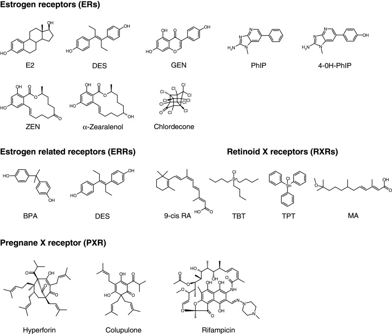
Endocrine-disrupting chemicals (EDCs) represent a broad class of exogenous substances that cause adverse effects in the endocrine system by interfering with hormone biosynthesis, metabolism, or action. The molecular mechanisms of EDCs involve different pathways including interactions with nuclear hormone receptors (NHRs) which are primary targets of a large variety of environmental contaminants. Here, based on the crystal structures currently available in the Protein Data Bank, we review recent studies showing the many ways in which EDCs interact with NHRs and impact their signaling pathways. Like the estrogenic chemical diethylstilbestrol, some EDCs mimic the natural hormones through conserved protein-ligand contacts, while others, such as organotins, employ radically different binding mechanisms. Such structure-based knowledge, in addition to providing a better understanding of EDC activities, can be used to predict the endocrine-disrupting potential of environmental pollutants and may have applications in drug discovery.
Figures
Similar articles
-
Structural and functional evidences for the interactions between nuclear hormone receptors and endocrine disruptors at low doses.C R Biol. 2017 Sep-Oct;340(9-10):414-420. doi: 10.1016/j.crvi.2017.08.002. C R Biol. 2017. PMID: 29126514
-
New modes of action for endocrine-disrupting chemicals.Mol Endocrinol. 2006 Mar;20(3):475-82. doi: 10.1210/me.2004-0513. Epub 2005 Jul 21. Mol Endocrinol. 2006. PMID: 16037129 Review.
-
Perturbation of Nuclear Hormone Receptors by Endocrine Disrupting Chemicals: Mechanisms and Pathological Consequences of Exposure.Cells. 2019 Dec 19;9(1):13. doi: 10.3390/cells9010013. Cells. 2019. PMID: 31861598 Free PMC article. Review.
-
Activation of steroid hormone receptors: Shed light on the in silico evaluation of endocrine disrupting chemicals.Sci Total Environ. 2018 Aug 1;631-632:27-39. doi: 10.1016/j.scitotenv.2018.03.003. Epub 2018 Mar 6. Sci Total Environ. 2018. PMID: 29522903 Review.
-
Possible relationship between endocrine disrupting chemicals and hormone dependent gynecologic cancers.Med Hypotheses. 2016 Jul;92:84-7. doi: 10.1016/j.mehy.2016.04.041. Epub 2016 Apr 25. Med Hypotheses. 2016. PMID: 27241264
Cited by
-
Mechanistic insights into the synergistic activation of the RXR-PXR heterodimer by endocrine disruptor mixtures.Proc Natl Acad Sci U S A. 2021 Jan 5;118(1):e2020551118. doi: 10.1073/pnas.2020551118. Proc Natl Acad Sci U S A. 2021. PMID: 33361153 Free PMC article.
-
Plastic Food Packaging from Five Countries Contains Endocrine- and Metabolism-Disrupting Chemicals.Environ Sci Technol. 2024 Mar 19;58(11):4859-4871. doi: 10.1021/acs.est.3c08250. Epub 2024 Mar 5. Environ Sci Technol. 2024. PMID: 38441001 Free PMC article.
-
Divergent teratogenicity of agonists of retinoid X receptors in embryos of zebrafish (Danio rerio).Ecotoxicology. 2012 Jul;21(5):1465-75. doi: 10.1007/s10646-012-0900-9. Epub 2012 Apr 10. Ecotoxicology. 2012. PMID: 22526925
-
In Silico Molecular Docking and In Vivo Validation with Caenorhabditis elegans to Discover Molecular Initiating Events in Adverse Outcome Pathway Framework: Case Study on Endocrine-Disrupting Chemicals with Estrogen and Androgen Receptors.Int J Mol Sci. 2019 Mar 10;20(5):1209. doi: 10.3390/ijms20051209. Int J Mol Sci. 2019. PMID: 30857347 Free PMC article.
-
Phthalate and novel plasticizer concentrations in food items from U.S. fast food chains: a preliminary analysis.J Expo Sci Environ Epidemiol. 2022 May;32(3):366-373. doi: 10.1038/s41370-021-00392-8. Epub 2021 Oct 27. J Expo Sci Environ Epidemiol. 2022. PMID: 34702987 Free PMC article.
References
-
- Swedenborg E, Ruegg J, Makela S, Pongratz I. Endocrine disruptive chemicals: mechanisms of action and involvement in metabolic disorders. J Mol Endocrinol. 2009;43:1–10. - PubMed
-
- Tabb MM, Blumberg B. New modes of action for endocrine-disrupting chemicals. Mol Endocrinol. 2006;20:475–482. - PubMed
-
- Janosek J, Hilscherova K, Blaha L, Holoubek I. Environmental xenobiotics and nuclear receptors: interactions, effects and in vitro assessment. Toxicol In Vitro. 2006;20:18–37. - PubMed
Publication types
MeSH terms
Substances
LinkOut - more resources
Full Text Sources
