. 2010 Mar 5;285(10):7645-56.
doi: 10.1074/jbc.M109.058990. Epub 2010 Jan 5.

Lysine 63-linked polyubiquitination of the dopamine transporter requires WW3 and WW4 domains of Nedd4-2 and UBE2D ubiquitin-conjugating enzymes

Affiliations

Lysine 63-linked polyubiquitination of the dopamine transporter requires WW3 and WW4 domains of Nedd4-2 and UBE2D ubiquitin-conjugating enzymes

Arnau Vina-Vilaseca et al. J Biol Chem. .

Abstract

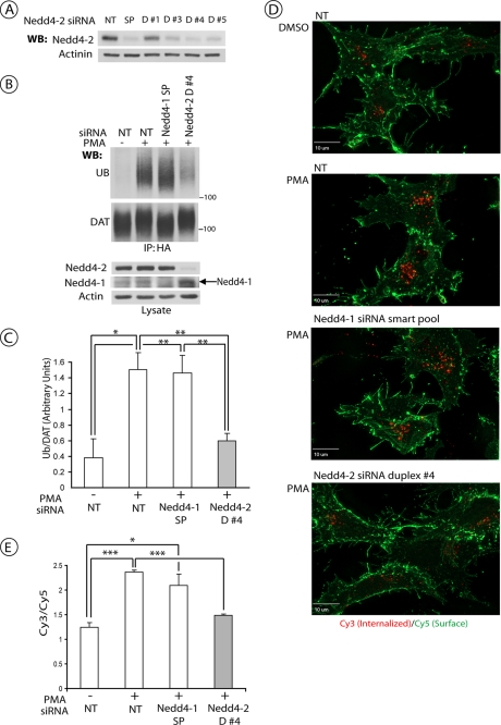
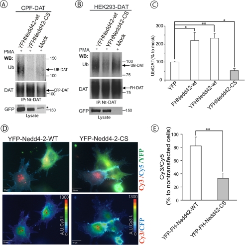
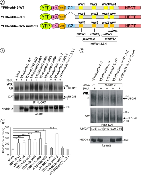
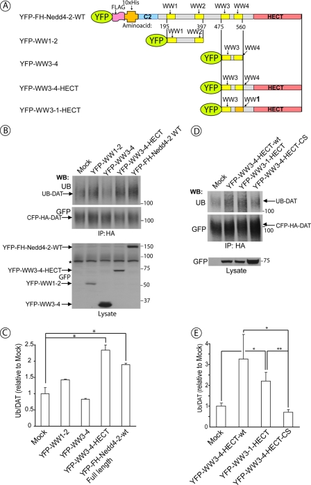
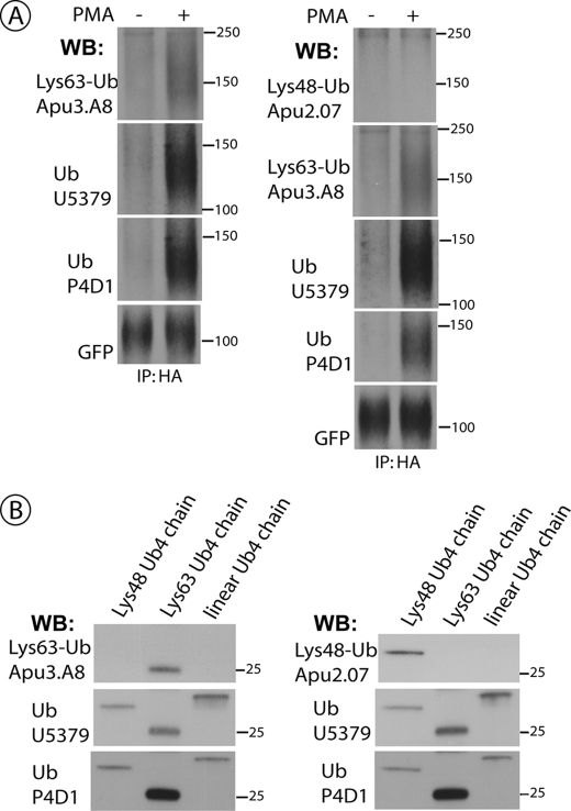
RNA interference screen previously revealed that a HECT-domain E3 ubiquitin ligase, neuronal precursor cell expressed, developmentally down-regulated 4-2 (Nedd4-2), is necessary for ubiquitination and endocytosis of the dopamine transporter (DAT) induced by the activation of protein kinase C (PKC). To further confirm the role of Nedd4-2 in DAT ubiquitination and endocytosis, we demonstrated that the depletion of Nedd4-2 by two different small interfering RNA (siRNA) duplexes suppressed PKC-dependent ubiquitination and endocytosis of DAT in human and porcine cells, whereas knock-down of a highly homologous E3 ligase, Nedd4-1, had no effect on DAT. The abolished DAT ubiquitination in Nedd4-2-depleted cells was rescued by expression of recombinant Nedd4-2. Moreover, overexpression of Nedd4-2 resulted in increased PKC-dependent ubiquitination of DAT. Mutational inactivation of the HECT domain of Nedd4-2 inhibited DAT ubiquitination and endocytosis. Structure-function analysis of Nedd4-2-mediated DAT ubiquitination revealed that the intact WW4 domain and to a lesser extent WW3 domain are necessary for PKC-dependent DAT ubiquitination. Moreover, a fragment of the Nedd4-2 molecule containing WW3, WW4, and HECT domains was sufficient for fully potentiating PKC-dependent ubiquitination of DAT. Analysis of DAT ubiquitination using polyubiquitin chain-specific antibodies showed that DAT is mainly conjugated with Lys(63)-linked ubiquitin chains. siRNA analysis demonstrated that this polyubiquitination is mediated by Nedd4-2 cooperation with UBE2D and UBE2L3 E2 ubiquitin-conjugating enzymes. The model is proposed whereby each ubiquitinated DAT molecule is modified by a single four-ubiquitin Lys(63)-linked chain that can be conjugated to various lysine residues of DAT.


Figures

FIGURE 1.
FIGURE 1.
Nedd4-2 siRNA inhibits PKC-dependent DAT ubiquitination and endocytosis in HEK293 cells. A, HEK293 cells stably expressing CFP-HA-DAT (HEK/CFP-HA-DAT) were transfected with non-targeting siRNA (NT), SmartPool™ (SP), or four individual siRNA duplexes targeting human Nedd4-2. Equal amounts of cell lysates were electrophoresed, and the amounts of Nedd4-2 and α-actinin (loading control) were determined by Western blotting (WB). B, HEK/CFP-HA-DAT cells were transfected with NT siRNA, SP targeting Nedd4-1, or duplex 4 (D#4) siRNA targeting Nedd4-2. Cells were treated with 1 μm PMA or Me2SO for 15 min, lysed, and DAT was precipitated with the HA11 antibody. Following electrophoresis and transfer, DAT immunoprecipitates were probed with monoclonal antibodies against ubiquitin (P4D1) and, consecutively, with the monoclonal GFP antibody. Total cell lysates were blotted with antibodies to Nedd4-2, Nedd4-1, and actin (loading control). C, quantification of three experiments performed identically to the experiment presented in B. Bars represent the mean amounts (±S.E.) of ubiquitinated DAT normalized to the amount of total DAT (arbitrary units). *, p < 0.05; **, p < 0.01. D, representative images of HEK/CFP-HA-DAT cells treated with NT, SP of siRNAs targeting Nedd4-1, or duplex 4 siRNA targeting Nedd4-2. HA11 endocytosis assay was performed as described under “Experimental Procedures.” Merged images of Cy5 immunofluorescence (green, surface CFP-HA-DAT) and Cy3 immunofluorescence (red, internalized CFP-HA-DAT) are presented. Identical intensity scales are used. Scale bars, 10 μm. E, quantification of the Cy3/Cy5 ratio in cells from the experiments represented in D. Bars represent mean values (±S.E.) of a minimum of 6 images per condition. *, p < 0.05; **, p < 0.001.
FIGURE 2.
FIGURE 2.
HECT domain activity of Nedd4-2 is necessary for PKC-dependent DAT ubiquitination and endocytosis. A, HEK293T cells were co-transfected with CFP-DAT and wild-type YFP-FH-Nedd4-2 (YFHNedd42-wt), HECT domain mutant of Nedd4-2 (YFHNedd42-CS), or empty vector (mock-transfected). After 2 days, the cells were treated with 1 μm PMA for 15 min, lysed, and CFP-DAT was immunoprecipitated with Nt-DAT antibody. The immunoprecipitates were probed by Western blotting (WB) with ubiquitin (P4D1) and, subsequently, Nt-DAT antibodies. YFP-tagged Nedd4-2 constructs were detected in cell lysates using GFP antibodies. Star indicates the nonspecific band. B, HEK293T cells stably expressing human DAT (HEK293/DAT) were transfected with YFHNedd42-wt, YFHNedd42-CS, or empty vector (mock). After 2 days the cells were treated with 1 μm PMA for 15 min and lysed. DAT was immunoprecipitated using Nt-DAT antibody, and the immunoprecipitates were probed with ubiquitin (P4D1) and, subsequently, Nt-DAT antibodies. YFP-tagged Nedd4-2 constructs were detected in cell lysates using GFP antibody. C, quantification of several experiments performed identically to the experiment presented in B. Bars represent the mean amounts (±S.E.) of ubiquitinated DAT normalized to the amount of total DAT in arbitrary units (Ub/DAT). All Ub/DAT values are calculated as percent of the Ub/DAT value in mock-transfected cells. *, p < 0.05; **, p < 0.01 (n = 3). D, HEK293 cells stably expressing CFP-HA-DAT (HEK/CFP-HA-DAT) were transfected with YFHNedd42-wt or the YFHNedd42-CS mutant. HA11 endocytosis assay was performed as described under “Experimental Procedures.” HA11 preincubated cells were exposed to 1 μm PMA for 30 min. Merged images (top row) of Cy5 immunofluorescence (blue, cell surface CFP-HA-DAT), Cy3 immunofluorescence (red, internalized CFP-HA-DAT), and YFP (green, YFP-FH-Nedd4-2) are presented. Pseudocolor images (Cy3/CFP) were generated as described under “Experimental Procedures” are shown in the bottom row. Identical intensity scales are used. Scale bars, 10 μm. E, quantification of the Cy3/Cy5 ratio in cells expressing and not expressing YFP-FH-Nedd4-2 (wild-type or CS mutant) from the experiments performed similarly to the experiment presented in D. Bars represent mean values (± S.E.) of a minimum of 6 cells from three independent experiments. **, p < 0.01 (n = 18).
FIGURE 3.
FIGURE 3.
WW3 and WW4 domains of Nedd4-2 are necessary for DAT ubiquitination. A, schematic representation of wild-type and mutant YFP-FH-Nedd4-2 constructs. B, HEK293T cells stably expressing FH-DAT (HEK293/FH-DAT) were transfected with empty vector (mock), wild-type, or mutant YFP-FH-Nedd4-2. After 2 days the cells were treated with 1 μm PMA for 15 min and lysed. DAT was immunoprecipitated using Nt-DAT antibody, and the immunoprecipitates were probed by Western blotting (WB) with ubiquitin (P4D1) and, subsequently, Nt-DAT antibodies. YFP-tagged Nedd4-2 constructs were detected in cell lysates using the Nedd4-2 antibody. C, quantification of several experiments performed identically to the experiment presented in B. Bars represent the mean values (±S.E.) of ubiquitinated DAT normalized to the amount of total DAT in arbitrary units (Ub/DAT). All Ub/DAT values are calculated as percent of the Ub/DAT value in mock-transfected cells. *, p < 0.05; **, p < 0.01; ***, p < 0.001 (n = 3). D, HEK/CFP-DAT cells were co-transfected with either non-targeting (NT) or duplex 4 siRNA (targeting Nedd4-2) and various DNA plasmids: wild-type YFP-FH-Nedd4-2, YFP-FH-Nedd4-2 resistant to siRNA duplex 4 (resistant form, rf), and YFP-FH-Nedd4-2-mWW1,2-rf or YFP-FH-Nedd4-2-mWW3,4-rf. After 2 days the cells were treated with 1 μm PMA for 15 min and lysed. DAT was immunoprecipitated using the Nt-DAT antibody, and the immunoprecipitates were probed with ubiquitin (P4D1) and, subsequently, Nt-DAT antibodies. YFP-tagged Nedd4-2 was detected in cell lysates using Nedd4-2 antibody. The amount of ubiquitinated DAT normalized to the amount of total DAT (Ub/DAT) is indicated for each experimental variant. The experiment is representative of two independent experiments with similar results.
FIGURE 4.
FIGURE 4.
WW3, WW4 and HECT domains of Nedd4-2 are sufficient for DAT ubiquitination. A, schematic representation of the deletion mutants of YFP-Nedd4-2 and a fragment of Nedd4-2 in which the WW4 domain was converted to the WW1 domain by multisite point mutagenesis. B, HEK293T/CFP-HA-DAT cells were transfected with empty vector (mock), wild-type YFP-FH-Nedd4-2 or deletion mutants of YFP-Nedd4-2 depicted in A. After 2 days the cells were treated with 1 μm PMA for 15 min and lysed. CFP-HA-DAT was immunoprecipitated using HA11 antibody, and the immunoprecipitates were probed with ubiquitin (P4D1) and, subsequently, GFP antibodies. YFP-tagged Nedd4-2 was detected in cell lysates using the GFP antibody. Star indicates the nonspecific band. C, quantification of several experiments performed identically to the experiment presented in B. Bars represent the mean values (±S.E., n = 3) of ubiquitinated DAT normalized to the amount of total DAT (arbitrary units). *, p < 0.05. D, HEK293T cells were co-transfected with CFP-HA-DAT and YFP-WW3-4-HECT, YFP-WW3-1-HECT, YFP-WW3-4-HECT-CS, or empty vector (mock-transfected). After 2 days the cells were treated with 1 μm PMA for 15 min and lysed. CFP-HA-DAT was immunoprecipitated using HA11 antibody, and the immunoprecipitates were probed with ubiquitin (P4D1) and, subsequently, GFP antibodies. YFP-tagged Nedd4-2 mutants were detected in cell lysates using the GFP antibody. E, quantification of several experiments performed identically to the experiment presented in A. Bars represent the mean values (±S.E., n = 5) of ubiquitinated DAT normalized to the amount of total DAT (arbitrary units). *, p < 0.05; **, p < 0.01. WB, Western blot.
FIGURE 5.
FIGURE 5.
UBE2D1-3 and UBE2L3 ubiquitin-conjugating enzymes are involved in DAT ubiquitination. A, HEK/CFP-HA-DAT cells were transfected with NT siRNA, Nedd4-2 siRNA, siRNA targeting UBE2D2-3, UBE2E3, UBE2L3, or a mixture of siRNAs targeting UBE2D2-3 and UBE2L3. Cells were treated with 1 μm PMA for 15 min, lysed, and DAT was precipitated with the HA11 antibody. Following electrophoresis and transfer, DAT immunoprecipitates (IP) were probed with antibodies against ubiquitin (P4D1) and, consecutively, with the monoclonal GFP antibody. Total cell lysates were blotted with antibodies to Nedd4-2, E2 enzymes, and actin (loading control). B, quantification of several siRNA experiments exemplified in A. Bars represent the mean values (±S.E., n = 3) of ubiquitinated DAT normalized to the amount of total DAT in arbitrary units (Ub/DAT). *, p < 0.05; **, p < 0.01. WB, Western blot.
FIGURE 6.
FIGURE 6.
DAT is ubiquitinated by Lys63-linked polyubiquitin chains. A, HEK/CFP-HA-DAT cells were treated with 1 μm PMA or Me2SO for 15 min, lysed, and DAT was precipitated with the HA11 antibody. Following electrophoresis and transfer, DAT immunoprecipitates (IP) were probed by Western blotting (WB) with antibodies against Lys63-Ub chains or Lys48-Ub chains, followed by goat polyclonal anti-ubiquitin (U5379), mouse monoclonal anti-ubiquitin (P4D1), and finally with the monoclonal GFP antibody. B, the recombinant four-ubiquitin Lys63-linked, Lys48-linked, and linear chains were electrophoresed and blotted with the same antibodies that were used to probe DAT immunoprecipitates in A.

Similar articles

Cited by

References

    1. Hicke L., Dunn R. (2003) Annu. Rev. Cell Dev. Biol. 19, 141–172 - PubMed
    1. Staub O., Rotin D. (2006) Physiol. Rev. 86, 669–707 - PubMed
    1. Mukhopadhyay D., Riezman H. (2007) Science 315, 201–205 - PubMed
    1. Woelk T., Oldrini B., Maspero E., Confalonieri S., Cavallaro E., Di Fiore P. P., Polo S. (2006) Nat. Cell Biol. 8, 1246–1254 - PubMed
    1. Mosesson Y., Yarden Y. (2006) Isr. Med. Assoc. J. 8, 233–237 - PubMed

Publication types

MeSH terms