

Type I interferons: crucial participants in disease amplification in autoimmunity
- PMID: 20046205
- PMCID: PMC3622245
- DOI: 10.1038/nrrheum.2009.237
Type I interferons: crucial participants in disease amplification in autoimmunity
Abstract
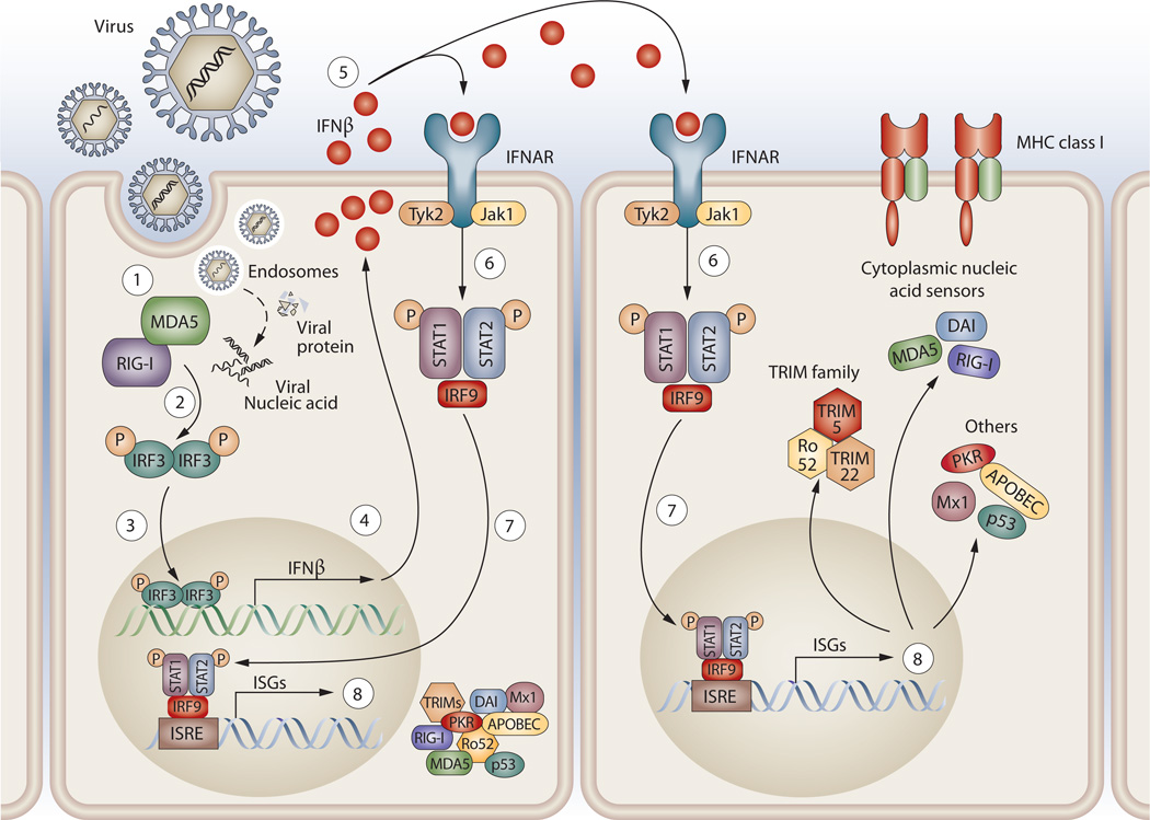
A significant body of data implicates the type I interferon (IFN) pathway in the pathogenesis of autoimmune rheumatic diseases. In these disorders, a self-reinforcing cycle of IFN production can contribute to immunopathology through multiple mechanisms. Type I IFN cytokines are pleiotropic in their effects, mediating antiviral and antitumor activities, and possess numerous immunomodulatory functions for both the innate and adaptive immune responses. A key principle of the type I IFN system is rapid induction and amplification of the signaling pathway, which generates a feed-forward loop of IFN production, ensuring that a vigorous antiviral immune response is mounted. Although such feed-forward pathways are highly adaptive when it comes to rapid and effective virus eradication, this amplification can be maladaptive in immune responses directed against host tissues. Such feed-forward loops, however, create special opportunities for therapy.
Figures

Similar articles
-
Type I Interferons in the Pathogenesis and Treatment of Autoimmune Diseases.Clin Rev Allergy Immunol. 2020 Oct;59(2):248-272. doi: 10.1007/s12016-020-08798-2. Clin Rev Allergy Immunol. 2020. PMID: 32557263 Review.
-
Too much of a good thing: Detrimental effects of interferon.Semin Immunol. 2019 Jun;43:101282. doi: 10.1016/j.smim.2019.101282. Semin Immunol. 2019. PMID: 31771763 Free PMC article. Review.
-
Plasmacytoid dendritic cells in antiviral immunity and autoimmunity.Sci China Life Sci. 2010 Feb;53(2):172-82. doi: 10.1007/s11427-010-0045-0. Epub 2010 Mar 7. Sci China Life Sci. 2010. PMID: 20596824 Free PMC article. Review.
-
Interferons in autoimmune and inflammatory diseases: regulation and roles.J Interferon Cytokine Res. 2011 Dec;31(12):857-65. doi: 10.1089/jir.2011.0101. J Interferon Cytokine Res. 2011. PMID: 22149411 Free PMC article. Review.
-
A central role for RNA in the induction and biological activities of type 1 interferons.Wiley Interdiscip Rev RNA. 2011 Jan-Feb;2(1):58-78. doi: 10.1002/wrna.32. Epub 2010 Aug 24. Wiley Interdiscip Rev RNA. 2011. PMID: 21956969 Review.
Cited by
-
Hemophagocytic lymphohistocytosis in trisomy 21: successful treatment with interferon inhibition.Pediatr Rheumatol Online J. 2022 Nov 18;20(1):104. doi: 10.1186/s12969-022-00764-w. Pediatr Rheumatol Online J. 2022. PMID: 36401314 Free PMC article.
-
GWAS implicates a role for quantitative immune traits and threshold effects in risk for human autoimmune disorders.Curr Opin Immunol. 2012 Oct;24(5):538-43. doi: 10.1016/j.coi.2012.09.003. Epub 2012 Sep 28. Curr Opin Immunol. 2012. PMID: 23026397 Free PMC article. Review.
-
Single-cell eQTL analysis of activated T cell subsets reveals activation and cell type-dependent effects of disease-risk variants.Sci Immunol. 2022 Feb 25;7(68):eabm2508. doi: 10.1126/sciimmunol.abm2508. Epub 2022 Feb 25. Sci Immunol. 2022. PMID: 35213211 Free PMC article.
-
Interleukin-1β Induces mtDNA Release to Activate Innate Immune Signaling via cGAS-STING.Mol Cell. 2019 May 16;74(4):801-815.e6. doi: 10.1016/j.molcel.2019.02.038. Epub 2019 Apr 2. Mol Cell. 2019. PMID: 30952515 Free PMC article.
-
Characterisation of anifrolumab, a fully human anti-interferon receptor antagonist antibody for the treatment of systemic lupus erythematosus.Lupus Sci Med. 2018 Apr 5;5(1):e000261. doi: 10.1136/lupus-2018-000261. eCollection 2018. Lupus Sci Med. 2018. PMID: 29644082 Free PMC article.
References
-
- Sheppard P, et al. IL-28, IL-29 and their class II cytokine receptor IL-28R. Nat. Immunol. 2003;4:63–68. - PubMed
-
- Kotenko SV, et al. IFN-lambdas mediate antiviral protection through a distinct class II cytokine receptor complex. Nat. Immunol. 2003;4:69–77. - PubMed
-
- Doyle SE, et al. Interleukin-29 uses a type 1 interferon-like program to promote antiviral responses in human hepatocytes. Hepatology. 2006;44:896–906. - PubMed
Publication types
MeSH terms
Substances
Grants and funding
LinkOut - more resources
Full Text Sources
Other Literature Sources
Medical
