. 2010 Apr;47(4):297-309.
doi: 10.1016/j.fgb.2009.12.012. Epub 2010 Jan 4.

PARP is involved in replicative aging in Neurospora crassa

Affiliations

PARP is involved in replicative aging in Neurospora crassa

Gregory O Kothe et al. Fungal Genet Biol. 2010 Apr.

Abstract

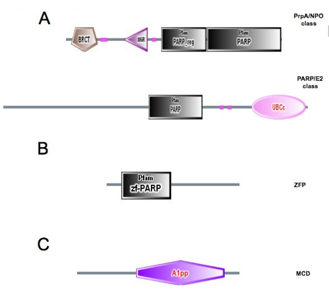
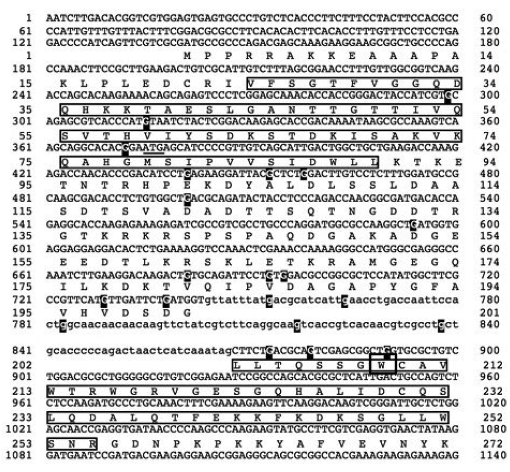
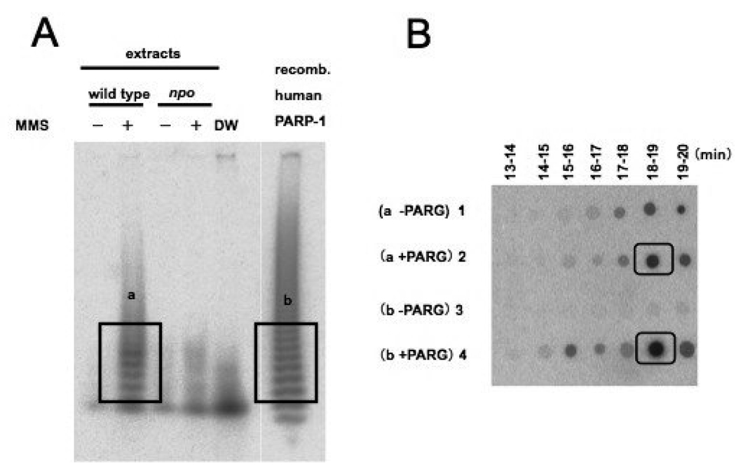
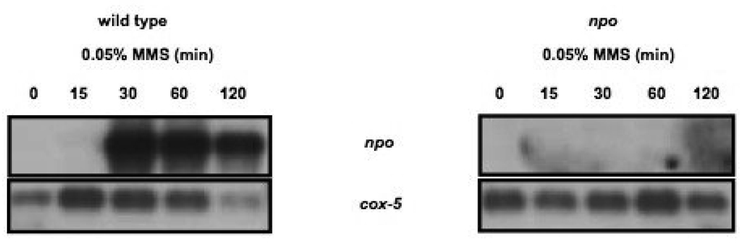
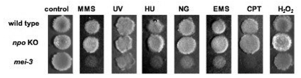
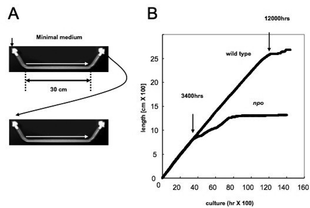
Modification of proteins by the addition of poly(ADP-ribose) is carried out by poly(ADP-ribose) polymerases (PARPs). PARPs have been implicated in a wide range of biological processes in eukaryotes, but no universal function has been established. A study of the Aspergillus nidulans PARP ortholog (PrpA) revealed that the protein is essential and involved in DNA repair, reminiscent of findings using mammalian systems. We found that a Neurospora PARP orthologue (NPO) is dispensable for cell survival, DNA repair and epigenetic silencing but that replicative aging of mycelia is accelerated in an npo mutant strain. We propose that PARPs may control aging as proposed for Sirtuins, which also consume NAD+ and function either as mono(ADP-ribose) transferases or protein deacetylases. PARPs may regulate aging by impacting NAD+/NAM availability, thereby influencing Sirtuin activity, or they may function in alternative NAD+-dependent or NAD+-independent aging pathways.


Figures

FIGURE 1
FIGURE 1. Domain organization of fungal PARP-like proteins and associated DNA and PAR binding proteins
(A) Schematic representation of the domain organization of the two classes of fungal PARP proteins. The complete amino acid sequences of NPO. Neurospora crassa PARP ortholog [CAD21266] and Aspergillus nidulans AN0482.2 [xm_652994.1] were used as queries to search the SMART database (smart.embl-heidelberg.de). Searching with NPO identified BRCT [IPR001357], WGR [IPR008893]. PARP-regulatory [PF02877], and PARP-catalytic [PF00644] domains, defining the PrpA/NPO class. Searching with AN0482.2 identified PARP-catalytic [PF00644] and Ubiquitin-conjugating enzyme E2 catalytic domains [SM00212], defining the PARP/E2 class. (B) Domain organization of the Neurospora MacroD protein. The complete amino acid sequence encoded by Neurospora ORF NCU07925.3 was used to search the SMART database identifying the A1pp domain [SM00506]. (C) Domain organization of the Neurospora zf-PARP protein. The complete amino acid sequence of a Neurospora hypothetical protein [Broad coordinates LGl, contig 2:447973-449833+] was used to search the SMART database identifying a single zf-PARP Pam domain [PF00645].
FIGURE 2
FIGURE 2. Confocal Images of GFP-tagged Neurospora proteins
Expression of heterochromatin protein 1 (HP1::GFP), a telomere repeat binding protein (RAP1::GFP), MacroD::GFP and NPO::GFP was driven by the ccg-1 promoter. An rDNA associated protein (YPH1::GFP) and zf-PARP::GFP were expressed via their endogenous promoters.
FIGURE 3
FIGURE 3. Sequence of the npoRIP1 allele
The NPO protein sequence is indicated beneath the upper case nucleotide coding sequence. Lower case nucleotides represent intron sequences. The boxed amino acids in the first exon indicate the BRCT domain and the boxed amino acids in the second exon indicate the WGR motif. Guanine residues mutated to adenines are highlighted, and the tryptophan codon that was mutated to a stop codon is boxed. The sequence of the npoRIP1 allele has been deposited in Genbank with the accession number EU869543.
FIGURE 4
FIGURE 4. Disruption of the N. crassa npo gene by homologous recombination
(A) Schematic illustration of knockout strategy for the npo gene. A white arrow box represents the npo gene and shows the direction of transcription. Stippled boxes indicate immediate flanking sequences. The knockout construct is shown below the genomic sequence with the E. coli hph gene represented by a black box. The genomic sequence with the E. coli hph gene represented by a black box. The genomic sequence resulting from correct replacement is shown beneath the knockout construct. Horizontal black arrows indicate the positions of PCR primers used to analyze the transformants. Vertical black arrows indicate restriction sites used to characterize the transformants by Southern hybridization A horizontal black line represents the probe used in the Southern blot.(B) The images labeled PCR6 and PCR7 are ethidium bromide-stained agarose gels with size markers run in the left-most lanes. The next three lanes contained PCR products that had been amplified from template DNAs isolated from the indicated transformants. The right-most lanes show PCR products amplified from wild type N.crasse DNA, as controls. The position of primers for the PCR6 and PCR7 reactions are shown in panel A. The right-most image shows an autoradiograph of a Southern blot probed with hph sequences. DNAs from the indicated transformations were digested with Ncol. DNA from wild type N.crassa was run in the right-most lane as a control.
FIGURE 5
FIGURE 5. Verification of NPO PARylation activity
(A) Autoradiogram of a 20% polyacrylamide gel showing 32P-PAR ladder. PARylation reactions with extracts from wild type N.crassa cells treated (+) or not treated (−) with MMS were run alongside reactions with extracts from npo KO cells treated (+) or not treated (−) with MMS. The lane labeled DW is a negative control reaction using distilled water in place of extract. The right most lane contains a positive control reaction with recombinant human PARP-1 expressed in E. coli. The boxed regions labeled a and b were excised and the radioactivity was eluted for analysis by HPLC.(B)An autoradiogram (BAS2500) of fractions from HPLC blotted onto DE81 paper (Whatman) with retention times indicated above and sample designations on the left. Eluents of 32P-PAR from these gel slices were either treated with recombinant PARG, or not, and fractionated by HPLC as described in Materials and Methods. The boxed regions show the peak signals eludated at the retention time for ADP-ribose.
FIGURE 6
FIGURE 6. Analysis of npo transcription by northern blot
The left panel shows an autoradiogram of a northern blot of RNAs extracted from wild type N. crassa after the indicated duration of MMS treatment. The upper panel shows results of probing the blot with npo sequence and in the lower panel shows results of probing with cox-5 sequences as a control for loading. The right panel shows the same for the npo strain.
FIGURE 7
FIGURE 7. Mutagen sensitivity of the npo KO strain
Spot-tests of conidia on FGS plates for wild type N. crassa (top), the npo (middle) and mel-3 strains (bottom) were done as described in Materials and Methods. The mel-3 strain was used as a positive control for mutagen sensitivity. Panels from left to right are as follows: no mutagen; 0.015% methyl methane sulfonate (MMS); conidia pretreated with 450 J/MF UV; 30 mM hydroxy urea (HU); 0.05µg/ml N-methyl-N-nitro-N-nitrosoguanidine (MNNG); 0.3% ethyl methane sulfonate (EMS); 0.3µg/ml camptothecin (CPT) and 0.0015% H2O2.
FIGURE 8
FIGURE 8. Telomere position effect assay
Spot-tests of conidia on FGS plates for wild type N. crassa, npoRIP1, and nst-3RIP1 strains on media with 1.5 mg/ml hygromycin or no hygromycin, as described in Materials and Methods.
FIGURE 9
FIGURE 9. Absence of effects of NAM treatment and npo mutation on DNA methylation
(A) Approximately 1 µg samples of chromosomal DNA, isolated from wild type N. crassa (N 150) grown for 3 days in Vogels minimal media with the indicated concentrations of NAM, were digested either with DpnII (D) or Sau3A (s) and fractionated on a 1× TAE/0.8% agarose gel containing 1 µg/ml ethidium bromide. The left-most lane contains 0.5 µg of 1 kilobase DNA ladder (Invitrogen). (B) A Southern blot of chromosomal DNAs from wild type and npoRP mutant strains digested with DpnII (D) or Sau3A (S), as described for panel A and in Materials and Methods. The Southern blot was probed with ψ63 sequences.
FIGURE 10
FIGURE 10. Method and results of senescence assay
(A) Schematic of race tube strategy for measuring long-term linear extension rate. (B) Plot of growth (cm/hr) for wild type N. crassa and npo strain. Arrows at 3,400 hrs and 12,000 hrs indicate entry into senescence for npo and wild type N. crassa strains, respectively.
FIGURE 11
FIGURE 11. Telomere stability in wild type and npo mutant strains
(A) Arrows on plot shows time points used in telomere erosion assay. (B) Chromosomal DNAs were isolated from wild type and the npo mutant at the time points indicated in panel A. The DNAs were digested with HaeIII, blotted as described in Materials and probed with telomere repeat sequences.

Similar articles

Cited by

References

    1. Ahel I, Ahel D, Matsusaka T, Clark AJ, Pines J, et al. Poly(ADP-ribose)-binding zinc finger motifs in DNA repair/checkpoint proteins. Nature. 2008;451:81–85. - PubMed
    1. Ame JC, Fouquerel E, Gauthier LR, Biard D, Boussin FD, et al. Radiation-induced mitotic catastrophe in PARG-deficient cells. Cell Sci. 2009a;122:1990–2002. - PubMed
    1. Ame JC, Hakme A, Quenet D, Fouquerel E, Dantzer F, et al. Detection of the Nuclear Poly(ADP-ribose)-Metabolizing Enzymes and Activities in Response to DNA Damage. Methods Mol Biol. 2009b;464:267–283. - PubMed
    1. Ame JC, Rolli V, Schreiber V, Niedergang C, Apiou F, et al. PARP-2, A novel mammalian DNA damage-dependent poly(ADP-ribose) polymerase. J Biol Chem. 1999;274:17860–17868. - PubMed
    1. Ame JC, Spenlehauer C, de Murcia G. The PARP superfamily. Bioessays. 2004;26:882–893. - PubMed

Publication types

Associated data

LinkOut - more resources