. 2010 Mar;30(5):1285-98.
doi: 10.1128/MCB.01190-09. Epub 2009 Dec 28.

The hsp90-FKBP52 complex links the mineralocorticoid receptor to motor proteins and persists bound to the receptor in early nuclear events

Affiliations

The hsp90-FKBP52 complex links the mineralocorticoid receptor to motor proteins and persists bound to the receptor in early nuclear events

Mario D Galigniana et al. Mol Cell Biol. 2010 Mar.

Abstract

In this study, we demonstrate that the subcellular localization of the mineralocorticoid receptor (MR) is regulated by tetratricopeptide domain (TPR) proteins. The high-molecular-weight immunophilin (IMM) FKBP52 links the MR-hsp90 complex to dynein/dynactin motors favoring the cytoplasmic transport of MR to the nucleus. Replacement of this hsp90-binding IMM by FKBP51 or the TPR peptide favored the cytoplasmic localization of MR. The complete movement machinery, including dynein and tubulin, could be recovered from paclitaxel/GTP-stabilized cytosol and was fully reassembled on stripped MR immune pellets. The whole MR-hsp90-based heterocomplex was transiently recovered in the soluble fraction of the nucleus after 10 min of incubation with aldosterone. Moreover, cross-linked MR-hsp90 heterocomplexes accumulated in the nucleus in a hormone-dependent manner, demonstrating that the heterocomplex can pass undissociated through the nuclear pore. On the other hand, a peptide that comprises the DNA-binding domain of MR impaired the nuclear export of MR, suggesting the involvement of this domain in the process. This study represents the first report describing the entire molecular system that commands MR nucleocytoplasmic trafficking and proposes that the MR-hsp90-TPR protein heterocomplex is dissociated in the nucleus rather than in the cytoplasm.


Figures
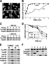
FIG. 1.
FIG. 1.
MR retrotransport requires a functional hsp90 complex. (A) Inhibitory effect of GA on MR nuclear accumulation. E82.A3 cells expressing Flag-MR were incubated on ice for 1 h with 0.1% ethanol (a and c) or 1 μM Aldo (b and d). Next, 2 μM GA (c and d) or 0.1% dimethyl sulfoxide (DMSO) (a and b) was added to the medium, and the incubation was continued on ice for 15 min. The temperature was shifted to 37°C (zero time), and MR was visualized by indirect immunofluorescence after 15 min. (B) The nuclear accumulation rate of MR was measured for the indicated incubation times at 37°C. Data are means ± standard errors of the means (SEM) (n = 3). (C) GA treatment of intact cells destabilizes MR by proteasome degradation. Flag-MR was immunoprecipitated from the cytosol of cells treated with 2 μM GA for the indicated times, in the presence or absence of 10 μM MG132. Coadsorbed hsp90 is also shown. (D) The relative amounts of MR and hsp90 shown in panel C were semiquantified by band density scanning (mean ± SEM; n = 3). (E) 293-T cells were treated with 0.1% DMSO (vehicle) or 2 μM GA for 3 h in the presence of 10 μM MG132 to prevent MR degradation. The receptor was immunoprecipitated with anti-Flag antibody (I; immune) or a nonimmune IgG (NI), and the associated proteins were resolved by Western blotting. (F) hsp90 was immunopurified from reticulocyte lysate, stripped of associated proteins by high ionic strength, and incubated with pure recombinant proteins. Stripped hsp90 was incubated with buffer (lane 1), 50 μg FKBP52 (lane 2), 50 μg Hop/p60 (lane 3), 50 μg Flag-TPR peptide (lane 4), 50 μg FKBP52 and 100 μg Hop/p60 (lane 5), or 50 μg FKBP52 and 100 μg Flag-TPR peptide (lane 6).
FIG. 2.
FIG. 2.
Swapping of TPR proteins. (A) Stripped hsp90 was incubated with hsp90-free reticulocyte lysate (source of endogenous TPR proteins and dynein) supplemented with buffer (lane 1), 50 μg Hop/p60 (lane 2), or 100 μg Hop/p60 (lane 3). (B) FKBPs compete for the TPR acceptor site of hsp90. Stripped hsp90 pellets were incubated with 50 μg FKBP51 in the presence of 100 μg FKBP52 or 100 μg FKBP52 and 100 μg Flag-TPR peptide. (C) Effect of hormone binding to MR on IMM recruitment to the complex. E82.A3 cells transfected with Flag-MR were incubated on ice for 1 h with vehicle (−Aldo) or 1 μM aldosterone (+Aldo) to allow steroid binding but not receptor translocation to the nucleus. MR was then immunoadsorbed, and the coadsorbed proteins were resolved by Western blotting. NI, nonimmune pellet.
FIG. 3.
FIG. 3.
MR heterocomplexes bind microtubules. (A) NIH 3T3 fibroblasts were transfected with Flag-MR and grown in a steroid-free medium. After 36 h, the cells were fixed and permeabilized in cold (−20°C) methanol (MeOH) for 10 min or in 4% p-formaldehyde (p-FA) for 1 h at room temperature and further permeabilized by immersing the coverslips in acetone at −20°C for 5 min. The cells were stained for MR with anti-Flag M2 antibody (red) and for tubulin with the TUB2.1 antibody (green). +Tub and +Flag, indirect immunofluorescence after preincubation of the primary antibody with an excess of pure tubulin or Flag peptide. (B) Close-up view of microtubules and MR. The areas defined by the white rectangles in panel A were enlarged to show the strong colocalization of MR with microtubules in p-formaldehyde-fixed cells (p-FA) compared with the diffuse distribution of MR observed in cells fixed with methanol (MeOH). (C) Taxol increases tubulin association with MR heterocomplexes. Flag-MR was immunoadsorbed with a nonimmune IgG antibody (NI) or the anti-Flag M2 IgG from E82.A3 standard cytosol (control) or supplemented with 20 μM Taxol and 100 μM GTP to stabilize microtubules (Tx/GTP). Tx/GTP+GA represents a Taxol/GTP-stabilized cytosol obtained from cells pretreated with 2 μM GA for 1 h. (D) FKBP52 domains prevent the assembly of the MR complex with tubulin. Flag-MR immune pellets were stripped of endogenous chaperones by high ionic strength and reincubated with 50 μl of E82.A3 cytosol containing an ATP-regenerating system and either 20 μl of buffer (native), 200 μg/20 μl PPIase domain (PPIase), or 200 μg/20 μl Flag-TPR domain. NI, nonimmune pellet. (E) Interaction of dynein with the PPIase domain of FKBP52. Bacterially expressed GST-FKBP52 was immobilized on GSH-agarose gel, stripped of associated proteins, and incubated for 20 min at 30°C with 60 μl of partially purified dynein from reticulocyte lysate in a medium containing 10 mM ATP and either 20 μl buffer (native), 200 μg Flag-TPR peptide/20 μl, 200 μg PPIase peptide/20 μl, or 200 μg FKBP12/20 μl.
FIG. 4.
FIG. 4.
Disruption of the dynein/dynactin complex impairs MR nuclear accumulation. (A) Renal duct cells were transfected with myc-p50/dynactin2 or the PPIase domain of FKBP52. Endogenous MR nuclear accumulation was achieved after 15 min of incubation with 1 μM Aldo. Cells were stained for MR (right) and either p50/dynactin2 or the PPIase domain (left). Arrowheads show the cytoplasmic localization of MR in transfected cells. (B) Nuclear accumulation rate of MR for cells transfected with PPIase domain or p50/Dyt. Note that MR nuclear accumulation was equally impaired in cells treated with the dynein inhibitor EHNA. The inset shows the expression of p50/Dyt or the PPIase domain compared to the endogenous level of FKBP52. (C) The PPIase domain, but not FKBP12, prevents dynein binding to the MR heterocomplex. Flag-MR was immunoprecipitated from 293-T cells cotransfected with vector, the PPIase domain, or FKBP12. Conditions were as follows (μg of plasmid are given in parentheses): lanes 1 and 6, nonimmune pellets; lanes 2 (2 μg), 4 (5 μg), and 7 (5 μg), immune pellets from cells cotransfected with empty vector; lanes 3 and 5 (2 μg and 5 μg), immune pellets from cells cotransfected with the PPIase domain of FKBP52; lane 8, immune pellet from cells cotransfected with FKBP12 (5 μg). (D) Nuclear accumulation was quantified in renal duct cells grown in steroid-free medium (no steroid) or incubated with Aldo for 20 min under the following conditions: 0.1% DMSO (control), 1 μM FK506, 2 μM radicicol (RAD), TPR domain, TPR mutant (R101A) unable to bind hsp90, or FKBP12. The inset shows the level of overexpression in FKBP12-transfected cells. Results in panels B and D are means ± SEM (n = 3).
FIG. 5.
FIG. 5.
Nuclear accumulation rate of MR in FKBP52 KO MEF cells. (A) MEF cells obtained from FKBP52 KO mice were cotransfected with GFP-MR and FKBP52. After 15 min of incubation with 1 μM Aldo, the cells were fixed and FKBP52 was visualized by indirect immunofluorescence (rhodamine-labeled cells). (B) The nuclear accumulation rate of GFP-MR was measured as described in the legend for Fig. 1. (C) GFP-MR was immunoprecipitated with an anti-MR rabbit serum, and the immune pellets were incubated with 5 nM [3H]Aldo (±1 μM radioinert Aldo). Bars show the specific binding for wild-type cells (WT), FKBP52 KO cells (KO), and FKBP52 KO cells transfected with FKBP52 (KO+FKBP52). Results are means ± SEM (n = 5). The Western blots show the coadsorption of FKBP52 with GFP-MR.
FIG. 6.
FIG. 6.
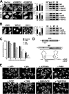
MR transformation is a nuclear event. (A) Primary renal duct cells grown in suspension were incubated with [3H]Aldo for 10 min, and nuclei were immediately isolated by a quick centrifugation at 2°C and lysed by three freeze-thaw cycles. Soluble nucleoplasmic MR isoforms were resolved in a continuous sucrose gradient before (gray dashed line) and after (gray continuous line) preincubation with anti-MR IgM. In the latter case, note the switch of the untransformed peak of MR from 9.4S to 11.4S. The column was calibrated with bovine serum albumin (BSA) (4.5S), β-amylase (β-AM) (8.9S), catalase (CA) (11.3S), untransformed MR (9.4S) obtained in a buffer supplemented with molybdate, and transformed MR (5.1S) obtained from cells incubated for 30 min with steroid and lysed in a buffer supplemented with 0.5 M KCl. (B) Untransformed (9.4S) peaks were pooled, and MR was immunoprecipitated. Coadsorbed proteins were resolved by Western blotting. Cyt, control of MR heterocomplex obtained from cell cytosol; Nuc, MR heterocomplexes from nucleoplasmic fractions. The bar graph shows a densitometric analysis of proteins bound to MR for the cytosolic (white bars) and nuclear (black bars) fractions. Results are means ± SEM for four independent assays. (C) Nucleoplasmic MR was immunoprecipitated after different periods of incubation with Aldo. Coadsorbed hsp90 is shown at the bottom, and MR associated with the insoluble pellet of chromatin is shown at the top. NI, nonimmune. (D) The optical densities of the bands shown in panel C were plotted against the times of incubation with hormone. ▪, MR in the insoluble pellet; ○, MR in the soluble nucleoplasm; •, hsp90 coimmunoadsorbed with soluble MR. Results are means ± SEM for six assays. (E) Cross-linked MR heterocomplexes were incubated with digitonin-permeabilized E82.A3 cells (a and b) or FKBP52 KO MEF cells (c to f). MR subcellular localization was visualized by indirect immunofluorescence in untreated cells (a and c) or cells incubated with 1 μM Aldo (b, d, and f). Conditions were as follows: a, E82 cells without steroid; b, E82 cells with steroid; c, MEF cells without steroid; d, MEF cells with steroid; e, permeabilization control of MEF cells incubated with Alexa Fluor 488-labeled albumin; f, control of MEF cells treated with steroid in a buffer not supplemented with cytosol and ATP. (F) Cross-linking controls. The Western blot shows the coimmunoprecipitation of hsp90, hsp70, and FKBP52 with Flag-MR from untreated cytosol (lysate) or from dithiosuccinimidyl propionate-cross-linked complexes boiled in sample buffer supplemented (+2-ME) or not (−2-ME) with β-mercaptoethanol. The plot on the right shows a sucrose gradient supplemented with 0.5 M KCl for cross-linked MR cytosol preincubated with [3H]aldosterone followed by treatment with (○) or without (•) β-mercaptoethanol.
FIG. 7.
FIG. 7.
FKBP52 expression favors steroid receptor nuclear retention. (A) 293-T cells and COS-7 cells were cotransfected with GFP-MR and either FKBP51 or FKBP52 and then cultured in a steroid-free medium. The primary subcellular localization of the receptor was visualized by indirect immunofluorescence without the addition of steroid. (B) Primary subcellular localization of endogenous GR, visualized in L929 (L), WCL2 (W), and HC11 (H) cells. The bar graphs shown in panels A and B represent the percentages of nuclear (white bars) and cytoplasmic (black bars) receptors for more than 100 cells counted in each experiment (mean ± SEM; n = 3). The Western blots on the right show the coadsorbed hsp90 and FKBPs for MR (A) and GR (B). Vec or V, cells transfected with vector only; NI, nonimmune pellet. (C) Nuclear export of GFP-MR was measured in E82.A3 cells preincubated with 10 nM Aldo, washed, and reincubated in a steroid-free medium with 0.1% DMSO (control), 10 ng/ml leptomycin B (LMB), or 2 μM geldanamycin (GA). TPR, cells cotransfected with the TPR domain of rat PP5 reincubated without additions after steroid withdrawal. The nuclear fraction of MR was measured for more than 100 cells per experiment (mean ± SEM; n = 4). (D) Schematic representation of recombinant TPR peptide from rat PP5, the DNA-binding peptide (DBP) (dotted box), and a control peptide (DCP) where the five amino acids next to the first loop were replaced by the DDAAA sequence. (E) GFP-MR was translocated to the nuclei of E82.A3 cells with Aldo. The coverslips were washed, permeabilized with digitonin, and incubated in Adam's buffer supplemented with 10 mM ATP, 20 mM molybdate (Mo), or 10 mM ATP plus 20 mM molybdate (all the other incubations), without further additions (ATP/Mo) or in the presence of a 1 mM concentration of each peptide or mixture of peptides (DBP, DCP, TPR, DBP and DCP, or DBP and TPR).

Similar articles

Cited by

References

    1. Adam, S. A., R. S. Marr, and L. Gerace. 1990. Nuclear protein import in permeabilized mammalian cells requires soluble cytoplasmic factors. J. Cell Biol. 111:807-816. - PMC - PubMed
    1. Akner, G., K. Mossberg, K. G. Sundqvist, J. A. Gustafsson, and A. C. Wikstrom. 1992. Evidence for reversible, non-microtubule and non-microfilament-dependent nuclear translocation of hsp90 after heat shock in human fibroblasts. Eur. J. Cell Biol. 58:356-364. - PubMed
    1. Black, B. E., J. M. Holaska, F. Rastinejad, and B. M. Paschal. 2001. DNA binding domains in diverse nuclear receptors function as nuclear export signals. Curr. Biol. 11:1749-1758. - PubMed
    1. Buchholz, I., K. Enss, C. Schafer, A. Schlune, V. Shahin, and H. Oberleithner. 2004. Transient permeability leak of nuclear envelope induced by aldosterone. J. Membr. Biol. 199:135-141. - PubMed
    1. Cheung, P. Y., Y. Zhang, J. Long, S. Lin, M. Zhang, Y. Jiang, and Z. Wu. 2004. p150(Glued), dynein, and microtubules are specifically required for activation of MKK3/6 and p38 MAPKs. J. Biol. Chem. 279:45308-45311. - PubMed

Publication types

MeSH terms