. 2010 Feb 26;285(9):6401-11.
doi: 10.1074/jbc.M109.064063. Epub 2009 Dec 27.

Transforming growth factor-beta-activated kinase 1 is an essential regulator of myogenic differentiation

Affiliations

Transforming growth factor-beta-activated kinase 1 is an essential regulator of myogenic differentiation

Shephali Bhatnagar et al. J Biol Chem. .

Abstract

Satellite cells/myoblasts account for the majority of muscle regenerative potential in response to injury and muscular adaptation to exercise. Although the ability to influence this process would provide valuable benefits for treating a variety of patients suffering from muscle loss, the regulatory mechanisms of myogenesis are not completely understood. We have tested the hypothesis that transforming growth factor-beta-activated kinase 1 (TAK1) is an important regulator of skeletal muscle formation. TAK1 is expressed in proliferating C2C12 myoblasts, and its levels are reduced upon differentiation of myoblasts into myotubes. In vivo, TAK1 is predominantly expressed in developing skeletal muscle of young mice. However, the expression of TAK1 was significantly up-regulated in regenerating skeletal muscle of adult mice. Overexpression of a dominant negative mutant of TAK1 or knockdown of TAK1 inhibited the proliferation and differentiation of C2C12 myoblasts. TAK1 was required for the expression of myogenic regulatory factors in differentiating myoblasts. Genetic ablation of TAK1 also inhibited the MyoD-driven transformation of mouse embryonic fibroblasts into myotubes. Inhibition of TAK1 suppressed the differentiation-associated activation of p38 mitogen-activated protein kinase (MAPK) and Akt kinase. Overexpression of a constitutively active mutant of MAPK kinase 6 (MKK6, an upstream activator of p38 MAPK) but not constitutive active Akt restored the myogenic differentiation in TAK1-deficient mouse embryonic fibroblasts. Insulin growth factor 1-induced myogenic differentiation was also found to involve TAK1. Collectively, our results suggest that TAK1 is an important upstream regulator of skeletal muscle cell differentiation.


Figures

FIGURE 1.
FIGURE 1.
Expression of TAK1 in cultured myoblasts and the skeletal muscle of mice. A, C2C12 myoblasts were incubated in DM (2% horse serum in Dulbecco's modified Eagle's medium) for the indicated time periods, and the levels of TAK1, phospho-TAK1, TAB1, TAB2, and MyHCf were measured by Western blot. Representative immunoblots and quantification from five independent experiments presented here show that TAK1, phosphorylated TAK1, TAB1, and TAB2 are highly expressed in C2C12 myoblasts, and their levels are reduced upon differentiation of myoblasts into myotubes. B, shown are protein levels of TAK1, phospho-TAK1, TAB1, and TAB2 in gastrocnemius muscle of mice of different ages. n = 4 for each age group.
FIGURE 2.
FIGURE 2.
Expression of TAK1 in regenerating TA muscle in vivo. A, TA muscle of 3-month-old C57BL6 mice was injected with saline alone or cardiotoxin as described under “Experimental Procedures.” After 5 days the TA muscle was isolated and processed for RNA isolation and measurement of mRNA levels for TAK1, myogenin, and MyoD by real-time-PCR. Data presented here show that the mRNA levels of TAK1 as well as myogenin and MyoD are significantly increased in cardiotoxin-injected regenerating TA muscle compared with contralateral saline-injected TA muscle (n = 3). *, p < 0.01, value significantly different from controls. B, representative immunoblots from two independent experiments presented here show that the protein levels of TAK1 are significantly increased in TA muscle 5 days after cardiotoxin injection. C, shown are protein levels of TAK1 in gastrocnemius muscle of 8-week-old wild-type and mdx mice measured by Western blot. The levels of TAK1 are noticeably higher in mdx mice compared with wild-type mice. There was no difference in the levels of unrelated protein actin.
FIGURE 3.
FIGURE 3.
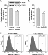
Role of TAK1 in proliferation of C2C12 myoblasts. A, C2C12 myoblasts (10,000 cells/well) were plated in 24-well plate and transfected with vector alone or TAK1 shRNA plasmid. Cells were selected in the presence of puromycin (1.5 μg/ml), and the proliferation of myoblasts was measured using the BrdUrd uptake method as described under “Experimental Procedures.” Data presented here show that transfection of C2C12 myoblasts with TAK1 shRNA significantly reduced its protein levels and inhibited proliferation. *, p < 0.01, values significantly different from control shRNA transfected cells. B, stable transfection of C2C12 myoblasts with a dominant negative TAK1 (dnTAK1) plasmid also inhibited the proliferation of C2C12 myoblasts. #, p < 0.01, values significantly different from control shRNA transfected myoblasts. C, knockdown of TAK1 using RNAi technique did not affect the percentage of apoptotic cells in C2C12 myoblasts assayed using the annexin V-EGFP apoptosis detection kit and fluorescence-activated cell sorting method.
FIGURE 4.
FIGURE 4.
Involvement of TAK1 in differentiation of C2C12 myoblasts. A, C2C12 myoblasts were transiently transfected with increasing amounts of dominant negative TAK1 (dnTAK1) plasmid along with either pSK-Luc or pMCK-Luc plasmid in a 1:10 ratio. After 24 h the cells were incubated in DM, and the luciferase activity in cell extracts was measured. Representative data from two independent experiments (each done in triplicate) presented here show that dnTAK1 inhibits the activation of both skeletal α actin and muscle creatine kinase promoters in a dose-dependent manner. *, p < 0.05, values significantly different from corresponding C2C12 cultures transfected with vector only. B, C2C12 myoblasts were transduced (multiplicity of infection 1:50) with control (Ad.Control) or dominant negative TAK1 (Ad.TAK1) adenoviral vectors for 24 h. The cells were then incubated in DM for indicated time intervals, and the expression of MyHCf was measured by Western blot. Representative immunoblots presented here show that dnTAK1 inhibits the expression of MyHCf without affecting the levels of an unrelated protein actin in C2C12 cultures. C, -fold difference is shown in the mRNA levels of Myf-5, MyoD, myogenin, and myocyte enhancer factor 2D (Mef2D) in Ad.control and Ad.dnTAK1-transduced C2C12 myoblasts 72 h after incubation in DM measured by real-time PCR technique. *, p < 0.01, values significantly different from C2C12 myoblasts transduced with Ad.control vector. D, C2C12 myoblasts were transfected with control or either of the two TAK1 shRNA plasmids, each containing a different target sequence for TAK1 knockdown. The cells were selected in the presence of puromycin (1.6 μg/ml) for 72–96 h followed by incubation in differentiation medium for 72h. CK activity in cell extracts was measured using the CK activity assay kit. The levels of MyHCf and TAK1 were measured by Western blot. Data presented here show that knockdown of TAK1 inhibits the expression of CK and MyHCf in C2C12 cultures. *, p < 0.01, values significantly different from control shRNA transfected C2C12 myoblasts.
FIGURE 5.
FIGURE 5.
Deletion of TAK1 inhibits MyoD-induced differentiation in fibroblasts. TAK1+/+ and TAK1−/− MEF were transduced with Ad.MyoD for 24 h at multiplicity of infection 50. The cells were then incubated in DM for different time intervals. A, myotube formation was measured after 48 h of incubation in DM by performing immunofluorescence using MF20 antibody. Nuclei were stained with 4′,6-diamidino-2-phenylindole. The top photomicrographs (green fluorescence protein) show equal transduction of TAK1+/+ and TAK1−/− MEF by Ad.MyoD vector. The bottom photomicrographs show that myotube formation (red color) is significantly reduced in TAK1−/− MEF cultures compared with TAK1+/+. B, Western blot showed that the levels of MyHCf were reduced in TAK1−/− MEF compared with TAK1+/+ after incubation in DM. Immunoblots also show equal levels of MyoD protein in both TAK1+/+ and TAK1−/− MEF and the presence of truncated TAK1 protein in TAK1−/− MEF. Wt, wild type. C, levels of CK were also found to be significantly reduced in TAK1−/− MEF compared with TAK1+/+ MEF after 24 and 48 h of incubation in DM. *, p < 0.01, values significantly different from TAK1+/+ MEF at corresponding time point. D, TAK1+/+ and TAK1−/− MEFs were transfected with pcDNA3-MyoD plasmid along with pSK-Luc plasmid in a 1:10 ratio for 24 h. Cells were then incubated in DM for 48 h, and the activation of skeletal α-actin promoter was monitored by measuring luciferase activity. Data presented here show a significant reduction in the activation of skeletal α actin promoter in TAK1−/− MEF compared with TAK1+/+ MEF. #, p < 0.01, value significantly different from TAK1−/− MEF.
FIGURE 6.
FIGURE 6.
TAK1 regulates myogenic differentiation through the activation of p38 MAPK. A, TAK1+/+ and TAK1−/− MEF were transduced with Ad.MyoD for 24h followed by incubation in DM for the indicated time intervals. Analysis of cell extracts by Western blot showed that the phosphorylation of p38 MAPK protein was completely blocked in TAK1−/− MEF compared with TAK1+/+. There was no difference in total p38, phosphorylated AMPK, and total or phosphorylated IκBα levels between TAK1+/+ and TAK1−/− MEFs. B, C2C12 myoblasts were stably transfected with vector alone or plasmid expressing either dominant negative TAK1 (dnTAK1) or TAK1 shRNAs and incubated in differentiation medium for 72 h. The levels of total and phospho-p38 MAPK and TAK1 were measured by Western blot. Representative immunoblots presented here show that overexpression of either dnTAK1 protein or TAK1 shRNAs inhibited the levels of phosphorylated p38 in C2C12 cultures. C, TAK1+/+ and TAK1−/− MEF were transiently transfected with either pcDNA3-MyoD alone or with pcDNA3-caMKK6 plasmid for 24 h followed by incubation in differentiation medium for additional 72 h. Myotube formation was monitored by immunocytochemistry using MF-20 antibody and 4′,6-diamidino-2-phenylindole. Representative photomicrographs presented here show that transfection with caMKK6 restored the myotube formation in TAK1−/− MEF cultures. D, shown is quantification of the differentiation index in TAK1+/+ and TAK1−/−MEF cultures transfected with MyoD along with caMKK6 or without caMKK6. *, p < 0.01, values significantly different from TAK1−/− MEF cultures without caMKK6. E, levels of CK measured using a CK activity assay kit and MyHCf and phospho-p38 protein (Western blot) were also found to be significantly increased in TAK1−/− MEF transfected with caMKK6. *, p < 0.05, values significantly different from TAK1+/+ MEFs without caMKK6. #, p < 0.05, values significantly different from TAK1−/− MEFs transfected with no caMKK6.
FIGURE 7.
FIGURE 7.
Role of TAK1 in activation of Akt during myogenic differentiation. A, TAK1+/+ and TAK1−/− MEF were transduced with Ad.MyoD for 24 h followed by incubation in DM for different time intervals. Levels of phosphorylated and total Akt were measured by Western blotting. Data presented here show that the phosphorylation of Akt is blocked in TAK1−/− MEF compared with TAK1+/+ MEF upon incubation in DM. B, C2C12 myoblasts were transfected with vector alone or dnTAK1 and incubated in differentiation medium for 72 h. Representative immunoblots show that the overexpression of dnTAK1 inhibits the levels of phosphorylated Akt in C2C12 cultures. C, TAK1+/+ and TAK1−/− MEF were transiently transfected with either pcDNA3-MyoD alone or with pUSEamp-caAkt plasmid for 24 h followed by incubation in DM for 72 h. Data presented here show that the expression of caAkt did not affect with the levels of CK or MyHCf in TAK1−/− MEF. D, TAK1+/+ and TAK1−/− MEF were transiently transfected with either pcDNA3-MyoD alone or with pcDNA3-caMKK6 plasmid followed by incubation in differentiation medium for 72 h. A representative immunoblot presented here shows that transfection with caMKK6 increased the levels of phosphorylated Akt in TAK1−/− MEF.
FIGURE 8.
FIGURE 8.
IGF-I augments myogenesis through the activation of TAK1. TAK1+/+ and TAK1−/− MEF were transduced with Ad.MyoD for 24 h followed by incubation in DM with or without IGF-I (100 ng/ml). A, data presented here show that IGF-I significantly increased the levels of CK in TAK1+/+ but not TAK1−/− MEF. *, p < 0.01, values significantly different compared with TAK1+/+ MEF incubated without IGF-I. B, Western blot analysis showed that there was no increase in the levels of either MyHCf or myogenin between IGF-1-treated or untreated TAK1−/− MEF. C, TAK1+/+ and TAK1−/− MEF were treated with IGF-I (100 ng/ml) for the indicated time periods, and the levels of phosphorylated or total Akt and p38 MAPK were measured by Western blot. Representative immunoblots presented here show that IGF-I-induced phosphorylation of p38 MAPK (but not Akt) was blocked in TAK1−/− MEF compared with TAK1+/+ MEF.

Similar articles

Cited by

References

    1. Chargé S. B., Rudnicki M. A. (2004) Physiol. Rev. 84, 209–238 - PubMed
    1. Perry R. L., Rudnick M. A. (2000) Front. Biosci. 5, D750–D767 - PubMed
    1. Sartorelli V., Caretti G. (2005) Curr. Opin. Genet. Dev. 15, 528–535 - PMC - PubMed
    1. Yun K., Wold B. (1996) Curr. Opin. Cell Biol. 8, 877–889 - PubMed
    1. Florini J. R., Ewton D. Z., Coolican S. A. (1996) Endocr. Rev. 17, 481–517 - PubMed

Publication types

LinkOut - more resources