. 2010 Feb;176(2):839-49.
doi: 10.2353/ajpath.2010.090570. Epub 2009 Dec 24.

Laminin alpha4-null mutant mice develop chronic kidney disease with persistent overexpression of platelet-derived growth factor

Affiliations

Laminin alpha4-null mutant mice develop chronic kidney disease with persistent overexpression of platelet-derived growth factor

Christine K Abrass et al. Am J Pathol. 2010 Feb.

Abstract

Each extracellular matrix compartment in the kidney has a unique composition, with regional specificity in the expression of various laminin isoforms. Although null mutations in the majority of laminin chains lead to specific developmental abnormalities in the kidney, Lama4-/- mice have progressive glomerular and tubulointerstitial fibrosis. These mice have a significant increase in expression of platelet-derived growth factor (PDGF)-BB, PDGF-DD, and PDGF receptor beta in association with immature glomerular and peritubular capillaries. In addition, mesangial cell exposure to alpha4-containing laminins, but not other isoforms, results in down-regulation of PDGF receptor mRNA and protein, suggesting a direct effect of LN411/LN421 on vessel maturation. Given the known role of overexpression of PDGF-BB and PDGF-DD on glomerular and tubulointerstitial fibrosis, these data suggest that failure of laminin alpha4-mediated down-regulation of PDGF activity contributes to the progressive renal lesions in this animal model. Given the recent demonstration that individuals with laminin alpha4 mutations develop cardiomyopathy, these findings may be relevant to kidney disease in humans.


Figures

Figure 1
Figure 1
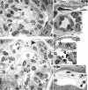
Light microscopy. Paraffin sections were stained with periodic acid–Schiff. Samples from wild-type (wt) (representative glomerulus shown in A) and lama4−/− (ko) mice (B and C) at 7 weeks of age and from lama4−/− animals at 6 (D), 9 (E), and 18 (F) months of age are shown. Dilated glomerular capillaries (B) and peritubular capillaries (B and C) are shown (arrowheads). Note the proliferative changes in the glomerulus in D (arrowhead), as well as the area of tubulointerstitial fibrosis (arrow). A perivascular inflammatory infiltrate is shown in E (arrowhead). Glomerulosclerosis (arrow) and tubular atrophy (arrowhead) are apparent in F. No glomerular or tubulointerstitial fibrosis was seen in wild-type mice up to 23 months in age.
Figure 2
Figure 2
Electron microscopy. Representative electron micrographs from wt (A–C) and (ko) (D–I) mice at 7–14 weeks of age are shown. Normal glomerular structure (A) including podocyte foot processes, GBM, and endothelial fenestrae (B) are shown in wild-type mice. The circumferential pattern of pericyte investment around an arteriole and a pericyte (arrowhead) adjacent to an erythrocyte-filled peritubular capillary is shown in (C). In lama4−/− mice dilated glomerular capillaries are present (D). Also shown in D (arrowhead) is a small arteriole with pericytes and abundant extracellular matrix. Lama4 null mice had normal regions of glomerular capillary wall with normal appearing GBM, podocyte foot processes and endothelial fenestrae similar to wild-type mice (E). Abnormal areas of GBM were also detected where out-pockets of GBM were associated with foot process effacement (F, asterisks). Although glomerular endothelial cells were abundant, apoptotic endothelial cells were also noted (G, arrowheads). Areas of mesangial interposition with split basement membranes were also seen in lama4−/− mice (H, arrowhead), as well as dilated peritubular capillaries with nearby and distant pericytes (I, arrowhead).
Figure 3
Figure 3
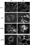
Immunofluorescence microscopy: identification of cells. Samples from wt and ko tissues were stained as indicated. Podocytes were identified by staining for WT-1. Endothelial cells were identified by staining for von Willebrand factor (vWF). Note increased staining with vWF in lama4−/− mice indicating endothelial cell activation. Vascular smooth muscle cells and MCs were identified by staining for α-smooth muscle actin (αSMA). Note increased αSMA expression in the glomerular hilum and discontinuous staining of arteriole walls of the lama4−/− mice.
Figure 4
Figure 4
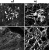
Endothelial cell staining. Endothelial cells were identified by staining with fluorescein-labeled isolectin B4. A and C: Wild-type mice. B and D: Lama4−/− mice.
Figure 5
Figure 5
Pericyte staining. Samples from wt (A, C, E) and ko (B, D, F) mice were stained with antibody to NG2. Note the increased staining in the extra-glomerular mesangium and around afferent and efferent arterioles in B. Note the enlarged vessels with discontinuous pericytes in D. Note the large number of pericytes that surround, but are distant from the vessel wall in F.
Figure 6
Figure 6
Immunofluorescence microscopy: patterns and transitions in laminin isoforms. Kidneys from wt and ko mice were stained with antibodies to the following proteins: A: LN111, LNα1, LNα2, LNα3. Anti-LN111 stains all kidney basement membranes showing overall structure. Typically this antibody stains mesangial matrix more brightly than GBM. This was true in wild-type and lama4−/− mice. Note the irregular organization of mesangial matrix and wide separation between adjacent tubular basement membranes, which represents the space occupied by dilated peritubular capillaries. In the mouse, staining for LNα1 is normally detected in GBM at early stages of glomerulogenesis, and disappears from this site as glomerulogenesis is completed, with detectable LNα1 remaining in the mesangium and TBM of proximal tubules. These transitions in LNα1 were identical in 7-to 14-week-old wild-type and lama4−/− mice. Note the absence of glomerular staining for LNα2 in lama4−/− mice (arrow) and the increase in staining around afferent and efferent arterioles at the glomerular hilum. LNα3 has a limited distribution on the endothelial side of GBM. No significant change in LNα3 was identified in lama4−/− mice. B: LNα4, LNα5, LNβ1, and LNβ2. LNα4 is detected in the mesangial matrix and surrounding peritubular capillaries in wild-type mice and is not detected in lama4−/− mice. During normal glomerulogenesis, LNα5 replaces LNα1 in the GBM and it facilitates maturation and condensation of the mesangium. LNα5 is a normal component of TBM and the peritubular capillary basement membrane. In lama4−/− mice, LNα5 staining of GBM, mesangial matrix, TBM and peritubular capillaries was not different from wild-type mice; however, structural differences in the mesangium were apparent with staining for this LN chain. The brightly stained, condensed mature mesangium was not uniformly present in lama4−/− mice. As glomeruli become fully mature, LNβ1 in GBM is replaced by LNβ2. This transition occurred normally in lama4−/− mice. In wild-type and lama4−/− mice, LNβ1 persists in other ECM compartments including the mesangium and TBM. Adjacent to TBM, a fibrillar matrix that supports peritubular capillaries normally contains LN411 and LN511. Of note, LNβ1 was not detected in this location, but LNβ2 was abundant, which indicates that peritubular capillary matrix is altered and contains only LN521 in lama4−/− mice.
Figure 7
Figure 7
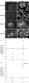
PDGF and PDGF-Rβ. Staining for PDGF-BB (A–D) and PDGF-Rβ (E–H) are shown in wt (A, C, E, G) and ko (B, D, F, H) mice. Note increased staining for both PDGF and PDGF-Rβ in lama4 mice. qPCR for PDGF-B mRNA (I) and PDGF-Rβ mRNA (J). mRNA in whole kidney cortex was determined by qPCR and corrected for 18S RNA. The line represents the upper value detected in normal mice. Note that mRNA for PDGF-Rβ was significantly increased (P < 0.05) in ko mice.
Figure 8
Figure 8
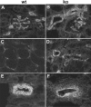
PDGF-DD. Staining for PDGF-DD shown in wt (A and C) and ko (B and D) mice. Note increased staining in lama4−/− mice. E: PDGF-D mRNA. mRNA in whole kidney cortex was determined by qPCR corrected for 18S RNA. The line represents the upper value detected in normal mice.
Figure 9
Figure 9
VEGF Staining. In lama4−/− mice (B, C, D) over 9 months of age, there was an increase in VEGF staining in both the glomerulus and peritubular capillaries as compared with wt mice (A).
Figure 10
Figure 10
Angiopoietin 2 Staining. In animals over 9 months of age, there was a modest increase in Ang2 staining in both the glomerulus and peritubular capillaries in ko mice (B and D) as compared with wt mice (A and C).
Figure 11
Figure 11
In vitro studies. Plating of MCs on LN411/421 as compared with LN111 was associated with a decrease in PDGF-Rβ mRNA (bar graph, left) and protein (Western blot, right).

Similar articles

Cited by

References

    1. Kleinman HK, Weeks BS, Schnaper HW, Kibbey MC, Yamamura K, Grant DS. The laminins: a family of basement membrane glycoproteins important in cell differentiation and tumor metastases. Vitamins Horm. 1993;47:161–186. - PubMed
    1. Goldfinger LE, Stack MS, Jones JCR. Processing of laminin-5 and its functional consequences: role of plasmin and tissue-type plasminogen activator. J Cell Biol. 1998;141:255–265. - PMC - PubMed
    1. Burgeson RE, Chiquet M, Deutzmann R, Ekblom P, Engel J, Kleinman H, Martin GR, Meneguzzi G, Paulsson M, Sanes J, Timpl R, Tryggvason K, Yamada Y, Yurchenco PD. A new nomenclature for the laminins. Matrix Biol. 1994;14:209–211. - PubMed
    1. Yamashita H, Beck K, Kitagawa Y. Heparin binds to the laminin α4 chain LG4 domain at a site different from that found for other laminins. J Mol Biol. 2004;335:1145–1149. - PubMed
    1. Talts JF, Sasaki T, Miosge N, Gohring W, Mann K, Mayne R, Timpl R. Structural and functional analysis of the recombinant G domain of the laminin α4 chain and its proteolytic processing in tissues. J Biol Chem. 2000;275:35192–35199. - PubMed

Publication types

MeSH terms