. 2010 Feb;48(2):106-13.
doi: 10.1002/dvg.20586.

A mouse model to dissect progesterone signaling in the female reproductive tract and mammary gland

Affiliations

A mouse model to dissect progesterone signaling in the female reproductive tract and mammary gland

Rodrigo Fernandez-Valdivia et al. Genesis. 2010 Feb.

Abstract

Considering the regulatory complexities of progesterone receptor (PR) action throughout the female reproductive axis and mammary gland, we generated a mouse model that enables conditional ablation of PR function in a spatiotemporal specific manner. Exon 2 of the murine PR gene was floxed to generate a conditional PR allele (PR(flox)) in mice. Crossing the PR(flox/flox) mouse with the ZP3-cre transgenic demonstrated that the PR(flox) allele recombines to a PR null allele (PR(d)). Mice homozygous for the recombined null PR allele (PR(d/d)) exhibit uterine, ovarian, and mammary gland defects that phenocopy those of our previously described PR knockout (PRKO) model. Therefore, this conditional mouse model for PR ablation represents an invaluable resource with which to further define in a developmental and/or reproductive stage-specific manner the individual and integrative roles of distinct PR populations resident in multiple progesterone-responsive target sites.


Figures

FIG. 1
FIG. 1
Generation of the PRflox/flox and PRd/d mouse. (a) Schematic shows the gene-targeting strategy and relevant mouse crosses to generate mice carrying the PRflox/flox and PRd allele. The targeting vector contains 7.9kb of PR homologous sequence in which the 5’ and 3’ arms of homology are 2.9kb and 5kb respectively. Within the 5’arm, exon 2 is engineered to be flanked by loxP sites (gray triangles) in which the 5’ loxP site also contains a novel Bam HI site (B*). The FRTNEOFRT cassette (NEO) is inserted in intron 2 in which the neomycin resistance gene (neo) is flanked by direct FRT repeats (black triangles). The HSV-TK gene is attached to the 3’ end of the targeting vector to allow negative selection. Homologous recombination with the PR allele in ES cells results in the generation of the PRflox(neo) targeted allele in which exon 2 is floxed and the FRTNEOFRT cassette is inserted in intron 2. Mice carrying the PRflox(neo) targeted allele were crossed with FLP deleter mice to remove the FRTNEOFRT cassette to generate mice harboring the PRflox allele in which exon 2 is floxed but phenotypically normal. The PRflox mouse was crossed with the Zp3 Cre mouse to excise exon 2. Following cre-mediated excision, a single loxP and FRT site remain in the intron of the PRd allele. For experiments described here, mice heterozygous for the PRd allele were crossed to generate mice homozygous for the PRd allele (PRd/d mice). Bam HI, Bgl II, and Xho I are denoted by B, b, and X respectively. The positions of the PCR primers used to identify mice carrying the WT and PRd allele are indicated by p, q, and r. (b) Southern analysis of mouse genomic DNA digested with Bam HI and hybridized with the 5’ probe yields a 9.7kb band for WT mice, a 9.7 kb and 2.9kb band for PRflox(neo)/+ mice, and a single 2.9kb for PRflox(neo)/flox(neo) mice (lanes 1, 2, and 3 respectively). Southern analysis using the 3’ probe yields a single 6.6kb hybridizing band for WT mice (lane 1), a 6.6kb and 8.4kb band for PRflox(neo)/+ mouse (lane 2), and a single 8.4kb hybridization band for PRflox(neo)/flox(neo) mice (lane 3). (c) Left panel shows a Southern analysis of genomic DNA digested with Bgl II and hybridized with the neo probe (see Materials & Methods). As expected, a hybridizing band is not detected in WT genomic DNA (lane 1); however, an 8.4kb band is detected in genomic DNA from the PRflox(neo)/+ mouse (lane 2). Lanes 3, 4, and 5 correspond to genomic DNA isolated from representative mice from three separate lines of PRflox/+ mice in which the FRTNEOFRT cassette is deleted following a cross with FLP deleter mice. The right panel shows a typical PCR result (using the primers p, q, and r (Fig 1a)) for the WT, PRd/+, and PRd/d genotype (lanes 1–3 respectively). The 226bp PCR band denotes the WT allele whereas the 381 bp band indicates the PRd allele; lane M signifies the marker lane. (d–f) show representative uterine sections stained for PR immunoreactivity from 10 week-old WT, PRKO, and PRd/d mice respectively. Note the absence of PR expression in the luminal epithelial (LE), glandular epithelial (GE), and stromal (S) compartments in the uterus of the PRKO and PRd/d mouse.
FIG. 2
FIG. 2
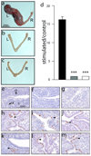
Uterine defects in the PRd/d mouse. (a) At the macro level, a decidual reaction is clearly evident in the left (L) uterine horn of the hormone-treated WT mouse following the decidual stimulus (Materials & Methods); a decidual reaction was not detected in the unstimulated right (R) or control horn. (b–c) show that similarly treated PRKO and PRd/d uteri respectively do not exhibit a decidual response in the mechanically stimulated left horn. (d) Histogram displays the average ratio of wet weights of stimulated horn over control horn (± SEM) for WT (black column); PRKO (grey column); and PRd/d (white column) mouse groups. ***, P < 0.001, one-way ANOVA followed by Tukey’s post-hoc multiple range test (N=4 per genotype). (e–g) show representative uterine sections stained for BrdU incorporation from sesame oil treated ovariectomized WT, PRKO, and PRd/d mice respectively. Note the absence of BrdU positive cells in the luminal epithelium (LE), glandular epithelium (GE), and stromal (S) compartments of the WT, PRKO, and PRd/d uterus. H, I, and J show representative uterine sections stained for BrdU incorporation from E2 treated WT, PRKO, and PRd/d mice respectively. Arrowheads indicate the position of BrdU positive cells in the luminal epithelial compartment of uteri from all three genotypes. (k–m) show representative uterine sections stained for BrdU incorporation from E2P4 treated WT, PRKO, and PRd/d mice. Note that BrdU positive cells are absent in the luminal epithelial compartment of the E2P4 treated WT uterus whereas a subset of stromal cells are positive for BrdU incorporation (arrowhead). However, only the luminal epithelial compartment is positive for BrdU incorporation in the E2P4 treated PRKO and PRd/d uterus (arrowhead in (l) and (m) respectively). Scale bar in (e) applies to (f–m).
FIG. 3
FIG. 3
Ovulation impairment in the gonadotropin treated PRd/d mouse. (a) Table summarizes the average number of oocytes (± SEM) retrieved per mouse following gonadotropin treatment (see Materials & Methods). Note that the WT ovary yields a significant number of oocytes whereas oocytes are absent from the similarly treated PRKO and PRd/d ovary. (b) Representative section (stained with H&E) of a WT ovary following gonadotropin treatment. Note the absence of intraovarian oocytes but the presence of a corpus luteum (CL). (c) and (d) show representative sections of an ovary derived from similarly treated PRKO and PRd/d mice respectively. Arrowhead indicates the position of an entrapped oocyte within CLs in both sections. Scale bar in (b) applies to (c) and (d) (N=5 per genotype).
FIG. 4
FIG. 4
A block in ductal-side branching and alveologenesis in the mammary gland of the hormone-treated PRd/d mouse. (a) Whole-mount of a mammary gland obtained from a WT mouse previously treated with E2P4 for three weeks; (b) higher magnification image of a region of this tissue. Note the widespread presence of extensive ductal side-branching and alveologenesis. (c), (d) and (e), (f), show low and high magnification images of mammary tissue derived from the similarly treated PRKO and PRd/d mouse respectively. Note the absence of ductal side branches and alveolar bodies in the PRKO and PRd/d mammary glands. (g) and (h) show numerous luminal epithelial cells scoring positive (arrowhead) in the alveolar and ductal compartment of the E2P4 treated WT mammary gland respectively. (i) and (j) show a single cell registering positive for BrdU incorporation (arrowhead) in the ductal compartment of the similarly treated PRKO and PRd/d mammary gland respectively. (k) A histogram displaying the percentage of BrdU positive luminal epithelial cells in the mammary gland of the hormone treated WT, PRKO, and PRd/d mouse. Scale bar in (a) and (b) applies to (c), (e), and (d), (f), respectively; LN denotes lymph node (a structural reference point).

Similar articles

Cited by

References

    1. Brisken C, Park S, Vass T, Lydon JP, O'Malley BW, Weinberg RA. A paracrine role for the epithelial progesterone receptor in mammary gland development. Proc Natl Acad Sci U S A. 1998;95:5076–5081. - PMC - PubMed
    1. Chen B, Pan H, Zhu L, Deng Y, Pollard JW. Progesterone inhibits the estrogen-induced phosphoinositide 3-kinase-->AKT-->GSK-3beta-->cyclin D1-->pRB pathway to block uterine epithelial cell proliferation. Mol Endocrinol. 2005;19:1978–1990. - PubMed
    1. Daikoku T, Hirota Y, Tranguch S, Joshi AR, DeMayo FJ, Lydon JP, Ellenson LH, Dey SK. Conditional loss of uterine Pten unfailingly and rapidly induces endometrial cancer in mice. Cancer Res. 2008;68:5619–5627. - PMC - PubMed
    1. Farley FW, Soriano P, Steffen LS, Dymecki SM. Widespread recombinase expression using FLPeR (flipper) mice. Genesis. 2000;28:106–110. - PubMed
    1. Hashimoto-Partyka MK, Lydon JP, Iruela-Arispe ML. Generation of a mouse for conditional excision of progesterone receptor. Genesis. 2006;44:391–395. - PubMed

Publication types

MeSH terms

LinkOut - more resources