. 2010 Feb 19;285(8):5713-25.
doi: 10.1074/jbc.M109.029462. Epub 2009 Dec 22.

Initiation factor eIF2-independent mode of c-Src mRNA translation occurs via an internal ribosome entry site

Affiliations

Initiation factor eIF2-independent mode of c-Src mRNA translation occurs via an internal ribosome entry site

Heba Allam et al. J Biol Chem. .

Abstract

Overexpression and activation of the c-Src protein have been linked to the development of a wide variety of cancers. The molecular mechanism(s) of c-Src overexpression in cancer cells is not clear. We report here an internal ribosome entry site (IRES) in the c-Src mRNA that is constituted by both 5'-noncoding and -coding regions. The inhibition of cap-dependent translation by m(7)GDP in the cell-free translation system or induction of endoplasmic reticulum stress in hepatoma-derived cells resulted in stimulation of the c-Src IRES activities. Sucrose density gradient analyses revealed formation of a stable binary complex between the c-Src IRES and purified HeLa 40 S ribosomal subunit in the absence of initiation factors. We further demonstrate eIF2-independent assembly of 80 S initiation complex on the c-Src IRES. These features of the c-Src IRES appear to be reminiscent of that of hepatitis C virus-like IRESs and translation initiation in prokaryotes. Transfection studies and genetic analysis revealed that the c-Src IRES permitted initiation at the authentic AUG351, which is also used for conventional translation initiation of the c-Src mRNA. Our studies unveiled a novel regulatory mechanism of c-Src synthesis mediated by an IRES element, which exhibits enhanced activity during cellular stress and is likely to cause c-Src overexpression during oncogenesis and metastasis.


Figures

FIGURE 1.
FIGURE 1.
A, organization of the c-src gene. Transcription from two promoters (indicated as P) and alternative splicing result in type 1α (GenBankTM accession number NM_005417) and type 1A (GenBankTM accession number NM_198291) mRNAs in liver cells. Both transcripts differ only in the 5′-distal region of the 5′NCR. The sequence of the exons 1B and 1C and ORF are shared in both transcripts (35). AUGi, initiator AUG codon. B, schematic of the type 1A 5′NCR. Two cryptic AUGs at nt 147 and 179, the initiator AUG at nt 351, and a pyrimidine tract (nt 330–344) located 6 nt upstream of the initiator AUG are shown. The gray lines with arrows on both sides show sequence locations in different putative domains, whereas similar arrows with black solid lines indicate position of the conserved stem-loop structures as shown in Fig. 2.
FIGURE 2.
FIGURE 2.
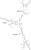
Computer-assisted folding of the type 1A c-Src mRNA sequence. The Zucker M-Fold program (version 3.2, 41) was used for prediction of the secondary structures representing various segments of the c-Src mRNA. Only a representative structure of the c-Src nt 1–383 that includes the entire 5′NCR followed by the 33-nt coding sequence is shown here. The initiator AUG at position 351 (arrow) and putative stem-loop (SL) structures, domains, and nt positions are indicated.
FIGURE 3.
FIGURE 3.
c-Src 5′NCR-mediated translation in cell-free lysates. A, organization of in vitro transcribed uncapped reporter RNAs. The 33-nt coding sequence (dotted box, C) and Kozak sequence are shown at translation initiation site. The solid line represents 5′NCR. An, poly(A) tail. B, 5 μg of uncapped (lanes 2 and 4) and capped (lanes 3 and 5) 5′Src-FLuc (lanes 2 and 3) or 5′PV(Δ286–605)-FLuc (lanes 4 and 5) RNAs were translated in RRL for 1.5 h in the presence of [35S]methionine. The FLuc protein bands were visualized by autoradiography after SDS-PAGE. Two μl of the translation lysates were assayed for enzymatic activity (shown as relative light units (RLU)) of FLuc using D-luciferin substrate. Lane 1, translation without exogenous RNA (control). C, translation of uncapped 5′Src-FLuc (lane 2), 5′PV(Δ286–605)-FLuc (lane 3), and 5′PV-FLuc (lane 4) RNAs in HeLa cell-free lysate as described above. The FLuc activity and the protein bands are shown.
FIGURE 4.
FIGURE 4.
c-Src 5′NCR-mediated translation in Huh7 cells. A, schematic of an in vitro transcribed dicistronic reporter mRNA (5′Src-RFLuc). B, Huh7 cultured cells (80% confluency, 50-mm dish) were transfected with 10 μg of capped 5′Src-RFLuc for 3 h, and Renilla and firefly luciferase activities were assayed in the cytoplasmic fractions. Each transfection was carried out in triplicate, and the experiment was repeated three times to confirm the results. The cytoplasmic fractions of untransfected cells were used as negative control. C, Northern blot analysis of total RNA isolated from 5′Src-RFLuc-transfected (lane 3) and untransfected (lane 4) Huh7 cells. Lanes 1 and 2 show position of monocistronic (5′Src-FLuc) and dicistronic (5′Src-RFLuc) RNAs, respectively, as RNA markers. The 32P-labeled oligonucleotide probe was derived from 3′ end of the FLuc ORF. Overexposure of the film during autoradiography (for more than a week) did not show any fragment of the dicistronic mRNA in lane 3.
FIGURE 5.
FIGURE 5.
Effect of deletion mutations on the c-Src sequence-controlled translation of reporter RNAs. A, organization of in vitro transcribed uncapped wild type reporter RNA (5′Src-RFLuc) and the mutants containing various lengths of deletions (Δ) in the c-Src sequences are shown; dashed line, extent of deletion. AUG, initiator codon is underlined. B, RNAs were translated for 1.5 h in HeLa lysates in the presence of [35S]methionine, and the FLuc protein bands were visualized by autoradiography after SDS-PAGE. Lane M, 14C-labeled protein markers. C, Stability of the reporter RNAs in HeLa translation lysates. The 32P-labeled reporter RNAs (∼1 × 10⁶ dpm) were added to a standard HeLa translation mixture for 1.5 h, and total RNAs were isolated by Qiagen RNeasy column method. Half of the eluted RNA samples (20 μl) were subjected to formaldehyde-agarose gel electrophoresis. The gel was photographed after ethidium bromide staining for detection of ribosomal RNAs (lower panel), dried, and autoradiographed (upper panel). Reporter RNAs are as indicated for each lane. D, comparison of the kinetics of translation between wild type 5′Src-FLuc and a deletion mutant (5′SrcΔ2-FLuc). The RNAs were translated in a standard HeLa lysates mixture, and FLuc activities were assayed with an aliquot (2 μl) of the reaction at various time points. Control, translation lysates without exogenous RNA. E, transfection of uncapped monocistronic c-Src mutant RNAs. The Huh7 cells (80% confluent in 60-mm culture plates) were transfected in triplicate with uncapped RNAs as indicated, and FLuc activities were assayed 3 h post-transfection in the total lysates. F, relative stability of mutant reporter RNAs in the transfected cells. The 32P-labeled mutant RNAs (as indicated) were transfected as above. The total RNAs were isolated, and the labeled RNAs were detected by agarose gel electrophoresis followed by autoradiography of the dried gel (upper panel). Lower panel, the same gel showing 18 S rRNA in each lane.
FIGURE 6.
FIGURE 6.
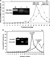
Assembly of translation initiation complexes on the c-Src IRES in HeLa cell-free lysates and analysis by sucrose density gradient centrifugation. A, translation mixtures were incubated with 1 mm GMP-PNP in and ice bath for 5 min and in vitro transcribed 32P-labeled RNA as follows: 5′Src-FLuc (squares and solid line) or capped FLuc (lacking IRES, triangles and solid line) or uncapped FLuc (triangles and broken line) were added to the translation reaction and incubated for 15 min at 30 °C. The complexes formed in the absence of exogenous ATP and GTP on the 5′Src-FLuc probe are shown as squares and dashed line. The lysates were separated by 10–30% sucrose density gradient centrifugation, and fractions (250 μl) were collected from the bottom of the tube to determine RNA contents. B, total RNAs from the peak fractions (Peak I or II) were isolated and analyzed by agarose gel electrophoresis. Marker lanes are as follows: 1, input 5′Src-FLuc RNA probe; 2, total RNA extracted from HeLa S10 translation lysate showing 18 S and 28 S rRNAs; 3, 18 S rRNA extracted from purified 40 S subunit. The total RNAs extracted from Peak I (fractions 5 and 6) for 5′Src-FLuc probe are shown in lane 4, and standard translation reactions are shown as squares and solid line (A), and lane 5, translation reaction deficient in exogenous ATP/GTP (squares and broken line shown in A). RNAs isolated from Peak II are shown in lanes 6 (triangles, broken line) and 7 (triangles, solid line). C, comparison of ribosomal complex formed at wild type c-Src IRES and a mutant PV 5′NCR used as scrambled IRES in the presence of 1 mm GMP-PNP. The 32P-labeled uncapped 5′Src-FLuc (squares and solid line) or 5′PV(Δ286–605)-FLuc (scrambled IRES, triangles and dashed line) RNAs were used in sucrose gradient centrifugation analysis as described above. D, total RNA isolated and resolved by agarose gel electrophoresis is shown. Lanes 1 and 2, rRNA (as markers) isolated from lysates and purified 40 S subunit, respectively; lanes 3 and 4, RNA isolated from Peak I and Peak II, respectively.
FIGURE 7.
FIGURE 7.
Direct binding of the c-Src IRES with purified HeLa 40 S ribosomal subunit. A, purified HeLa 40 S subunit was mixed with 32P-labeled RNA representing c-Src nt 1–383 (solid line) and subjected to sucrose density gradient centrifugation. A separate sample without 40 S was run to locate position of the free probe (broken line) during sedimentation. Peak fractions were analyzed for RNA contents (inset). Inset, lane M, 18 S rRNA as marker; lane 1, RNA extracted from 40 S plus probe peak; lane 2, RNA from free probe peak. B, experiment similar to that described in A but repeated with a scrambled IRES (5′PV286–605)) as indicated. Inset, lane M, 18 S rRNA; RNA isolated from c-Src IRES probe (lane 1) and scrambled IRES probe (lane 2).
FIGURE 8.
FIGURE 8.
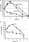
Stimulation of the c-Src IRES-controlled mRNA translation when eIF4E function is inhibited. A, capped RNAs as follows: 5′Src-FLuc (triangle) and FLuc (5′Cap-FLuc, circle) or uncapped 5′HCV-FLuc (square) RNAs were translated in triplicate in the presence of increasing amounts of m7GDP in RRL for 1 h, and one-tenth of each reaction mixture was assayed for FLuc activity. Average FLuc activity of three reactions is shown for each m7GDP concentration. The FLuc activity in samples without m7GDP was considered as 100% translation and compared with those containing the cap analogue (inhibitor). Similar translation reactions were carried out twice to confirm the results. B, translation of a capped Dual-Luciferase RNA construct (5′Src-RFLuc) in RRL in the presence of increasing amounts of m7GDP as described above. Relative cap-dependent translations of RLuc and c-Src IRES-dependent FLuc synthesis are shown. Each translation mixture was carried out in triplicate. The results were confirmed by three independent experiments.
FIGURE 9.
FIGURE 9.
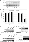
c-Src IRES-controlled translation is not inhibited during cellular stress. A, phosphorylation of eIF2α by TG treatment. The cytoplasmic lysates (40 μg) from Huh7 cells that were treated with 1 μm TG for 0.5, 1, 2, 4, and 6 h (lanes 2–6) were subjected to Western blot analysis using anti-(phospho-eIF2α(Ser-51)) antibody (Cell Signaling, upper panel) and anti-eIF2α antibodies (Sigma, lower panel). Lane 1, 0-min treatment. B, Huh7 cells in triplicate were treated with DMSO alone or 1 μm thapsigargin dissolved in DMSO (DMSO-TG) for 3 h followed by transfection with in vitro transcribed capped 5′Src-RFLuc RNA (solid black bar) or RL-HCV1b (solid gray bar). The upstream RLuc in RL-HCV1b RNA is translated by cap-dependent mechanism, whereas HCV IRES mediates downstream FLuc translation. The cytoplasmic lysates were assayed for FLuc (IRES-dependent translation) and RLuc (cap-dependent translation) activities 3 h post-transfection. The activities of FLuc and RLuc in untreated (control) samples were considered as 100% and compared with the solvent alone (DMSO) or TG-treated cells. C, cytoplasmic lysates from experiments described in B (above) were subjected to Western blot for the total c-Src protein with monoclonal anti-Src antibody (clone 327, Santa Cruz Biotechnology, upper panel) or anti-actin antibodies (lower panel). D, Northern blot analysis of total RNA extracted from Huh7 cells described in B and probed with 32P-labeled oligonucleotide corresponding to nt 320–350 of the c-Src 5′NCR (upper panel). Lower panel shows 18 S rRNA in the same samples. E, total c-Src level in Huh7 cell lysates cultured for 72 h in serum-deprived (indicated as minus) or 10% serum containing (plus) media. Western blot was carried out with anti-Src antibody as described above. F, Northern blot for probing c-Src mRNA (as described above) in total RNA extracted from Huh7 cells cultured in serum-starved (minus) and serum supplemented (plus) regular media. The 18 S rRNAs in each lane is shown in the lower panel.

Similar articles

Cited by

References

    1. Sonenberg N., Hinnebusch A. G. (2007) Mol. Cell 28, 721–729 - PubMed
    1. Holcik M., Sonenberg N. (2005) Nat. Rev. Mol. Cell Biol. 6, 318–327 - PubMed
    1. Baird S. D., Turcotte M., Korneluk R. G., Holcik M. (2006) RNA 12, 1755–1785 - PMC - PubMed
    1. Wek R. C., Jiang H. Y., Anthony T. G. (2006) Biochem. Soc. Trans. 34, 7–11 - PubMed
    1. Hellen C. U., Sarnow P. (2001) Genes Dev. 15, 1593–1612 - PubMed

Publication types

MeSH terms

LinkOut - more resources