

Sentinel node detection in head and neck malignancies: innovations in radioguided surgery
- PMID: 20016804
- PMCID: PMC2792958
- DOI: 10.1155/2009/681746
Sentinel node detection in head and neck malignancies: innovations in radioguided surgery
Abstract
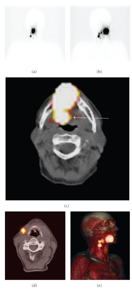
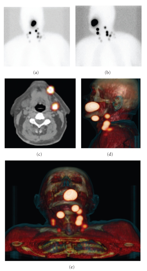
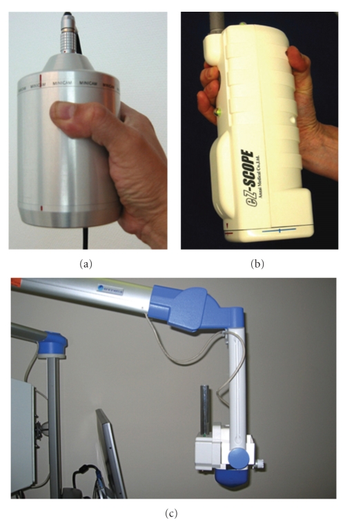
Sentinel node mapping is becoming a routine procedure for staging of various malignancies, because it can determine lymph node status more precisely. Due to anatomical problems, localizing sentinel nodes in the head and neck region on the basis of conventional images can be difficult. New diagnostic tools can provide better visualization of sentinel nodes. In an attempt to keep up with possible scientific progress, this article reviews new and innovative tools for sentinel node localization in this specific area. The overview comprises a short introduction of the sentinel node procedure as well as indications in the head and neck region. Then the results of SPECT/CT for sentinel node detection are described. Finally, a portable gamma camera to enable intraoperative real-time imaging with improved sentinel node detection is described.
Figures

Similar articles
-
An innovative multimodality approach for sentinel node mapping and biopsy in head and neck malignancies.Rev Esp Med Nucl Imagen Mol. 2014 Sep-Oct;33(5):274-9. doi: 10.1016/j.remn.2013.11.005. Epub 2014 May 16. Rev Esp Med Nucl Imagen Mol. 2014. PMID: 24842707
-
SPECT-CT for topographic mapping of sentinel lymph nodes prior to gamma probe-guided biopsy in head and neck squamous cell carcinoma.J Craniomaxillofac Surg. 2004 Dec;32(6):343-9. doi: 10.1016/j.jcms.2004.05.008. J Craniomaxillofac Surg. 2004. PMID: 15555515
-
Paraaortic sentinel lymph nodes: toward optimal detection and intraoperative localization using SPECT/CT and intraoperative real-time imaging.J Nucl Med. 2010 Mar;51(3):376-82. doi: 10.2967/jnumed.109.071779. Epub 2010 Feb 11. J Nucl Med. 2010. PMID: 20150260
-
Contribution of SPECT/CT imaging to radioguided sentinel lymph node biopsy in breast cancer, melanoma, and other solid cancers: from "open and see" to "see and open".Q J Nucl Med Mol Imaging. 2014 Jun;58(2):127-39. Q J Nucl Med Mol Imaging. 2014. PMID: 24835289 Review.
-
Evaluation and localization of lymphatic drainage and sentinel lymph nodes in patients with head and neck melanomas by hybrid SPECT/CT lymphoscintigraphic imaging.J Nucl Med Technol. 2007 Mar;35(1):10-6; quiz 17-20. J Nucl Med Technol. 2007. PMID: 17337652 Review.
Cited by
-
Feasibility of sentinel node biopsy in head and neck melanoma using a hybrid radioactive and fluorescent tracer.Ann Surg Oncol. 2012 Jun;19(6):1988-94. doi: 10.1245/s10434-011-2180-7. Epub 2011 Dec 30. Ann Surg Oncol. 2012. PMID: 22207047 Free PMC article.
-
SPECT/CT for Lymphatic Mapping of Sentinel Nodes in Early Squamous Cell Carcinoma of the Oral Cavity and Oropharynx.Int J Mol Imaging. 2011;2011:106068. doi: 10.1155/2011/106068. Epub 2010 Sep 6. Int J Mol Imaging. 2011. PMID: 21490726 Free PMC article.
-
Molecular imaging agents for SPECT (and SPECT/CT).Eur J Nucl Med Mol Imaging. 2014 May;41 Suppl 1:S26-35. doi: 10.1007/s00259-013-2643-0. Epub 2013 Dec 7. Eur J Nucl Med Mol Imaging. 2014. PMID: 24318159 Review.
-
eTFC-01: a dual-labeled chelate-bridged tracer for SSTR2-positive tumors.EJNMMI Radiopharm Chem. 2024 May 22;9(1):44. doi: 10.1186/s41181-024-00272-0. EJNMMI Radiopharm Chem. 2024. PMID: 38775990 Free PMC article.
-
Design and implementation of a prototype head and neck phantom for the performance evaluation of gamma imaging systems.EJNMMI Phys. 2017 Dec;4(1):19. doi: 10.1186/s40658-017-0186-3. Epub 2017 Jul 6. EJNMMI Phys. 2017. PMID: 28685477 Free PMC article.
References
-
- Nieweg OE, Tanis PJ, Kroon BR. The definition of a sentinel node. Annals of Surgical Oncology. 2001;8(6):538–541. - PubMed
-
- Gershenwald JE, Thompson W, Mansfield PF, et al. Multi-institutional melanoma lymphatic mapping experience: the prognostic value of sentinel lymph node status in 612 stage I or II melanoma patients. Journal of Clinical Oncology. 1999;17(3):976–983. - PubMed
-
- Kovács AF, Stefenelli U, Seitz O, et al. Positive sentinel lymph nodes are a negative prognostic factor for survival in T1-2 oral/oropharyngeal cancer-a long-term study on 103 patients. Annals of Surgical Oncology. 2009;16(2):233–239. - PubMed
-
- Civantos F, Jr., Zitsch R, Bared A, Amin A. Sentinel node biopsy for squamous cell carcinoma of the head and neck. Journal of Surgical Oncology. 2008;97(8):683–690. - PubMed
-
- Paleri V, Rees G, Arullendran P, Shoaib T, Krishman S. Sentinel node biopsy in squamous cell cancer of the oral cavity and oral pharynx: a diagnostic meta-analysis. Head and Neck. 2005;27(9):739–747. - PubMed
LinkOut - more resources
Full Text Sources
