. 2010 Feb 15;21(4):562-71.
doi: 10.1091/mbc.e09-03-0217. Epub 2009 Dec 16.

GRASP and IPCEF promote ARF-to-Rac signaling and cell migration by coordinating the association of ARNO/cytohesin 2 with Dock180

Affiliations

GRASP and IPCEF promote ARF-to-Rac signaling and cell migration by coordinating the association of ARNO/cytohesin 2 with Dock180

David T White et al. Mol Biol Cell. .

Abstract

ARFs are small GTPases that regulate vesicular trafficking, cell shape, and movement. ARFs are subject to extensive regulation by a large number of accessory proteins. The many different accessory proteins are likely specialized to regulate ARF signaling during particular processes. ARNO/cytohesin 2 is an ARF-activating protein that promotes cell migration and cell shape changes. We report here that protein-protein interactions mediated by the coiled-coil domain of ARNO are required for ARNO induced motility. ARNO lacking the coiled-coil domain does not promote migration and does not induce ARF-dependent Rac activation. We find that the coiled-coil domain promotes the assembly of a multiprotein complex containing both ARNO and the Rac-activating protein Dock180. Knockdown of either GRASP/Tamalin or IPCEF, two proteins known to bind to the coiled-coil of ARNO, prevents the association of ARNO and Dock180 and prevents ARNO-induced Rac activation. These data suggest that scaffold proteins can regulate ARF dependent processes by biasing ARF signaling toward particular outputs.


Figures

Figure 1.
Figure 1.
Structure of ARNO constructs used in this study. The domain structures of full-length, Δcoiled-coil, and ΔPH ARNO are depicted. The coiled-coil domain is involved in protein–protein interactions. The Sec7 domain is the catalytic ARF GEF domain. The PH and polybasic domains interact with phosphoinositides and acidic phospholipids, respectively.
Figure 2.
Figure 2.
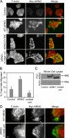
The coiled-coil domain of ARNO is necessary for the induction of epithelial motility. (A) MDCK cells were infected with adenoviruses encoding the indicated ARNO constructs for 3 h. Cells were then fixed and stained with mouse anti-myc followed by Alexa-488–conjugated anti-mouse antibody and rhodamine-phalloidin. Control cells were infected with adenovirus encoding WT ARNO in the presence of doxycycline to suppress transgene expression. Bar, 50 μm. (B) Motility of cells expressing the indicated constructs was tested using the transwell assay as described in Materials and Methods. The percent of cells migrating through the filter in 18 h are indicated. Data shown are mean ± SD of triplicate samples. (C) Expression levels of the myc-tagged ARNO constructs and actin in the cells subjected to the transwell assay shown in B were visualized by Western blot of saved cell samples with mouse anti-myc and mouse anti-actin antibodies. (D) MDCK cells were processed for immunofluorescence as in A. Bar, 25 μM.
Figure 3.
Figure 3.
ARNO lacking the coiled-coil domain is deficient at inducing Rac activation. MDCK cells were infected with adenovirus encoding indicated ARNO constructs in the absence of doxycycline or with WT ARNO in the presence of doxycycline (control) for 3 h. Cells were then lysed and active ARF6 or Rac isolated by pulldown as described in Materials and Methods. (A and B) Activation of ARF 6 (A) or Rac1 (B) in cells expressing full-length or Δcoiled-coil ARNO. Data shown are mean ± SE of eight independent experiments. Data were analyzed for statistically significant differences using a paired t test. *p < 0.05; **p < 0.01; n.s.; not significant. (C) Representative gels from the pulldown experiments presented in A and B.
Figure 4.
Figure 4.
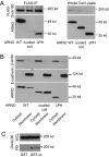
The coiled-coil domain of ARNO is required for interaction with Dock180. (A) MDCK cells expressing flag-Dock180 and the indicated myc-tagged ARNO constructs were cross-linked, lysed, and incubated with M2 anti-flag resin as described in Materials and Methods. The immunoprecipitates were blotted with goat anti-Dock180 and rabbit anti-ARNO. Saved samples of the starting lysate were blotted with goat anti-Dock180 and mouse anti-myc antibodies. (B) Subcellular location of full-length ARNO and ARNO truncation mutants. MDCK cells were infected adenovirus encoding the indicated myc-tagged ARNO constructs for 3 h. Cells were then lysed and separated into membrane and cytosol fractions by ultracentrifugation as described in Materials and Methods. Equal proportions of the membrane and cytosol fractions were Western-blotted with mouse anti E-cadherin (membrane marker), mouse anti B-actin (cytosol marker), and mouse anti-myc to detect the location of the ARNO proteins. (C) Dock180 can be isolated from a cell lysate by the ARNO coiled-coil domain. MDCK cells expressing Dock180 were lysed and the postnuclear supernatant incubated with glutathione-Sepharose and either 30 μg of GST or 30 μg of GST fused to the ARNO coiled-coil domain. Starting lysates (WCL) and the isolated pulldown samples (PD) were blotted with goat anti-Dock180.
Figure 5.
Figure 5.
The N-terminus of Dock180 is required for interaction with ARNO. (A) Dock180 truncation constructs used in these experiments. All constructs are Flag-tagged at the N-terminus. (B) MDCK cells were cotransfected with the indicated Dock180 and pTRE-ARNO constructs in the presence of 2 ng/ml doxycycline to repress ARNO expression. After 18 h the doxycycline was removed, and ARNO allowed to express for 4 h. Cells were then treated with cross-linker, lysed, and incubated with M2 anti-flag as described in Materials and Methods. The immunoprecipitates were blotted with rabbit anti-ARNO and goat anti-Dock180 antibodies. A mixture of Dock180 antibodies directed against both N- and C-terminal epitopes was used to allow visualization of all truncations. (C) MDCK cells were infected with adenoviruses encoding Dock180, ARNO, and the indicated Elmo constructs for 4 h (WT Elmo) or for 18 h in the presence of 1 ng/ml doxycycline to moderate expression (T625). Cells were cross-linked, lysed, and immunoprecipitated as above. Immunoprecipitates and aliquots of the starting lysate were blotted with goat anti-Dock180, rabbit anti-ARNO, and rabbit anti-GFP.
Figure 6.
Figure 6.
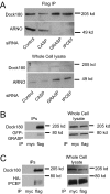
Knockdown of GRASP or IPCEF impairs interaction of ARNO and Dock180. (A) MCF-7 cells were transfected with siRNAs targeting firefly luciferase (control) or the indicated proteins as described in Materials and Methods. After 48 h the cells were then infected with adenoviruses encoding ARNO and Dock180 for 18 h. Cells were then cross-linked, lysed, and incubated with M2 anti-flag resin as described in Materials and Methods. The immunoprecipitates were blotted with goat anti-Dock180 and rabbit anti-ARNO. Samples of the starting lysates were blotted with goat anti-Dock180 and mouse anti-myc. (B and C) GRASP (B) and IPCEF (C) are coimmunoprecipitated with Dock180. MDCK cells were infected with adenoviruses encoding flag-Dock180 and either GFP-GRASP or HA-IPCEF for 18 h. Cells were treated with cross-linker, lysed, and incubated with mouse anti-myc or M2 anti-flag as indicated. Immunoprecipitates and samples of starting lysates were Western-blotted with goat anti-Dock180 and mouse anti-GFP or mouse anti-HA.
Figure 7.
Figure 7.
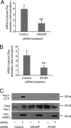
Knockdown of GRASP or IPCEF impairs ARNO-induced Rac activation. MDCK cells were transfected with the indicated siRNAs for 48 h. The cells were then split onto duplicate plates. After 18 h of recovery they were infected with adenovirus encoding ARNO in the presence or absence of doxycycline to repress transgene expression. After 3 h of expression cells were lysed, and active Rac was isolated by pulldown as described in Materials and Methods. Levels of active GTPase were normalized to the level of total GTPase in the starting lysate. (A and B) Activation of Rac by ARNO is impaired by knockdown of GRASP (A) or IPCEF (B). Data shown are the mean ± SE of the fold activation of Rac in ARNO-expressing cells compared with control cells. The effect of knockdown on ARNO-induced Rac activation was analyzed using a paired t test on multiple independent knockdown experiments. **p < 0.01, n = 14 (GRASP knockdowns) or n = 12 (IPCEF knockdowns). (C) Representative gel from the pulldown experiments quantified in A and B.
Figure 8.
Figure 8.
Knockdown of GRASP or IPCEF prevents ARNO from scattering MDCK cells. MDCK cells were transfected with 80 pmol of the indicated siRNA plus 20 pmol of Block-It Alexafluor red, fluorescent control siRNA, as a transfection marker. Forty-eight hours after transfection cells were replated onto coverslips and infected with adenovirus encoding ARNO in the presence of doxycycline to prevent transgene expression. After 18 h of recovery the doxycycline was removed, and the cells were allowed to express ARNO for 3 h. Cells were then fixed and stained with mouse anti-ARNO followed by Alexa-488–conjugated anti-mouse secondary antibody and Alexa-647–conjugated phalloidin. In the merge ARNO is psuedocolored green, f-actin is psuedocolored red, and the fluorescent siRNA is psuedocolored blue. Bar, 25 μM.
Figure 9.
Figure 9.
Model for the duel role of GRASP and IPCEF in promoting ARF-to-Rac cross-talk. See Discussion for details.

Similar articles

Cited by

References

    1. Abramoff M. D., Magelhaes P. J., Ram S. J. Image processing with Image. J. Biophotonics Int. 2004;11:36–42.
    1. Balasubramanian N., Scott D. W., Castle J. D., Casanova J. E., Schwartz M. A. Arf6 and microtubules in adhesion-dependent trafficking of lipid rafts. Nat. Cell Biol. 2007;9:1381–1391. - PMC - PubMed
    1. Beemiller P., Hoppe A. D., Swanson J. A. A phosphatidylinositol-3-kinase-dependent signal transition regulates ARF1 and ARF6 during Fcgamma receptor-mediated phagocytosis. PLoS Biol. 2006;4:e162. - PMC - PubMed
    1. Boehm T., et al. Attenuation of cell adhesion in lymphocytes is regulated by CYTIP, a protein which mediates signal complex sequestration. EMBO J. 2003;22:1014–1024. - PMC - PubMed
    1. Boshans R. L., Szanto S., van Aelst L., D'Souza-Schorey C. ADP-ribosylation factor 6 regulates actin cytoskeleton remodeling in coordination with Rac1 and RhoA. Mol. Cell. Biol. 2000;20:3685–3694. - PMC - PubMed

Publication types

MeSH terms

LinkOut - more resources