. 2010 Feb 19;285(8):5405-16.
doi: 10.1074/jbc.M109.041418. Epub 2009 Dec 11.

Protein kinase C zeta mediates cigarette smoke/aldehyde- and lipopolysaccharide-induced lung inflammation and histone modifications

Affiliations

Protein kinase C zeta mediates cigarette smoke/aldehyde- and lipopolysaccharide-induced lung inflammation and histone modifications

Hongwei Yao et al. J Biol Chem. .

Abstract

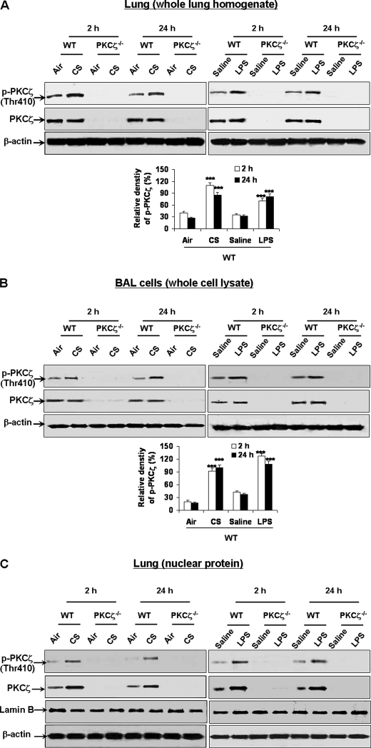
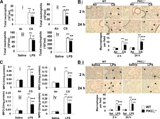
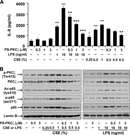
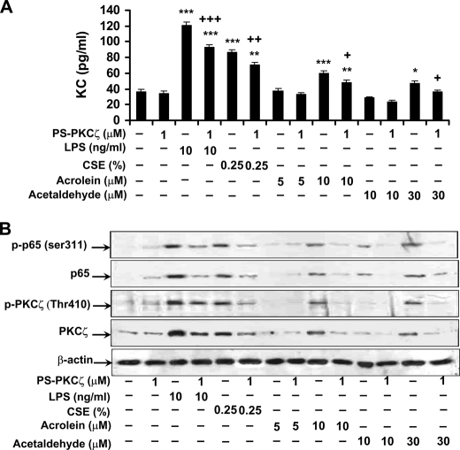
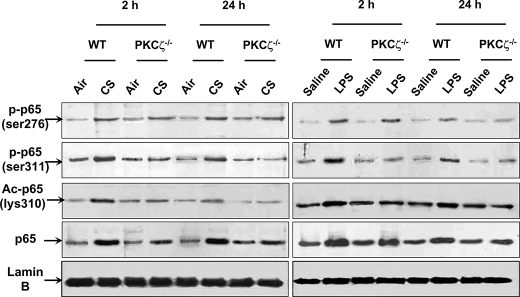
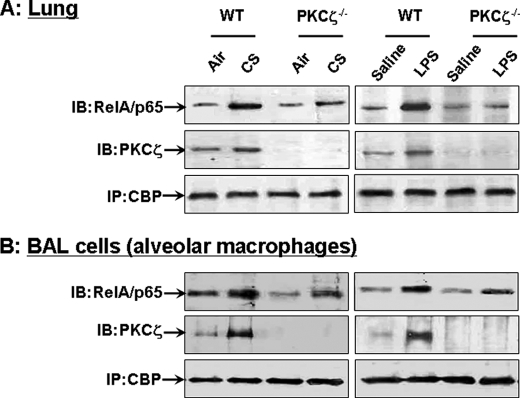
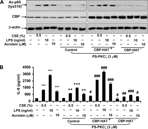
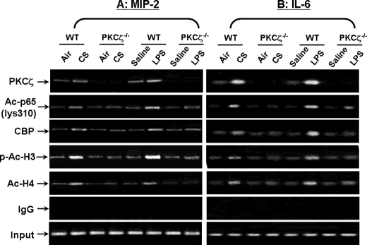
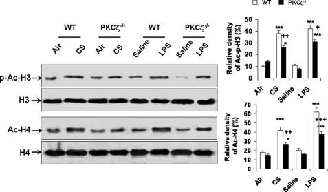
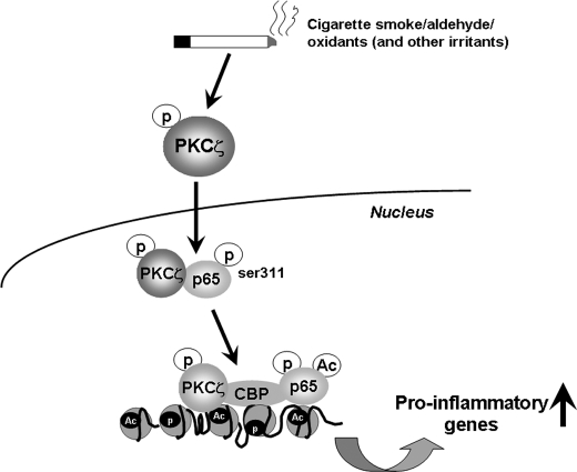
Atypical protein kinase C (PKC) zeta is an important regulator of inflammation through activation of the nuclear factor-kappaB (NF-kappaB) pathway. Chromatin remodeling on pro-inflammatory genes plays a pivotal role in cigarette smoke (CS)- and lipopolysaccharide (LPS)-induced abnormal lung inflammation. However, the signaling mechanism whereby chromatin remodeling occurs in CS- and LPS-induced lung inflammation is not known. We hypothesized that PKCzeta is an important regulator of chromatin remodeling, and down-regulation of PKCzeta ameliorates lung inflammation by CS and LPS exposures. We determined the role and molecular mechanism of PKCzeta in abnormal lung inflammatory response to CS and LPS exposures in PKCzeta-deficient (PKCzeta(-/-)) and wild-type mice. Lung inflammatory response was decreased in PKCzeta(-/-) mice compared with WT mice exposed to CS and LPS. Moreover, inhibition of PKCzeta by a specific pharmacological PKCzeta inhibitor attenuated CS extract-, reactive aldehydes (present in CS)-, and LPS-mediated pro-inflammatory mediator release from macrophages. The mechanism underlying these findings is associated with decreased RelA/p65 phosphorylation (Ser(311)) and translocation of the RelA/p65 subunit of NF-kappaB into the nucleus. Furthermore, CS/reactive aldehydes and LPS exposures led to activation and translocation of PKCzeta into the nucleus where it forms a complex with CREB-binding protein (CBP) and acetylated RelA/p65 causing histone phosphorylation and acetylation on promoters of pro-inflammatory genes. Taken together, these data suggest that PKCzeta plays an important role in CS/aldehyde- and LPS-induced lung inflammation through acetylation of RelA/p65 and histone modifications via CBP. These data provide new insights into the molecular mechanisms underlying the pathogenesis of chronic inflammatory lung diseases.


Figures

FIGURE 1.
FIGURE 1.
PKCζ was activated and translocated into nucleus in lungs of WT mice exposed to CS and LPS. WT and PKCζ-deficient mice were exposed to CS and LPS and were sacrificed at 2 and 24 h of post-last exposures. Protein levels of PKCζ and its phosphorylation on the Thr410 residue were analyzed by immunoblot in lung tissue (A) and BAL cell pellets (B) of whole cell lysates and in lung nuclear protein (C) of WT and PKCζ-deficient mice exposed to CS and LPS. PKCζ was activated and translocated into the nucleus in lungs and BAL cells of WT mice exposed to CS and LPS. There was no expression of PKCζ and its phosphorylated form in lungs and BAL cells of PKCζ-deficient mice. Blots are representative of three separate experiments. ***, p < 0.001, significant compared with respective air- or saline-exposed group.
FIGURE 2.
FIGURE 2.
Lung inflammation was decreased in PKCζ−/− mice in response to CS exposure and LPS aerosolization. Neutrophil influx in BAL fluid of mice was determined on cytospin-prepared slides stained with Diff-Quik (A). Immunohistochemical detection of macrophages was performed using rat anti-mouse Mac-3 antibody on mouse lung sections (B). Myeloperoxidase activity in lung tissue was determined spectrophotometrically (C). Number of neutrophils in BAL fluid (A) was decreased in PKCζ-deficient mice in response to CS exposure (panels i and ii) and LPS aerosolization (panels iii and iv) compared with respective WT mice at 2 h (panels i and iii) and 24 h (panels ii and iv) of post-last exposure. Deficiency of PKCζ decreased CS (panel i)- and LPS (panel ii)-induced macrophage influx into the lungs (B) at both the 2- and 24-h time points. PKCζ-deficient mice showed decreased myeloperoxidase activity in lung (C) at both 2-h (panels i and iii) and 24-h (panels ii and iv) time points as compared with WT mice exposed to CS (panels i and ii) and LPS (panels iii and iv). Data are shown as mean ± S.E. (n = 4 to 5 per group). **, p < 0.01; and ***, p < 0.001, significant compared with respective air- or saline-exposed groups; +, p < 0.05; ++, p < 0.01; and +++, p < 0.001, significant compared with respective CS- or LPS-exposed WT mice. Immunohistochemical pictures represent three separate experiments. Arrows indicate macrophages. Original magnification, ×200.
FIGURE 3.
FIGURE 3.
Inhibition of PKCζ-attenuated CSE- and LPS-induced IL-8 release and NF-κB activation in MonoMac6 cells. MonoMac6 cells were pretreated with PKCζ inhibitor, PS-PKCζ, for 2 h prior to CSE (0.25, 0.5, and 1%) or LPS (1 and 10 ng/ml) treatments for 12 h. Pretreatment with PS-PKCζ (0.3–3 μm) significantly decreased CSE- and LPS-induced release of IL-8 (A) and PKCζ activation (B) in MonoMac6 cells. CSE- and LPS-induced phosphorylated (Ser311), acetylated (Lys310), and total levels of RelA/p65 were also significantly reduced in the nucleus of MonoMac6 cells pretreated with PS-PKCζ (1 and 3 μm) (B). Data are shown as mean ± S.E. (n = 4–5 per group). Each lane represents results of three independent experiments. *, p < 0.05; ***, p < 0.001, significant compared with control group; ++, p < 0.01; +++, p < 0.001, significant as compared with respective CSE (0.5%)- or LPS (10 ng/ml)-treated group. p-PKCζ (Thr410), phosphorylated PKCζ on Thr410; Ac-p65 (lys310), acetylated RelA/p65 on Lys310; p-p65 (ser311), phosphorylated RelA/p65 on Ser311; p65, RelA/p65.
FIGURE 4.
FIGURE 4.
PKCζ inhibitor pretreatment decreased KC release and RelA/p65 phosphorylation induced by CSE, LPS, acrolein, and acetaldehyde in primary mouse alveolar macrophages. Alveolar macrophages were isolated from C57BL/6J mice and were pretreated with PS-PKCζ for 2 h prior to CSE (0.25%), LPS (10 ng/ml), acrolein (5 and 10 μm), or acetaldehyde (10 and 30 μm) treatments for 12 h. Pretreatment with PS-PKCζ (1 μm) significantly decreased CSE-, LPS-, acrolein-, and acetaldehyde-induced KC release from mouse alveolar macrophages (A). Nuclear levels of phosphorylated RelA/p65 (Ser311) and PKCζ (Thr410) and total levels of RelA/p65 and PKCζ were decreased by PS-PKCζ (1 μm) pretreatment in primary mouse alveolar macrophages exposed to CSE, LPS, acrolein, and acetaldehyde (B). Data are shown as mean ± S.E. (n = 3–4 per group). Each lane represents results of three independent experiments. *, p < 0.05; **, p < 0.01; ***, p < 0.001; significant compared with control groups; +, p < 0.01; ++, p < 0.01; +++, p < 0.001, significant as compared with respective CSE-, LPS-, acrolein (10 μm)- and acetaldehyde (30 μm)-treated groups. p-PKCζ (Thr410), phosphorylated PKCζ on Thr410; p-p65 (ser311), phosphorylated RelA/p65 on Ser311; p65, RelA/p65.
FIGURE 5.
FIGURE 5.
Phosphorylated, acetylated, and total levels of RelA/p65 were decreased in lung nuclear proteins of PKCζ−/− mice in response to CS and LPS exposure. Levels of phosphorylated (Ser276 and Ser311 residues), acetylated (Lys310), and total RelA/p65 in lung nuclear proteins were analyzed by immunoblotting. Lamin B was used as a nuclear marker, and no bands of α-tubulin were observed in nuclear fractions. Levels of phosphorylated RelA/p65 on Ser311 was decreased, whereas no alteration in phosphorylated RelA/p65 on Ser276 was observed in lungs of PKCζ-deficient mice as compared with WT mice exposed to CS and LPS at 2 and 24 h of post-last exposures. Knock-out of PKCζ decreased the levels of acetylated (Lys310) and total RelA/p65 in lung nuclear protein of mice exposed to CS and LPS. Each lane represents results of three independent experiments. p-p65 (ser276), phosphorylated RelA/p65 on Ser276; p-p65 (ser311), phosphorylated RelA/p65 on Ser311; Ac-p65 (lys310), acetylated RelA/p65 at Lys310; p65, RelA/p65.
FIGURE 6.
FIGURE 6.
Increased interaction of CBP with PKCζ in lungs and BAL cells of CS- and LPS-exposed WT mice, and disruption of PKCζ decreased the interaction of CBP with RelA/p65 in lungs and BAL cells of mice exposed to CS and LPS. The interaction of CBP with PKCζ and RelA/p65 in lungs (A) and BAL cells (B) was analyzed using immunoprecipitation (IP) assay. Both CS and LPS exposures increased the interaction of CBP with PKCζ in lungs and BAL cells of WT mice after 2 h post-last exposure. Disruption of PKCζ decreased the interaction of CBP with RelA/p65 induced by CS and LPS exposures in lungs and BAL cells at 2 h post-last exposure. Gel pictures shown are representative of at least three separate experiments. IB, immunoblot.
FIGURE 7.
FIGURE 7.
CSE and LPS induced the interaction of CBP with PKCζ and RelA/p65, which was decreased in MonoMac6 cells pretreated with PS-PKCζ. CSE- and LPS-induced interaction of CBP with PKCζ and RelA/p65 in MonoMac6 cells in the absence or presence of PS-PKCζ was assessed using immunoprecipitation (IP) assay. The interaction of CBP with PKCζ and RelA/p65 was increased in response to CSE (0.5 and 1%) and LPS (1 and 10 ng/ml) treatments for 12 h in MonoMac6 cells. Pretreatment of PS-PKCζ (1 and 3 μm for 2 h) attenuated CSE- and LPS-mediated interaction of CBP with PKCζ and RelA/p65 in MonoMac6 cells. Gel pictures shown are representative of at least three separate experiments. IB, immunoblot.
FIGURE 8.
FIGURE 8.
CBP was required for PKCζ-mediated RelA/p65 acetylation and IL-8 release in MonoMac6 cells in response to CSE, acrolein, and LPS treatments. MonoMac6 cells were transfected with WT CBP or CBP-HAT mutants for 24 h before PKCζ inhibitor pretreatment and subsequent CSE, acrolein, and LPS exposures. HAT activity of CBP was required for PKCζ-mediated RelA/p65 acetylation (A) and IL-8 release (B) induced by CSE, acrolein, and LPS in MonoMac6 cells. Gel pictures shown are representative of at least three separate experiments. Data are shown as mean ± S.E. (n = 3–4 per group). ***, p < 0.001, significant compared with control groups; ++, p < 0.01; +++, p < 0.001, significant as compared with respective CSE-, LPS-, and acrolein-treated groups; #, p < 0.05; ##, p < 0.01; ###, p < 0.001, significant as compared with respective PS-PKCζ treated but without transfected groups.
FIGURE 9.
FIGURE 9.
PKCζ recruited CBP and RelA/p65 on pro-inflammatory gene promoters in mouse lungs in response to CS and LPS exposures. The recruitment of PKCζ, acetylated RelA/p65 (Lys310), CBP, phosphorylated/acetylated histone H3 (Ser10/Lys9), and acetylated histone H4 on the promoters mip-2 and il-6 in mouse lung was analyzed by ChIP assay. Lung homogenates were cross-linked and immunoprecipitated with relevant antibodies against the aforementioned proteins, and then chromatin modification on the promoter regions of mip-2 (A) and il-6 (B) was detected by PCR. Both CS and LPS exposures caused the recruitment of acetylated RelA/p65 (Lys310), CBP, phosphorylated/acetylated histone H3 (Ser10/Lys9), and acetylated histone H4 on the promoters mip-2 and il-6 in mouse lung, which was attenuated in PKCζ-deficient mice at 2 h of post-last exposures. Furthermore, PKCζ was also recruited on the promoters of mip-2 and il-6 in lungs of WT mice, but not in PKCζ deficient mice exposed to CS and LPS. IgG was used as a negative control. Gel pictures shown are representative of at least three separate experiments. Ac-p65 (lys310), acetylated RelA/p65 on Lys310; p-Ac-H3, phosphorylated/acetylated histone H3 at Ser10/Lys9; Ac-H4, acetylated histone H4.
FIGURE 10.
FIGURE 10.
Disruption of PKCζ decreased the phosphorylation/acetylation of histone H3 and acetylation of histone H4 in mouse lung exposed to CS and LPS. Acid-extracted histone proteins in lungs of WT and PKCζ-deficient mice were used for immunoblotting against anti-phosphorylated/acetylated (Ser10/Lys9) histone H3 and acetylated histone H4. Levels of phosphorylated/acetylated (Ser10/Lys9) histone H3 and acetylated histone H4 were decreased in lungs of PKCζ−/− mice as compared with WT mice exposed to CS and LPS at 2 h of post-last exposure. Gel pictures shown are representative of at least three separate experiments. *, p < 0.05; ***, p < 0.001, significant compared with respective air- or saline-exposed groups; +, p < 0.05; ++, p < 0.01; +++, p < 0.001, significant compared with respective CS- and LPS-exposed WT mice. p-Ac-H3, phosphorylated/acetylated histone H3 on Ser10/Lys9; H3, histone H3; Ac-H4, acetylated histone H4; H4, histone H4.
FIGURE 11.
FIGURE 11.
Summarized schematic model of the regulation of PKCζ in CS/aldehyde- and LPS-induced lung inflammatory response and chromatin remodeling. PKCζ was activated and translocated into the nucleus in response to CS and LPS exposures. Activated PKCζ interacted and phosphorylated RelA/p65 on Ser311, leading to increased NF-κB transactivation. Furthermore, PKCζ functions as a master regulator and facilitates the assembly of RelA/p65 with coactivator CBP in transcriptional machinery, thereby increasing acetylation of RelA/p65 and histones on the promoters of pro-inflammatory genes in response to CS and LPS exposures in lungs.

Similar articles

Cited by

References

    1. Yang S. R., Valvo S., Yao H., Kode A., Rajendrasozhan S., Edirisinghe I., Caito S., Adenuga D., Henry R., Fromm G., Maggirwar S., Li J. D., Bulger M., Rahman I. (2008) Am. J. Respir. Cell Mol. Biol. 38, 689–698 - PMC - PubMed
    1. Yang S. R., Wright J., Bauter M., Seweryniak K., Kode A., Rahman I. (2007) Am. J. Physiol. Lung Cell Mol. Physiol. 292, L567–L576 - PubMed
    1. Yao H., Edirisinghe I., Rajendrasozhan S., Yang S. R., Caito S., Adenuga D., Rahman I. (2008) Am. J. Physiol. Lung Cell Mol. Physiol. 294, L1174–L1186 - PubMed
    1. Yao H., Yang S. R., Edirisinghe I., Rajendrasozhan S., Caito S., Adenuga D., O'Reilly M. A., Rahman I. (2008) Am. J. Respir. Cell Mol. Biol. 39, 7–18 - PMC - PubMed
    1. Hogg J. C., Chu F., Utokaparch S., Woods R., Elliott W. M., Buzatu L., Cherniack R. M., Rogers R. M., Sciurba F. C., Coxson H. O., Paré P. D. (2004) N. Engl. J. Med. 350, 2645–2653 - PubMed

Publication types

MeSH terms