. 2010 Feb;84(4):1752-63.
doi: 10.1128/JVI.01758-09. Epub 2009 Dec 9.

The murine coronavirus nucleocapsid gene is a determinant of virulence

Affiliations

The murine coronavirus nucleocapsid gene is a determinant of virulence

Timothy J Cowley et al. J Virol. 2010 Feb.

Abstract

The murine coronavirus, mouse hepatitis virus (MHV) strain A59, causes acute encephalitis and chronic demyelinating disease as well as hepatitis in mice. The JHM strain (also called MHV-4 or JHM.SD) causes fatal encephalitis and only minimal hepatitis. Previous analysis of chimeric recombinant MHVs in which the spike gene, encoding the protein that mediates viral entry and cell-to-cell fusion, was exchanged between JHM and A59 showed that the spike plays a major role in determining organ tropism and neurovirulence but that other genes also play important roles in pathogenic outcome. Here, we have investigated the role of the nucleocapsid protein in MHV-induced disease. The multifunctional nucleocapsid protein is complexed with the genomic RNA, interacts with the viral membrane protein during virion assembly, and plays an import role in enhancing the efficiency of transcription. A pair of chimeric recombinant viruses in which the nucleocapsid gene was exchanged between JHM and A59 was selected and compared to wild-type parental strains in terms of virulence. Importantly, expression of the JHM nucleocapsid in the context of the A59 genome conferred increased mortality and spread of viral antigen in the mouse central nervous system compared to the parental A59 strain, while having little effect on the induction of hepatitis. While the JHM nucleocapsid did not appear to enhance neuron-to-neuron spread in primary neuronal cultures, the increased neurovirulence it conferred may be due in part to the induction of a less robust T-cell response than that induced by strain A59.


Figures

FIG. 1.
FIG. 1.
Schematic representation of the virions and corresponding genomes of recombinant viruses. Shown on the left are A59 background viruses and, on the right, the JHM background viruses. A59 components are in white, and JHM components are in black. The positions of the spike (S) and nucleocapsid (N) genes are noted. The 5′ replicase gene is not shown to scale, as indicated by hash marks.
FIG. 2.
FIG. 2.
Replication of recombinant viruses. L2 cells were infected (in duplicate) at an MOI of 1 with wild-type (A) and N exchange viruses or with S exchange viruses with and without N exchange (B). At the times indicated, virus was harvested from combined supernatants and cells, and titers were determined on L2 cell monolayers as described in Materials and Methods. Duplicate samples were averaged. The data are from one representative experiment of two.
FIG. 3.
FIG. 3.
Survival curves of infected 4-week-old C57BL/6 mice. Mice were infected with recombinant viruses at the doses indicated and observed over 21 days for mortality. (A) Infection with rA59 (500 PFU and 5 × 10³ PFU) and rA59/NJHM (10 PFU, 100 PFU, and 1 × 10³ PFU). rA59/NJHM at a dose of 100 PFU is significantly more virulent than rA59 at a dose of 500 (n = 5 each; P = 0.0155). (B) Infection with rJHM and rJHM/NA59 at 10 PFU. rJHM/NA59 is significantly less virulent than rJHM (n = 10 and 5, respectively; P = 0.0272). (C) Infection with rJHM, rA59/NJHM, rA59/SJHM, and rA59/SJHM/NJHM (10 PFU). rA59/SJHM/NJHM is more virulent than rA59/NJHM (n = 10 each; P = 0.0030) and rA59/SJHM (n = 10 and 9, respectively; P = 0.0016), and rJHM is more virulent than rA59/SJHM/NJHM (n = 10 each; P = 0.0319). (D) Infection with rJHM/SA59/NA59 (3 × 10⁵ PFU) and rJHM/SA59 (3 × 10³ PFU, 3 × 10⁴ PFU, and 3 × 10⁵ PFU). rJHM/SA59 is more virulent than rJHM/SA59/NA59 (n = 5 each; P = 0.0041). The data are from one representative experiment of two or more.
FIG. 4.
FIG. 4.
Replication of chimeric viruses in the brain. C57BL/6 mice (four or more per virus) were inoculated i.c. with 50 PFU of the indicated viruses. At 5 days postinfection mice were sacrificed, and virus titers from brain lysates were determined by plaque assay on L2 cells. Symbols represent individual animals, and the lines represent the mean and the standard error. *, P = 0.01; **, P = 0.0001. Data are from one representative experiment of two or more.
FIG. 5.
FIG. 5.
Viral antigen expression in the brains of infected mice. C57BL/6 mice were inoculated i.c. with 50 PFU of virus and sacrificed at 5 days postinfection. Brains were harvested, sectioned sagitally, and stained for viral antigen expression with anti-N MAb as described in Materials Methods. (A) Frame i, rA59; frame ii, rJHM; frame iii, rA59/NJHM; frame iv, rA59/SJHM; frame v, rA59/SJHM//NJHM. (B) Frame i, rA59; frame ii, JHM; frame iii, rJHM/NA59; frame iv, rJHM/SA59; frame v, rJHM/SA59/NA59. Panels C (A59 background viruses from panel A) and D (JHM background viruses from panel B) show quantification of antigen staining using the color-cubed segmentation function of Image Pro, version 5.0, software. The y axis represents the area of antigen stain relative to rJHM, which is set to 100. Data shown represent the mean and standard error and are from one representative of two or more experiments. *, P < 0.05; **, P ≤ 0.0001; #, P = 0.08.
FIG. 6.
FIG. 6.
Virus spread in primary neuronal cultures. Neuronal cultures were infected with recombinant viruses as indicated at an MOI of 1 and were fixed and stained with anti-N MAb using immunoperoxidase at the times postinfection as indicated. (A) frame i, rA59 at 2 days postinfection (dpi) (magnification, ×20); frame ii, rA59 at 4 dpi (×20); frame iii, rA59/NJHM at 2 dpi (×20); frame iv, rA59/NJHM at 4 dpi (×20). (B) Frame i, rJHM at 1 dpi (×4); frame ii, rJHM at 3 dpi (×4); frame iii, rJHM/NA59 at 1 dpi (×4); frame iv, rJHM/NA59 at 3 dpi (×4). (C and D) Quantification of neuronal spread for A59 background viruses (C) and JHM background viruses (D) using the color-cubed segmentation function of Image Pro, version 5.0, software. The y axis shows arbitrary units that represent the average area of infection foci; differences in scales between panels C and D are due to differences in magnifications of analyzed photos. Data shown represent the mean and standard error and are from one representative experiment of five.
FIG. 7.
FIG. 7.
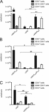
T-cell response to recombinant viruses in the brain. Mice were infected with recombinant viruses and sacrificed at 7 days postinfection. Mononuclear cells were isolated from the brains of infected animals, stained with T-cell type-specific antibodies and either S510-specific tetramers (A and B) or assayed for secretion of IFN-γ in response to S598 peptide (C), and analyzed by flow cytometry as described in Materials and Methods. The total number of each T-cell population in the brain was determined by multiplying the percentage of each cell type by the total number of cells isolated per brain. Shown are the means and standard errors for the total CD8 T, epitope-specific CD8 T, and CD4 T cells in the brains of mice infected with rJHM/NA59, rA59/SJHM, and rJHM (A); rA59/SJHM/NJHM, rA59/SJHM, and rJHM (B); and rA59 and rA59/NJHM (C). *, P < 0.05; **, P ≤ 0.0005; #, P = 0.15; ##, P ≤ 0.09. Each panel is representative of at least two experiments.
FIG. 8.
FIG. 8.
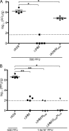
Replication of chimeric viruses in the liver. C57BL/6 mice were inoculated intrahepatically with viruses at the doses as indicated. Mice were sacrificed at 5 days postinfection, and virus titers were determined from liver lysates. (A) rA59, rJHM, and rA59/NJHM (500 PFU). (B) rA59 (500 PFU) and rJHM, rJHM/NA59, and rJHM/SA59/NA59 (1.6 × 10⁴ PFU). Symbols represent individual animals, and the lines represent the mean and the standard error. The dotted lines represent the limits of detection. These data are from one representative experiment of two or more. *, P < 0.02; **, P < 0.0001.
FIG. 9.
FIG. 9.
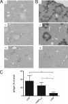
Pathology and viral antigen expression in the livers of mice infected with chimeric viruses. C57BL/6 mice were inoculated intrahepatically with 500 PFU of rA59 and rA59/NJHM and 1.8 × 10⁴ PFU of rJHM. Mice were sacrificed at 5 days postinfection. Livers were harvested, sectioned, and stained either for pathology or viral antigen expression. (A) Hematoxylin- and eosin-stained sections: frame I, rA59; frame ii, rA59/NJHM; frame iii, rJHM. (B) Immunoperoxidase-stained sections using anti-N MAb: frame i, rA59; frame ii, rA59/NJHM; frame iii, rJHM. (C) Quantification of antigen staining using the color cube-based segmentation function of Image Pro, version 5.0, software. The y axis represents the area of antigen stain over total area of the liver section, and values are shown as mean and standard error. The data shown in all panels are representative of four or five mice per group; two adjacent sections were stained per animal. *, P < 0.05.

Similar articles

Cited by

References

    1. Armstrong, J., S. Smeekens, and P. Rottier. 1983. Sequence of the nucleocapsid gene from murine coronavirus MHV-A59. Nucleic Acids Res. 11:883-891. - PMC - PubMed
    1. Baric, R. S., G. W. Nelson, J. O. Fleming, R. J. Deans, J. G. Keck, N. Casteel, and S. A. Stohlman. 1988. Interactions between coronavirus nucleocapsid protein and viral RNAs: implications for viral transcription. J. Virol. 62:4280-4287. - PMC - PubMed
    1. Batts, K. P., and J. Ludwig. 1995. Chronic hepatitis. An update on terminology and reporting. Am. J. Surg. Pathol. 19:1409-1417. - PubMed
    1. Cervantes-Barragan, L., R. Zust, F. Weber, M. Spiegel, K. S. Lang, S. Akira, V. Thiel, and B. Ludewig. 2007. Control of coronavirus infection through plasmacytoid dendritic-cell-derived type I interferon. Blood 109:1131-1137. - PMC - PubMed
    1. Dalziel, R. G., P. W. Lampert, P. J. Talbot, and M. J. Buchmeier. 1986. Site-specific alteration of murine hepatitis virus type 4 peplomer glycoprotein E2 results in reduced neurovirulence. J. Virol. 59:463-471. - PMC - PubMed

Publication types

MeSH terms

Substances

LinkOut - more resources