. 2010 Feb;84(4):1764-70.
doi: 10.1128/JVI.01682-09. Epub 2009 Dec 9.

A rotavirus spike protein conformational intermediate binds lipid bilayers

Affiliations

A rotavirus spike protein conformational intermediate binds lipid bilayers

Shane D Trask et al. J Virol. 2010 Feb.

Abstract

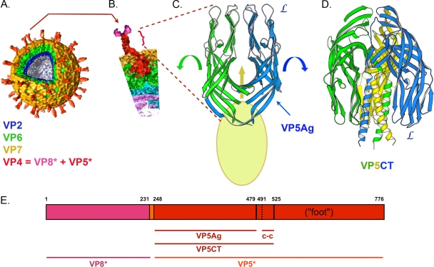
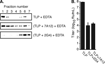
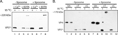
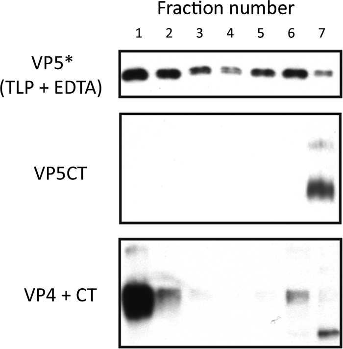
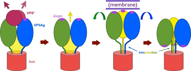
During rotavirus entry, a virion penetrates a host cell membrane, sheds its outer capsid proteins, and releases a transcriptionally active subviral particle into the cytoplasm. VP5, the rotavirus protein believed to interact with the membrane bilayer, is a tryptic cleavage product of the outer capsid spike protein, VP4. When a rotavirus particle uncoats, VP5 folds back, in a rearrangement that resembles the fusogenic conformational changes in enveloped-virus fusion proteins. We present direct experimental evidence that this rearrangement leads to membrane binding. VP5 does not associate with liposomes when mounted as part of the trypsin-primed spikes on intact virions, nor does it do so after it has folded back into a stably trimeric, low-energy state. But it does bind liposomes when they are added to virions before uncoating, and VP5 rearrangement is then triggered by addition of EDTA. The presence of liposomes during the rearrangement enhances the otherwise inefficient VP5 conformational change. A VP5 fragment, VP5CT, produced from monomeric recombinant VP4 by successive treatments with chymotrypsin and trypsin, also binds liposomes only when the proteolysis proceeds in their presence. A monoclonal antibody that neutralizes infectivity by blocking a postattachment entry event also blocks VP5 liposome association. We propose that VP5 binds lipid bilayers in an intermediate conformational state, analogous to the extended intermediate conformation of enveloped-virus fusion proteins.


Figures

FIG. 1.
FIG. 1.
Conformational states of rotavirus VP4. (A) Multilayered structure of a rotavirus particle, based on cryo-EM three-dimensional image reconstructions (4, 15). Color coding for the structural proteins is indicated by labels. (B) Detail of a VP4 spike. The VP5* fragment is in bold red; the VP8* fragment is in pink. (C) Structure of the dimer-clustered, projecting portion of VP5*, as determined by comparison of the crystal structure of the VP5Ag fragment (PDB accession code 2B4H) and cryo-EM images (9, 26). The presence of a third VP5Ag domain is indicated by the yellow oval. “L” labels the hydrophobic loops at the apex of the VP5Ag domain. Arrows suggest the transition to the folded-back conformation. (D) Folded-back structure of VP5CT (9) (PDB accession code 1SLQ). This conformation probably represents the final, post-membrane-penetration state of VP5*. A “foot” would be appended to each of the α-helices in the central coiled coil. (E) Position in the RRV VP4 polypeptide chain of various proteolytic fragments. Numbers above the bar are the initial and final residues in each segment. c-c, coiled-coil.
FIG. 2.
FIG. 2.
Interaction of rotavirus proteins with liposomes. Virions were incubated in 1 mM CaCl2 or in 1 mM EDTA in the presence or absence of liposomes, and the mixtures were analyzed on sucrose gradients (25% to 70% [wt/vol] in HN buffer). (A to D) Immunoblots of fractions from top (1) to bottom (10) of the gradients, using HS2 to detect VP5*, m60 to detect VP7, and guinea-pig anti-VP6 (1:5,000 dilution) to detect VP6 (DLPs). Refractive index measurements (not shown) for the four gradients in panels A to D confirmed that all four had indistinguishable density profiles.
FIG. 3.
FIG. 3.
Requirement of VP4 cleavage for membrane association. (A) Virions and recoated particles were treated as for Fig. 2 and analyzed on discontinuous sucrose gradients (5% to 40% [wt/vol]). Immunoblots with HS2 were used to detected VP5* in gradient fractions from top (1) to bottom (7). (B) Infectivity of recoated particles following treatment with trypsin (T), chymotrypsin (C), or chymotrypsin followed by trypsin (CT), normalized to trypsin-treated recoated particles as 100%. Uncl, mock digested.
FIG. 4.
FIG. 4.
Liposome association of VP5* requires release in their presence. In each of the three experiments shown, the two components indicated in parentheses were mixed first, followed by addition of the third component 30 min later. Samples were then incubated for an additional 30 min (1 h total) before density gradient separation. Immunoblots with HS2 detected the positions of VP5* in fractions from top (1) to bottom (7) of discontinuous sucrose gradients (5% to 40% [wt/vol]).
FIG. 5.
FIG. 5.
MAb 2G4 blocks VP5* interaction with liposomes. (A) TLPs, either mock incubated or incubated with MAb 2G4 or 7A12, were uncoated in the presence of liposomes by the addition of 1 mM EDTA and separated over a discontinuous sucrose gradient. The fractions from top (1) to bottom (7) were heated to 95°C, analyzed by SDS-PAGE, and immunoblotted with horseradish peroxidase-conjugated HS2. (B) Neutralization of virus by the MAbs was determined by infectious focus assay. Error bars represent standard deviations of three titrations.
FIG. 6.
FIG. 6.
Anomalous migration of VP5*. (A) Uncoating experiment. Virions were uncoated (by addition of 1 mM EDTA) in the presence or absence of liposomes, with addition of 1 mM CaCl2 as a control. The products were analyzed by SDS-PAGE and immunoblotting with HS2, with (+) or without (−) heating of the samples at 95°C for 10 min. When virions (lanes 2 and 6) are not uncoated by addition of EDTA or by heating in SDS-PAGE sample buffer, VP5* is not detected, presumably because it remains on intact virions and does not enter the gel. (B) Importance of VP4 proteolytic cleavage for SDS-resistant trimer formation. VP4 on recoated DLPs was cleaved with trypsin (T) or chymotrypsin (C) or left uncleaved (Un), and the particles were then uncoated with 1 mM EDTA in the presence or absence of liposomes. The products were analyzed by SDS-PAGE and immunoblotting with HS2, with (+) or without (−) heating of the samples at 95°C for 10 min. The slowly migrating VP5* band (lane 10) is seen only in unheated samples following conventional tryptic cleavage. In panel A (compare lanes 4 and 8) and panel B (compare lanes 4 and 10), the presence of liposomes enhances the yield of the SDS-resistant species.
FIG. 7.
FIG. 7.
Association of VP5CT with liposomes, analyzed by immunoblotting of sucrose gradient fractions that have been separated by SDS-PAGE. Top panel: virions were uncoated with EDTA in the presence of liposomes before analysis. VP5* was detected by HS2 immunoblotting (as a standard). Middle panel: addition of purified VP5CT to liposomes, followed by analysis. Bottom panel: digestion of VP4 successively with chymotrypsin and trypsin in the presence of liposomes before analysis. Samples in middle and bottom panels were not heated and were probed with MAb 4D8.
FIG. 8.
FIG. 8.
Model for membrane association of VP5*. The sequence of conformational rearrangements in the diagram begins with dissociation of VP8*, followed by formation of a transient, extended intermediate. The coiled coil (see Fig. 1) may form at this stage. The surface presented by the exposed hydrophobic loops allows the extended trimer to bind firmly to a membrane. This interaction enhances the yield of SDS-resistant VP5* trimers in our experiments. Folding back to the stable state represented by the VP5CT crystal structure (Fig. 1D)—the process proposed to drive membrane disruption (9)—results in a trimer conformation that can no longer associate with liposomes.

Similar articles

Cited by

References

    1. Aoki, S. T., E. C. Settembre, S. D. Trask, H. B. Greenberg, S. C. Harrison, and P. R. Dormitzer. 2009. Structure of rotavirus outer-layer protein VP7 bound with a neutralizing Fab. Science 324:1444-1447. - PMC - PubMed
    1. Arias, C. F., P. Romero, V. Alvarez, and S. Lopez. 1996. Trypsin activation pathway of rotavirus infectivity. J. Virol. 70:5832-5839. - PMC - PubMed
    1. Barnett, B. B., R. S. Spendlove, and M. L. Clark. 1979. Effect of enzymes on rotavirus infectivity. J. Clin. Microbiol. 10:111-113. - PMC - PubMed
    1. Chen, J. Z., E. C. Settembre, S. T. Aoki, X. Zhang, A. R. Bellamy, P. R. Dormitzer, S. C. Harrison, and N. Grigorieff. 2009. Molecular interactions in rotavirus assembly and uncoating seen by high-resolution cryo-EM. Proc. Natl. Acad. Sci. U. S. A. 106:10644-10648. - PMC - PubMed
    1. Ciarlet, M., J. E. Ludert, M. Iturriza-Gomara, F. Liprandi, J. J. Gray, U. Desselberger, and M. K. Estes. 2002. The initial interaction of rotavirus strains with N-acetyl-neuraminic (sialic) acid residues on the cell surface correlates with VP4 genotype, not species of origin. J. Virol. 76:4087-4095. - PMC - PubMed

Publication types

MeSH terms

LinkOut - more resources