

Notch and the skeleton
- PMID: 19995916
- PMCID: PMC2815558
- DOI: 10.1128/MCB.01285-09
Notch and the skeleton
Abstract
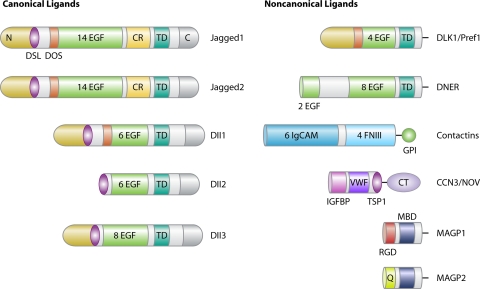
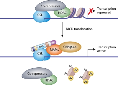
Notch receptors are transmembrane receptors that regulate cell fate decisions. There are four Notch receptors in mammals. Upon binding to members of the Delta and Jagged family of transmembrane proteins, Notch is cleaved and the Notch intracellular domain (NICD) is released. NICD then translocates to the nucleus, where it associates with the CBF-1, Suppressor of Hairless, and Lag-2 (CSL) and Mastermind-Like (MAML) proteins. This complex activates the transcription of Notch target genes, such as Hairy Enhancer of Split (Hes) and Hes-related with YRPF motif (Hey). Notch signaling is critical for the regulation of mesenchymal stem cell differentiation. Misexpression of Notch in skeletal tissue indicates a role as an inhibitor of skeletal development and postnatal bone formation. Overexpression of Notch inhibits endochondral bone formation and osteoblastic differentiation, causing severe osteopenia. Conditional inactivation of Notch in the skeleton causes an increase in cancellous bone volume and enhanced osteoblastic differentiation. Notch ligands are expressed in the hematopoietic stem cell niche and are critical for the regulation of hematopoietic stem cell self-renewal. Dysregulation of Notch signaling is the underlying cause of diseases affecting the skeletal tissue, including Alagille syndrome, spondylocostal dysostosis, and possibly, osteosarcoma.
Figures


Similar articles
-
Notch regulation of bone development and remodeling and related skeletal disorders.Calcif Tissue Int. 2012 Feb;90(2):69-75. doi: 10.1007/s00223-011-9541-x. Epub 2011 Oct 16. Calcif Tissue Int. 2012. PMID: 22002679 Free PMC article. Review.
-
Notch Signaling and the Skeleton.Endocr Rev. 2016 Jun;37(3):223-53. doi: 10.1210/er.2016-1002. Epub 2016 Apr 13. Endocr Rev. 2016. PMID: 27074349 Free PMC article. Review.
-
Notch signaling in skeletal health and disease.Eur J Endocrinol. 2013 May 8;168(6):R95-103. doi: 10.1530/EJE-13-0115. Print 2013 Jun. Eur J Endocrinol. 2013. PMID: 23554451 Free PMC article. Review.
-
Role of the RAM domain and ankyrin repeats on notch signaling and activity in cells of osteoblastic lineage.J Bone Miner Res. 2006 Aug;21(8):1317-26. doi: 10.1359/jbmr.060505. J Bone Miner Res. 2006. PMID: 16869730
-
HES and HERP families: multiple effectors of the Notch signaling pathway.J Cell Physiol. 2003 Mar;194(3):237-55. doi: 10.1002/jcp.10208. J Cell Physiol. 2003. PMID: 12548545 Review.
Cited by
-
Jagged1 expression by osteoblast-lineage cells regulates trabecular bone mass and periosteal expansion in mice.Bone. 2016 Oct;91:64-74. doi: 10.1016/j.bone.2016.07.006. Epub 2016 Jul 12. Bone. 2016. PMID: 27416809 Free PMC article.
-
Breast tumor cells promotes the horizontal propagation of EMT, stemness, and metastasis by transferring the MAP17 protein between subsets of neoplastic cells.Oncogenesis. 2020 Oct 26;9(10):96. doi: 10.1038/s41389-020-00280-0. Oncogenesis. 2020. PMID: 33106480 Free PMC article.
-
CCN3 and DLL1 co-regulate osteogenic differentiation of mouse embryonic fibroblasts in a Hey1-dependent manner.Cell Death Dis. 2018 Dec 11;9(12):1188. doi: 10.1038/s41419-018-1234-1. Cell Death Dis. 2018. PMID: 30538222 Free PMC article.
-
Notch regulation of bone development and remodeling and related skeletal disorders.Calcif Tissue Int. 2012 Feb;90(2):69-75. doi: 10.1007/s00223-011-9541-x. Epub 2011 Oct 16. Calcif Tissue Int. 2012. PMID: 22002679 Free PMC article. Review.
-
A Novel Organ Culture Model of Mouse Intervertebral Disc Tissues.Cells Tissues Organs. 2016;201(1):38-50. doi: 10.1159/000439268. Epub 2015 Oct 9. Cells Tissues Organs. 2016. PMID: 26447649 Free PMC article.
References
-
- Androutsellis-Theotokis, A., R. R. Leker, F. Soldner, D. J. Hoeppner, R. Ravin, S. W. Poser, M. A. Rueger, S. K. Bae, R. Kittappa, and R. D. McKay. 2006. Notch signalling regulates stem cell numbers in vitro and in vivo. Nature 442:823-826. - PubMed
-
- Artavanis-Tsakonas, S., M. D. Rand, and R. J. Lake. 1999. Notch signaling: cell fate control and signal integration in development. Science 284:770-776. - PubMed
-
- Bae, S., Y. Bessho, M. Hojo, and R. Kageyama. 2000. The bHLH gene Hes6, an inhibitor of Hes1, promotes neuronal differentiation. Development 127:2933-2943. - PubMed
-
- Bai, S., R. Kopan, W. Zou, M. J. Hilton, C. T. Ong, F. Long, F. P. Ross, and S. L. Teitelbaum. 2008. NOTCH1 regulates osteoclastogenesis directly in osteoclast precursors and indirectly via osteoblast lineage cells. J. Biol. Chem. 283:6509-6518. - PubMed
Publication types
MeSH terms
Substances
Grants and funding
LinkOut - more resources
Full Text Sources
Other Literature Sources