. 2010 Feb;84(4):2001-12.
doi: 10.1128/JVI.01791-09. Epub 2009 Nov 25.

Fusion-deficient insertion mutants of herpes simplex virus type 1 glycoprotein B adopt the trimeric postfusion conformation

Affiliations

Fusion-deficient insertion mutants of herpes simplex virus type 1 glycoprotein B adopt the trimeric postfusion conformation

Jessica L Silverman et al. J Virol. 2010 Feb.

Abstract

Glycoprotein B (gB) enables the fusion of viral and cell membranes during entry of herpesviruses. However, gB alone is insufficient for membrane fusion; the gH/gL heterodimer is also required. The crystal structure of the herpes simplex virus type 1 (HSV-1) gB ectodomain, gB730, has demonstrated similarities between gB and other viral fusion proteins, leading to the hypothesis that gB is a fusogen, presumably directly involved in bringing the membranes together by refolding from its initial or prefusion form to its final or postfusion form. The only available crystal structure likely represents the postfusion form of gB; the prefusion form has not yet been determined. Previously, a panel of HSV-1 gB mutants was generated by using random 5-amino-acid-linker insertion mutagenesis. Several mutants were unable to mediate cell-cell fusion despite being expressed on the cell surface. Mapping of the insertion sites onto the crystal structure of gB730 suggested that several insertions might not be accommodated in the postfusion form. Thus, we hypothesized that some insertion mutants were nonfunctional due to being "trapped" in a prefusion form. Here, we generated five insertion mutants as soluble ectodomains and characterized them biochemically. We show that the ectodomains of all five mutants assume conformations similar to that of the wild-type gB730. Four mutants have biochemical properties and overall structures that are indistinguishable from those of the wild-type gB730. We conclude that these mutants undergo only minor local conformational changes to relieve the steric strain resulting from the presence of 5 extra amino acids. Interestingly, one mutant, while able to adopt the overall postfusion structure, displays significant conformational differences in the vicinity of fusion loops, relative to wild-type gB730. Moreover, this mutant has a diminished ability to associate with liposomes, suggesting that the fusion loops in this mutant have decreased functional activity. We propose that these insertions cause a fusion-deficient phenotype not by preventing conversion of gB to a postfusion-like conformation but rather by interfering with other gB functions.


Figures
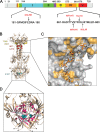
FIG. 1.
FIG. 1.
Location of the insertion sites in the sequence of gB and the structure of the postfusion form of its ectodomain. (A) Linear diagram of the full-length gB with functional domains highlighted (as in reference 18). Domain I is shown in cyan, domain II in green, domain III in yellow, domain IV in orange, domain V in red, and the disordered region between domains II and III in purple. Regions absent from the crystal structure of gB730 are shown in gray. Sequences in the region of 5-amino-acid insertions (residues 181 to 190 and residues 661 to 680) are shown in black. Arrows mark the locations of 5-amino-acid insertions, shown as red text. (B) Crystal structure of gB730 (18). Residues preceding the 5-amino-acid insertions in mutants studied here are shown as spheres colored by domain, consistent with panel A. Boxes delineate the hinge region, enlarged in panel C, and the cavity region, enlarged in panel D. (C) Close-up view of the hinge region shown in molecular surface representation, with residues 663 to 675 displayed as sticks. Hydrophobic residues are colored orange. Residues preceding the 5-amino-acid insertions in mutants studied here are labeled with asterisks; remaining labels correspond to additional hydrophobic residues in the 663-675 region. (D) Enlarged view of the cavity region. Residues that line the cavity and are not solvent exposed are colored magenta. Residue E187 of each protomer is colored teal and shown as spheres. Fusion loops for two protomers are marked with asterisks; the third pair of fusion loops lies behind the crystal structure and is not visible. Panels B, C, and D were made by using Pymol (http://www.pymol.org/).
FIG. 2.
FIG. 2.
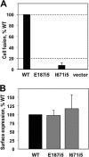
Characterization of the fusion phenotype of the insertion mutants. (A) Quantitative cell-cell fusion assay. Target CHO-K1 cells, expressing the luciferase protein and the HSV receptor HVEM, were cocultivated with effector cells expressing T7 polymerase, gD, gH, gL, and either wild-type (WT) gB, mutant, or the empty vector, and assayed for light production 20 h later. Extent of fusion is expressed as percentage WT. (B) FACS assay to determine surface expression of full-length WT and mutant gB proteins. Mouse melanoma C10 cells were transfected with either WT gB, or mutant, or the empty vector. Cells were stained with a primary antibody R68, a secondary antibody goat anti-rabbit-phycoerythrin, fixed with paraformaldehyde, and subjected to FACS analysis. In the above graph, the signal from C10 cells that were transfected with empty vector DNA was subtracted out. Error bars represent the standard error. The mutant gB proteins I671i5 and E187i5 were expressed on the cell surface, as well as the wild-type gB protein.
FIG. 3.
FIG. 3.
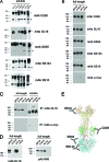
gB insertion mutant proteins are similarly recognized by different antibodies, with the exception of E187i5. (A) Purified protein samples were resolved on mildly denaturing 4 to 15% Tris gradient gels and visualized by Western blot analysis using different anti-gB MAbs, as indicated under each blot. Wild-type gB ectodomain, gB730, was used as a positive control in all blots. For each blot and gel in this figure, molecular mass markers are indicated on the left in kilodaltons. “T” stands for trimeric and “M” stands for monomeric gB species. (B) WT gB and the E187i5 and I671i5 mutants were expressed as full-length proteins in C10 cells and immunoprecipitated (IP) with different anti-gB MAbs. IP samples were then resolved on 10% polyacrylamide gels and visualized by Western blot analysis with a polyclonal anti-gB antibody, R68. IP results for anti-myc antibody are shown as a negative control. (C) Recognition of full-length gB versus soluble gB ectodomains by MAb DL16 was tested by IP. IP samples of full-length proteins were prepared as in panel B. Purified soluble ectodomains were diluted in IP binding buffer and immunoprecipitated with DL16. The results of the IP were analyzed by Western blot analysis with R68, as in panel B. IP results for anti-myc antibody are shown as a negative control. (D) Recognition of full-length gB by MAb DL16 was tested by Western blotting. WT gB and the E187i5 and I671i5 mutants were expressed as full-length proteins in C10 cells, and whole lysates were resolved on a 10% polyacrylamide gel under mildly denaturing conditions, blotted, and probed with MAb DL16. Equivalent expression of full-length gB proteins in C10 cells was verified by Western blot analysis with PAb R68 using SDS-10% PAGE under reducing, denaturing conditions. (E) Approximate locations of the epitopes of four of the MAbs used in the present study in the crystal structure of gB730. The location of the epitope for the trimer-specific antibody DL16 is unknown.
FIG. 4.
FIG. 4.
E187i5 mutant protein elutes completely in the void volume of the size exclusion column. An overlay of the size exclusion chromatograms for the mutant and the wild-type ectodomain proteins is shown. Elution volumes of size exclusion standards are indicated with arrows above the traces.
FIG. 5.
FIG. 5.
Negative-stain EM images of wild-type gB730, T665i5 mutant, and E187i5 mutant proteins. (A) Representative particles of WT gB730 and an insertion mutant, T665i5, are shown. Two distinct “crown” and “base” ends of particles are labeled with red arrows in one representative particle. The scale bar for these images is the same as for panel C. (B) The structure of gB730 in surface representation is colored by domain as in Fig. 1A. The crown and base regions are labeled. The approximate location of residue E187 is marked with a white star. (C) Representative particles of two types are shown for the E187i5 mutant protein. Crown regions of individual trimers are labeled with red arrows. (D) Model of how higher-order oligomers are formed from gB730 trimers in the E187i5 mutant, as depicted in panel C. Dimers of trimers and trimers of trimers are shown. The approximate locations of residue E187 in all structures are marked with a white star, as in panel B.
FIG. 6.
FIG. 6.
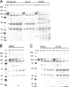
Limited proteolysis reveals a new chymotrypsin site in the E187i5 mutant. Wild-type gB730 and five insertion mutants were digested with chymotrypsin at decreasing mass ratios of substrate to protease, indicated above each lane. ND, nondigested. For each gel in this figure, molecular mass markers are indicated on the left in kilodaltons. (A) Digestion pattern of wild-type gB730, the I671i5 mutant, and the E187i5 mutant. (B) Digestion pattern of the T665i5 mutant. (C) Digestion pattern of the V667i5 and L673i5 mutants. Asterisks indicate proteolytic products that are seen in the wild-type gB730 and all of the mutants. Arrows in the E187i5 digestion pattern indicate new bands absent from the digests of the wild-type gB730 and the rest of the mutants. Weak bands corresponding to chymotrypsin autoproteolysis products are observed at 15 and 10 kDa in some of the digests at 25:1 and 10:1 substrate/protease ratios.
FIG. 7.
FIG. 7.
Newly identified proteolytic cleavage site in E187i5 mutant is not exposed in the wild-type gB730. (A) Crystal structure of gB730 is shown as a beige ribbon with residues E187 in all three protomers shown as orange spheres. Residues F223 and H224, the site of chymotrypsin cleavage, are shown as pink sticks. The box delineates the region enlarged in panel B. (B) Close-up view of the region boxed in panel A, rotated 90° clockwise relative to the view in panel A and shown in molecular surface representation. Residues E187 for two protomers are shown as orange spheres and labeled with arrows; the third E187 residue is situated behind the molecule and is not visible. Residues F223 and H224 for one protomer are shown as magenta sticks. Only the tip of the F223 side chain is exposed, shown as magenta surface. The figure was prepared by using Pymol (http://www.pymol.org/).
FIG. 8.
FIG. 8.
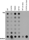
Binding of insertion mutants to liposomes. Purified soluble wild-type (WT) or mutant proteins were incubated with phosphatidylcholine/cholesterol liposomes at 37°C for 1 h. Each sample was then incubated in 1 M KCl for 15 min, adjusted to 40% sucrose, and layered beneath a sucrose step gradient. Gradients were centrifuged for 3 h, fractionated, and analyzed by immuno-dot blot using anti-gB rabbit polyclonal antibody R69. The E187i5 mutant has impaired liposome binding, whereas the two hinge mutants (T665i5 and I671i5) cofloat with liposomes as efficiently as wild-type gB730.

Similar articles

Cited by

References

    1. Atanasiu, D., J. C. Whitbeck, T. M. Cairns, B. Reilly, G. H. Cohen, and R. J. Eisenberg. 2007. Bimolecular complementation reveals that glycoproteins gB and gH/gL of herpes simplex virus interact with each other during cell fusion. Proc. Natl. Acad. Sci. U. S. A. 104:18718-18723. - PMC - PubMed
    1. Avitabile, E., C. Forghieri, and G. Campadelli-Fiume. 2007. Complexes between herpes simplex virus glycoproteins gD, gB, and gH detected in cells by complementation of split enhanced green fluorescent protein. J. Virol. 81:11532-11537. - PMC - PubMed
    1. Backovic, M., and T. S. Jardetzky. 2009. Class III viral membrane fusion proteins. Curr. Opin. Struct. Biol. 19:189-196. - PMC - PubMed
    1. Backovic, M., R. Longnecker, and T. S. Jardetzky. 2009. Structure of a trimeric variant of the Epstein-Barr virus glycoprotein B. Proc. Natl. Acad. Sci. U. S. A. 106:2880-2885. - PMC - PubMed
    1. Bender, F. C., M. Samanta, E. E. Heldwein, M. P. de Leon, E. Bilman, H. Lou, J. C. Whitbeck, R. J. Eisenberg, and G. H. Cohen. 2007. Antigenic and mutational analyses of herpes simplex virus glycoprotein B reveal four functional regions. J. Virol. 81:3827-3841. - PMC - PubMed

Publication types

MeSH terms

Substances

LinkOut - more resources