. 2009 Nov 18;4(11):e7913.
doi: 10.1371/journal.pone.0007913.

Anthrax lethal toxin induced lysosomal membrane permeabilization and cytosolic cathepsin release is Nlrp1b/Nalp1b-dependent

Affiliations

Anthrax lethal toxin induced lysosomal membrane permeabilization and cytosolic cathepsin release is Nlrp1b/Nalp1b-dependent

Kathleen M Averette et al. PLoS One. .

Abstract

NOD-like receptors (NLRs) are a group of cytoplasmic molecules that recognize microbial invasion or 'danger signals'. Activation of NLRs can induce rapid caspase-1 dependent cell death termed pyroptosis, or a caspase-1 independent cell death termed pyronecrosis. Bacillus anthracis lethal toxin (LT), is recognized by a subset of alleles of the NLR protein Nlrp1b, resulting in pyroptotic cell death of macrophages and dendritic cells. Here we show that LT induces lysosomal membrane permeabilization (LMP). The presentation of LMP requires expression of an LT-responsive allele of Nlrp1b, and is blocked by proteasome inhibitors and heat shock, both of which prevent LT-mediated pyroptosis. Further the lysosomal protease cathepsin B is released into the cell cytosol and cathepsin inhibitors block LT-mediated cell death. These data reveal a role for lysosomal membrane permeabilization in the cellular response to bacterial pathogens and demonstrate a shared requirement for cytosolic relocalization of cathepsins in pyroptosis and pyronecrosis.


Conflict of interest statement

Competing Interests: The authors have declared that no competing interests exist.

Figures

Figure 1
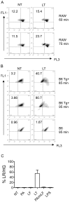
Figure 1. LT causes LMP in LTS macrophages.
(A) RAW 264.7 (RAW) cells pretreated with AO and subjected to LT (3 µg/mL LF and 1 µg/mL PA) for 60 or 75 minutes or media alone (NT). Cells were analyzed by flow cytometry, live cells were gated based on forward and side scatter and cells were analyzed for red (FL3) and green (FL1) fluorescence. Cells are depicted here as a density plot. Upper left quadrants represent cells with low red and high green fluorescence (LR/HG). Numbers correspond to percent of cell population in LR/HG quadrant. (B) C57BL/6Nlrp1b(129S1) BMDMs (B6 Tg+) or littermate controls (B6) were pretreated with AO and subjected to either LT (1 µg/mL LF and 1 µg/mL PA) for 85 or 95 minutes or media alone (NT). Cells were analyzed as in (A). Density plot represent BMDMs from one of three C57BL/6Nlrp1b(129S1) or C57BL/6 littermate controls and are representative of results obtained. (C) C57BL/6Nlrp1b(129S1) BMDMs were treated with 1 µg/mL of LF, PA, LF and PA (LT), PA and LF-H719C (PA/mLF), or 10 ng/mL of lipopolysaccharide (LPS) for 90 min. Cells were collected and analyzed for red and green fluorescence as in (A). BMDMs from three C57BL/6Nlrp1b(129S1) were used for each condition and error bars represent standard deviation.
Figure 2
Figure 2. Lysosome ultrastructure appears unaltered during Nlrp1b-mediated pyroptosis.
(A) RAW 264.7 cells were pre-stained with Lysotracker Red DND-99 followed by LT or untreated (NT) for 75 min and imaged on glass slides at 40× magnification. Black arrows correspond to condensed nuclear DNA observed in pyroptotic cells.
Figure 3
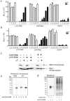
Figure 3. CtsB is active in the cytosol and inhibition of cathepsin activity blocks LT-mediated pyroptosis.
(A) C57BL/6Nlrp1b(129S1) BMDMs were pre-treated with varying concentrations of z-FA-FMK, CA074Me, or CA074 for 4 hours, followed by addition of 400 ng/mL of PA and 300 ng/mL of LF (LT), or no treatment (NT), for an additional 3.5 hours. Cytotoxicity was measured using ATP-lite. BMDMs from three C57BL/6Nlrp1b(129S1) were used with each condition tested in quadruplicate. Error bars represent standard deviation. (B) RAW 264.7 cells were pre-treated with varying concentrations of z-FA-FMK, CA074Me, or CA074 for 4 hours, followed by addition of 400 ng/mL of PA and 300 ng/mL of LF (LT), or no treatment (NT), for an additional 3.5 hours. Cytotoxicity was measured using ATP-lite. The data presented here are representative of three or more independent experiments. Each point represents the mean of triplicate or quadruplicate samples from a single experiment, with error bars representing standard error. (C) RAW 264.7 cells were pre-treated with 100 µM z-FA-FMK or 50 µM CA074Me for 4 hours followed by LT (400 ng/mL PA and 300 ng/mL LF; +) or no toxin (−) for 2.5 hours. Cell lysates were subjected to western blot analysis and probed with an antibody that recognizes the N-terminus of MEK2. (D) Specificity of the ctsB probe Ak-EVD-AMK was determined by treating RAW 264.7 or NIH 3T3 cells with or without high concentration (5 or 10 µM) of probe. Cycloaddition assays were performed on Ak-EVD-AMK labeled cellular lysates with azido-rhodamine, and Ak-EVD-AMK labeled proteins were analyzed by in-gel fluorescence. The pro-form of ctsB is 43 kDa, whereas active ctsB is seen as either 31 kDa or 25 kDa. (E) In-gel fluorescence showing electrophoretically separated proteins from RAW 264.7 cells treated with 400 ng/mL of PA and 300 ng/mL of LF and/or, low dose (625 nM) Ak-EVD-AMK. Under these conditions, cytosolic ctsB is preferentially labeled. Cycloaddition was preformed as in (D). The gel was then stained with coomassie to indicate equal sample loading.
Figure 4
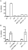
Figure 4. Heat shock, proteasome and ctsB/L inhibition, but not potassium chloride, prevents LT-induced LMP.
(A) C57BL/6Nlrp1b(129S1) BMDMs were heat shocked at 42°C (HS) for 15 min prior to addition of LT (1 µg/mL of PA and 500 ng/mL of LF) for 90 min. Proteasome inhibition was accomplished with co-incubation of cells with 10 µM MG-132 and either LT (1 µg/mL of PA and 500 ng/mL of LF) or PA only for 90 min. Cells were collected for flow cytometry and % LR/HG was determined as in Figure 1A. Experiments were preformed using BMDMs from three C57BL/6Nlrp1b(129S1) and samples were collected in triplicate. Error bars represent standard deviation. (B) In a separate experiment C57BL/6Nlrp1b(129S1) BMDMs were pretreated with LT (400 ng/mL PA and 300 ng/mL LF) or without toxin (NT) for 2.5 hours. Cells were also either pre-incubated with 50 µM CA074Me for 4 hours or co-treated with 150 mM potassium chloride (KCl) followed by LT. Cells were collected for flow cytometry and % LR/HG was determined as in Figure 1A. Experiments were preformed using BMDMs from three C57BL/6Nlrp1b(129S1) and samples were collected in triplicate. Error bars represent standard deviation.
Figure 5
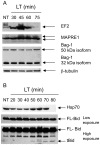
Figure 5. Proteomic changes in LT-treated cells.
Western blots showing electrophoretically separated proteins from RAW 264.7 cells treated with LT for various time points, or untreated (NT). Arrows indicate protein isoforms detected using protein-specific primary antibodies and fluorescently labeled secondary antibodies. (A) Identical cellular lysates were subjected to SDS-PAGE, transferred to PVDF and probed with different primary and secondary antibodies. β-tubulin was used as an equal loading control in each experiment. This blot represents one of three independent experiments showing similar results. (B) Identical cellular lysates were subjected to SDS-PAGE, transferred to PVDF and probed with different primary and secondary antibodies. Anti-Bid antibody recognizes both full-length Bid (FL-Bid) and the truncated form (tBid). The membrane was exposed to film for 1 sec (low exposure) or 45 sec (high exposure) to detect both FL-Bid and tBid. tBid protein surfaces at 50 min following LT treatment at high toxin concentrations. This blot represents one of three independent experiments showing similar results.
Figure 6
Figure 6. Model of LF internalization and activation of LMP and MOMP.
LF binds oligomerized PA pre-pore and is internalized into endosomes where acidic pH triggers PA to form a pore in the endosomal membrane and translocate LF into the host cytosol. Following translocation, LF cleaves MKKs and induces Nlrp1-dependent pyroptosis. LF directly, or indirectly, causes LMP, resulting in release of cathepsins into the cytosol. Cathepsins or LMP-mediated signaling may directly activate the inflammasome. Alternatively, LMP may occur downstream of inflammasome activation, potentially through caspase-1 mediated cleavage of Bid. Activation of caspase-1 or cytosolic release of cathepsins can result in cleavage of Bid and a positive feedback amplification of LMP, inflammasome activation and mitochondrial outer membrane permeabilization (MOMP).

Similar articles

Cited by

References

    1. Franchi L, Warner N, Viani K, Nunez G. Function of Nod-like receptors in microbial recognition and host defense. Immunol Rev. 2009;227:106–128. - PMC - PubMed
    1. Franchi L, Eigenbrod T, Munoz-Planillo R, Nunez G. The inflammasome: a caspase-1-activation platform that regulates immune responses and disease pathogenesis. Nat Immunol. 2009;10:241–247. - PMC - PubMed
    1. McIntire CR, Yeretssian G, Saleh M. Inflammasomes in infection and inflammation. Apoptosis 2009 - PubMed
    1. Bergsbaken T, Fink SL, Cookson BT. Pyroptosis: host cell death and inflammation. Nat Rev Microbiol. 2009;7:99–109. - PMC - PubMed
    1. Suzuki T, Franchi L, Toma C, Ashida H, Ogawa M, et al. Differential regulation of caspase-1 activation, pyroptosis, and autophagy via Ipaf and ASC in Shigella-infected macrophages. PLoS Pathog. 2007;3:e111. - PMC - PubMed

Publication types