. 2009 Dec 16;28(24):3910-20.
doi: 10.1038/emboj.2009.338.

Structure of the zinc-bound amino-terminal domain of the NMDA receptor NR2B subunit

Affiliations

Structure of the zinc-bound amino-terminal domain of the NMDA receptor NR2B subunit

Erkan Karakas et al. EMBO J. .

Abstract

N-methyl-D-aspartate (NMDA) receptors belong to the family of ionotropic glutamate receptors (iGluRs) that mediate the majority of fast excitatory synaptic transmission in the mammalian brain. One of the hallmarks for the function of NMDA receptors is that their ion channel activity is allosterically regulated by binding of modulator compounds to the extracellular amino-terminal domain (ATD) distinct from the L-glutamate-binding domain. The molecular basis for the ATD-mediated allosteric regulation has been enigmatic because of a complete lack of structural information on NMDA receptor ATDs. Here, we report the crystal structures of ATD from the NR2B NMDA receptor subunit in the zinc-free and zinc-bound states. The structures reveal the overall clamshell-like architecture distinct from the non-NMDA receptor ATDs and molecular determinants for the zinc-binding site, ion-binding sites, and the architecture of the putative phenylethanolamine-binding site.


Conflict of interest statement

The authors declare that they have no conflict of interest.

Figures

Figure 1
Figure 1
Isolated NR2B ATD proteins bind zinc. (A) Domain organization of the NR2B subunit. ATD in cyan (R1 domain) and yellow (R2 domain) binds allosteric modulators including zinc and phenylethanolamine, S1S2 binds neurotransmitter L-glutamate, the transmembrane domain (TM) and the P-loop (arrow) form the ion channel pore, and the C-terminal domain (CTD) binds postsynaptic molecules and mediates intracellular signalling. NR2B ATD can be isolated and recombinantly expressed in insect cells. (B) ITC analysis of zinc binding to the NR2B ATD protein. Upper panel, calorimetric titration of 0.8 mM ZnCl2 into 0.02 mM NR2B ATD. Lower panel, integrated heat as a function of Zn2+/protein molar ratio with experimental data (filled squares) and the best fit (solid line). The Kd of zinc binding is calculated to be 5.5 μM.
Figure 2
Figure 2
Overall architecture of NR2B ATD. (A) Ribbon representation of the NR2B ATD structure in complex with zinc. The clamshell-like architecture of NR2B ATD is composed of two domains, R1 (cyan) and R2 (yellow), defined as residues 32–147 and 287–359 and residues 148–286 and 360–394, respectively. Hypervariable loop (HVL; in magenta) is oriented by a disulfide bond so that it covers the ‘top' of the R1 domain. The disordered region (residues 208–214) between β7 and α5 is indicated with a dashed line. Stick representations are used to show the disulfide bond (Cys 86–Cys 321), and sugar molecules attached to Asn 74 and Asn 341. Residues involved in zinc inhibition, phenylethanolamine inhibition, and ion binding are represented by balls and sticks in blue, orange, and green, respectively. (B) View of the NR2B ATD structure from the ‘entrance' of the clamshell.
Figure 3
Figure 3
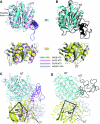
NR2B ATD has a different bi-lobe orientation from non-NMDA receptor ATD or mGluR1 LBD. (A) Superposition of the R1 (cyan, upper panel) and R2 (yellow, lower panel) domains of NR2B ATD with the equivalent domains of GluR2 ATD (pink, PDB code: 3H5V, chain A) and GluR6 ATD (light blue, PDB code: 3H6G, chain B). The regions of GluR2 ATD (Helix 8) and GluR6 ATD (Helix L), which are absent in NR2B ATD, are highlighted in magenta and blue, respectively. Disulfide bonds are shown in sticks (arrow). The root-mean-square deviations are 2.37 Å (R1, 152 Cαs) and 2.17 Å (R2, 142 Cαs) and 2.18 Å (R1, 152 Cαs) and 1.85 Å (R2, 142 Cαs) for GluR2 and GluR6 ATDs, respectively. (B) Superposition of NR2B ATD domains with the equivalent domains of mGluR1 LBD (grey, PDB code: 1EWK, chain A). The regions of mGluR1 LBD, which are absent in NR2B ATD, are coloured in black. The root-mean-square deviations are 2.4 Å (R1, 150 Cαs) and 2.0 Å (R2, 143 Cαs). (C) Superposition of the R1 domains from GluR2 and GluR6 ATDs onto the R1 domain of NR2B ATD illustrates a large difference in the pattern of the R1–R2 inter-domain orientation. The R2 domain of NR2B ATD is rotated by ∼45 and ∼54° relative to the R2 domains of GluR2 and GluR6 ATDs, respectively. The reference rods (in black) for the angle measurement are drawn from the pivotal point of the rotation (NR2B Pro 148, GluR2 Asp 131 and GluR6 Asp 152) to the last residue of β7 in all three structures. (D) The same manipulation as in (C) illustrates the ‘twisted' R1–R2 inter-domain orientation of NR2B ATD compared with mGluR1 LBD by ∼50o. The two reference rods for the angle measurements in mGluR1 LBD are drawn from Pro 206 to the end of β8.
Figure 4
Figure 4
Surface presentation of NR2B ATD and non-NMDA receptor ATDs. Surface presentation is coloured by hydrophobicity. (A) The R1 domains of NR2B, GluR2, and GluR6 ATDs are superimposed and viewed onto the dimer interface from the same angle. GluR6 ATD has a notable hydrophobic patch composed of five hydrophobic residues in R2, whereas NR2B ATD has a smaller hydrophobic patch formed by Tyr 175, Tyr 179, and Leu 204. (B) The surface presentation viewed from the ‘bottom' of the R2 domain. Unlike non-NMDA receptor ATDs, NR2B ATD contains a large hydrophobic patch composed of Tyr 164, Trp 166, Ile 168, Phe 194, Val 195, Pro 226, Ile 227, Tyr 389, Val 390, and Trp 391. Hydrophobicity was calculated using the hotpatch server (http://hotpatch.mbi.ucla.edu/). The surface was coloured from hydrophobic to hydrophilic in a dark orange to white gradient.
Figure 5
Figure 5
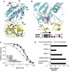
Zinc-binding site. (A) Crystal structure of NR2B ATD in complex with zinc coloured as cyan (R1) and yellow (R2). The mesh in magenta represents the zinc anomalous difference map contoured at 6.0 σ. There are five zinc molecules identified per asymmetric unit (Zn1–5). Zn4 and Zn4′ are related by crystallographic symmetry. (B) Zoom up view of the Zn1 site. Zn1 is directly coordinated by the His 127 and Glu 284 side chains. Residues including Glu 47 and Asp 265 are too far from Zn1 (>5 Å) to make direct contacts but may be involved in placing water molecules around Zn1 through hydrogen bonds. Side chain of Asp 102 beyond β carbon is disordered in the zinc-bound structure. (C) Mutagenesis and the zinc dose–response curve showing an involvement of His 127 and Glu 284 in zinc sensitivity. Mutation of residues directly involved in the Zn1 coordination (His 127 and Glu 284) has significant effect on zinc sensitivity, whereas that of non-contacting residue, Asp 283, has minor effect. All of the recordings were done at the holding potential of −40 mV. The wild-type response is shown by a dotted line. The error bars represent standard deviation for recordings from at least four different oocytes. (D) Fold difference in zinc and ifenprodil sensitivities in each mutant. Mutation of the Zn1 site residues (His 127 and Glu 284) has a major effect on zinc sensitivity but little or no effect on ifenprodil sensitivity.
Figure 6
Figure 6
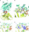
Structure of NR2B ATD in the zinc-free form. (A) The crystal structure of NR2B ATD in the absence of zinc at 2.8 Å. The structure is coloured as in Figure 2. The red mesh represents Fo-Fc omit map at 5.0 σ for one Na ion and three chloride ions (Cl1–3) at the clamshell cleft. (B) The zoom up view of the ion-binding site at the clamshell cleft. Na and Cl ions are coloured as purple and green, respectively. Residues involved in ion binding, zinc binding, and phenylethanolamine binding are coloured in green, blue, and orange, respectively. (C) Anomalous difference map at 3.3 σ calculated from data collected on Rb soaked crystals showing that the density has an overlap with the Na-binding site. (D) Anomalous difference map at 4.2 σ calculated from data collected on Br soaked crystals showing that Br can substitute all of the Cl sites at the cleft.
Figure 7
Figure 7
Hydrophobic pocket is the critical structural locus for ifenprodil sensitivity. (A) Hydrophobic residues responsible for ifenprodil sensitivity are clustered at the inner core of the NR2B ATD clamshell (in orange). Residues that have been mutated but had only minor effect are coloured in grey. (B) Dose response of ifenprodil showing the critical involvement of Ile 133 in ifenprodil inhibition. Mutation of Ile 133 to alanine or serine causes dramatic shift in ifenprodil sensitivity, whereas mutation of Tyr 356 to alanine has only minor effect. Data points for both wild type and Tyr356Ala are fit to a single-site model, whereas those for the Ile 133 mutants are fit to a two-site model described in ‘Materials and methods.' The IC50 values calculated by the fits are listed in Supplementary Table SIII. The wild-type response is shown as a dotted line. All the currents were measured at the holding potential of −20 mV to minimize voltage-dependent pore block by ifenprodil.

Similar articles

Cited by

References

    1. Adams PD, Grosse-Kunstleve RW, Hung LW, Ioerger TR, McCoy AJ, Moriarty NW, Read RJ, Sacchettini JC, Sauter NK, Terwilliger TC (2002) PHENIX: building new software for automated crystallographic structure determination. Acta Crystallogr D Biol Crystallogr 58: 1948–1954 - PubMed
    1. Alarcon K, Martz A, Mony L, Neyton J, Paoletti P, Goeldner M, Foucaud B (2008) Reactive derivatives for affinity labeling in the ifenprodil site of NMDA receptors. Bioorg Med Chem Lett 18: 2765–2770 - PubMed
    1. Armstrong N, Gouaux E (2000) Mechanisms for activation and antagonism of an AMPA-sensitive glutamate receptor: crystal structures of the GluR2 ligand binding core. Neuron 28: 165–181 - PubMed
    1. Choi YB, Lipton SA (1999) Identification and mechanism of action of two histidine residues underlying high-affinity Zn2+ inhibition of the NMDA receptor. Neuron 23: 171–180 - PubMed
    1. Clayton A, Siebold C, Gilbert RJ, Sutton GC, Harlos K, McIlhinney RA, Jones EY, Aricescu AR (2009) Crystal structure of the GluR2 amino-terminal domain provides insights into the architecture and assembly of ionotropic glutamate receptors. J Mol Biol 392: 1125–1132 - PubMed

Publication types